Part of the Master Guide

Complex Revision Total Knee Arthroplasty: Addressing Instability, Loosening & Wear

Periprosthetic Joint Infection in Total Knee Arthroplasty: Epidemiology, Risk Factors, and Foundational Principles

17 Apr 2026 16 min read 191 Views
infected TKR

Key Takeaway

Periprosthetic joint infection (PJI) following total knee arthroplasty (TKA) is a devastating complication characterized by microorganisms in periprosthetic tissue. Its incidence ranges from 0.5-2%, escalating with revision. PJI is influenced by patient, intraoperative, and postoperative risk factors, necessitating a deep understanding of knee anatomy and biomechanics for accurate diagnosis and management.
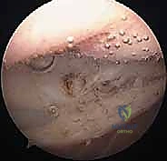
Introduction and Epidemiology

Periprosthetic joint infection following total knee arthroplasty represents one of the most devastating complications in orthopedic surgery, significantly impacting patient quality of life, functional outcomes, and healthcare costs. Despite advancements in surgical technique, sterile protocols, and antibiotic prophylaxis, the incidence of periprosthetic joint infection following primary total knee arthroplasty ranges from 0.5% to 2%, with rates escalating to 3% to 5% for revision arthroplasty. The global burden is substantial, with projections indicating a continued exponential rise in total knee arthroplasty procedures and, consequently, a proportional increase in infection cases.

Periprosthetic joint infection is characterized by the presence of microorganisms in the periprosthetic tissue, leading to an inflammatory response and potential implant loosening or failure. The pathogenesis is fundamentally driven by biofilm formation. Planktonic bacteria adhere to the inert implant surface and rapidly transition into a sessile state, encasing themselves in a protective extracellular polymeric substance or glycocalyx. This biofilm confers immense resistance to both host immune responses and systemic antimicrobial therapy, rendering minimal inhibitory concentration values derived from standard in vitro testing largely obsolete for eradication.

The infection can manifest acutely, often within weeks to months post-surgery, or chronically, sometimes years after the index procedure. The Tsukayama classification categorizes these clinical presentations into four distinct types: Type I (positive intraoperative cultures during presumed aseptic revision), Type II (early postoperative infection within four weeks), Type III (acute hematogenous infection in a previously well-functioning joint), and Type IV (late chronic infection). Differentiating periprosthetic joint infection from aseptic loosening, inflammatory arthropathy, or other pain etiologies is notoriously challenging yet paramount, as misdiagnosis leads to inappropriate and often detrimental management strategies.

Risk factors for periprosthetic joint infection are multifactorial and broadly categorized into patient-related, intraoperative, and postoperative factors.

Patient-related factors include older age, obesity with a body mass index greater than 30, diabetes mellitus (especially uncontrolled with an HbA1c greater than 7%), rheumatoid arthritis, psoriasis, chronic kidney disease, malnutrition, immunodeficiency (such as HIV or chronic corticosteroid use), prior history of infection, peripheral vascular disease, smoking, and nasal colonization with Staphylococcus aureus. Intraoperative factors encompass prolonged surgical time exceeding two hours, excessive blood loss, allogeneic blood transfusion, poor aseptic technique, large skin incisions, inadequate debridement of devitalized tissue, and the use of cementless components. Postoperative factors include superficial wound infection, hematoma formation, wound dehiscence, surgical site contamination, and remote infections such as urinary tract infections or dental abscesses that precipitate hematogenous spread.

The economic implications are staggering, with treatment costs estimated to be two to five times higher than aseptic revisions, often exceeding $100,000 per case. This financial strain, coupled with prolonged hospitalization, multiple surgical interventions, and the potential for permanent disability, underscores the critical need for early and accurate diagnostic criteria to guide effective management and improve patient outcomes.

Surgical Anatomy and Biomechanics

Understanding the surgical anatomy and biomechanics of the knee joint, particularly in the context of a total knee arthroplasty, is crucial for interpreting diagnostic findings, planning interventions, and comprehending the pathogenesis of infection.

Anatomical Considerations in Infection Pathogenesis

The knee joint capsule, lined by the synovial membrane, is the primary site of early bacterial colonization and biofilm formation. Inflammation within the synovium leads to effusion, pain, and eventual degradation of periprosthetic tissues. The complex topography of the knee capsule includes multiple recesses, such as the suprapatellar pouch, the medial and lateral gutters, and the popliteal hiatus. These dependent areas frequently harbor loculated purulence and require meticulous, systematic debridement during surgical intervention. Synovial fluid analysis and synovial tissue biopsy are cornerstone diagnostic tests, requiring precise anatomical knowledge for accurate aspiration or arthroscopic biopsy.

The periprosthetic bone-implant interface represents the critical zone where implants integrate with host bone. Infection thrives in this microenvironment, instigating a host-mediated inflammatory cascade dominated by macrophages and osteoclasts. This leads to aggressive osteolysis, loosening of components, and severe compromise of structural integrity. The resulting bone loss is often asymmetric and cavitary, necessitating advanced reconstructive techniques during revision surgery.

The extensor mechanism, comprising the quadriceps muscle, patella, patellar tendon, and tibial tubercle, is frequently involved in the infectious process, either directly through contiguous spread or indirectly through multiple surgical exposures. This leads to scar tissue formation, fibrosis, and potential rupture, which drastically complicates revision efforts. Preservation of the extensor mechanism vascularity, primarily supplied by the genicular anastomosis, is paramount. Previous surgical incisions often disrupt this vascular network, increasing the risk of skin necrosis and delayed wound healing during subsequent interventions.

Biomechanics of the Infected Arthroplasty

From a biomechanical perspective, the presence of infection alters the load-sharing characteristics of the knee. As osteolysis progresses, the implant subsides, leading to varus or valgus instability and catastrophic failure of the polyethylene bearing surface. During a two-stage exchange, the placement of an antibiotic-loaded polymethylmethacrylate spacer alters joint kinematics. Articulating spacers attempt to mimic native biomechanics, maintaining collateral ligament tension and facilitating early range of motion, which prevents profound quadriceps contracture. Conversely, static spacers act essentially as a temporary arthrodesis, providing absolute stability in the setting of massive bone loss or extensor mechanism deficiency but resulting in profound soft tissue contracture that complicates the eventual second-stage reimplantation.

Indications and Contraindications

The management of periprosthetic joint infection is dictated by the chronicity of the infection, the stability of the implant, the virulence of the infecting organism, and the physiological status of the host. The McPherson staging system is frequently utilized to categorize the host (A: uncompromised, B: compromised, C: severely compromised) and the local wound environment, which guides surgical decision-making.

Surgical intervention is the definitive standard of care, as medical management alone is universally inadequate for eradicating biofilm-associated infections. The primary surgical modalities include Debridement Antibiotics and Implant Retention, single-stage exchange arthroplasty, two-stage exchange arthroplasty, and salvage procedures.

Treatment Modalities and Patient Selection

Intervention Strategy Primary Indications Contraindications
Debridement Antibiotics and Implant Retention Acute postoperative infection (less than 4 weeks); Acute hematogenous infection (less than 3 weeks of symptoms); Well-fixed implants; Intact soft tissue envelope. Chronic infections; Loose implants; Sinus tract presence; Highly virulent or resistant organisms (e.g., MRSA, VRE); Poor host immune status.
Single Stage Exchange Arthroplasty Chronic infection with a known, highly susceptible organism; Healthy host (McPherson Type A); Adequate bone stock; Intact soft tissues. Unknown organism preoperatively; Polymicrobial infection; Resistant organisms; Severe immunocompromise; Massive uncontained bone loss; Sinus tract.
Two Stage Exchange Arthroplasty Chronic periprosthetic joint infection; Loose implants; Unknown or resistant organisms; Presence of a sinus tract; Failed prior Debridement Antibiotics and Implant Retention. Medically unstable patient unfit for multiple surgeries; Severe peripheral vascular disease precluding wound healing.
Chronic Antibiotic Suppression Patient medically unfit for any surgical intervention; Well-fixed implants; Highly susceptible organism with available oral, well-tolerated antibiotics. Loose implants causing severe mechanical pain; Resistant organisms; Medication toxicity or non-compliance.
Salvage Procedures (Arthrodesis or Amputation) Recurrent, recalcitrant infection failing multiple two-stage exchanges; Massive, un-reconstructible bone loss; Complete loss of the extensor mechanism; Life-threatening sepsis. Viable reconstructive options remaining; Patient refusal; Contralateral severe limb impairment (relative contraindication for amputation).

Pre Operative Planning and Patient Positioning

Thorough preoperative planning is critical to the success of revision surgery for periprosthetic joint infection. This phase requires a multidisciplinary approach involving orthopedic surgery, infectious disease specialists, and internal medicine.

Host Optimization and Diagnostic Workup

Host optimization is the first mandatory step. Modifiable risk factors must be rigorously addressed. Glycemic control must be optimized, targeting a preoperative HbA1c of less than 7%. Nutritional status should be evaluated via serum albumin, prealbumin, and total lymphocyte count, with aggressive supplementation instituted for malnourished patients. Smoking cessation is mandatory, ideally confirmed with cotinine levels, to mitigate the risk of catastrophic wound failure.

The diagnostic workup must confirm the diagnosis and identify the offending pathogen. The Musculoskeletal Infection Society and International Consensus Meeting criteria are the gold standards. A preoperative joint aspiration is mandatory to obtain synovial fluid for cell count, differential, and aerobic, anaerobic, and fungal cultures. Advanced biomarkers such as synovial alpha-defensin and leukocyte esterase provide high sensitivity and specificity. If the patient is on systemic antibiotics, a washout period of at least two weeks is required prior to aspiration to minimize the risk of false-negative cultures.

Imaging and Templating

Radiographic evaluation begins with orthogonal weight-bearing views of the knee, a sunrise view of the patella, and full-length standing lower extremity radiographs to assess mechanical alignment and extra-articular deformities. Computed tomography is highly recommended to quantify osteolysis, evaluate the structural integrity of the epicondyles and tibial plateau, and assess the rotational profile of the existing components.

Templating involves anticipating the required level of constraint (e.g., condylar constrained knee or rotating hinge) and planning for bone loss management using the Anderson Orthopaedic Research Institute classification. The surgeon must ensure the availability of various reconstructive options, including metaphyseal cones, diaphyseal engaging stems, and structural allografts.

Patient Positioning and Preparation

The patient is positioned supine on a radiolucent operating table. A bump is placed under the ipsilateral hip to control external rotation, and a lateral post or foot positioner is utilized to allow dynamic manipulation of the knee through a full arc of motion. A pneumatic tourniquet is placed proximally on the thigh. While the use of a tourniquet is standard for visualization during explantation, it should be deflated prior to final debridement to ensure adequate assessment of tissue viability and bleeding bone.

Skin preparation must be meticulous and extensive, extending from the toes to the proximal thigh, utilizing an alcohol-based chlorhexidine solution unless contraindicated. Previous surgical incisions must be carefully marked. The surgical approach should ideally utilize the most lateral previous anterior incision to preserve the vascular supply to the anterior skin flap, as the cutaneous blood supply to the knee is predominantly medial to lateral.

Detailed Surgical Approach and Technique

The surgical execution of periprosthetic joint infection management is demanding, requiring adherence to oncologic principles of resection. The infected arthroplasty must be treated as a localized malignancy, demanding radical excision of all devitalized tissue, pseudocapsule, and foreign material.

Surgical Exposure and Internervous Planes

The standard approach is a longitudinal midline incision utilizing a medial parapatellar arthrotomy. Because the soft tissues are often scarred, inelastic, and contracted, standard exposure is frequently inadequate and risks avulsion of the patellar tendon from the tibial tubercle.

The surgeon must be facile with extensile exposures. The quadriceps snip (rectus snip) is the most common first-line extensile maneuver, involving an oblique incision directed superiorly and laterally through the rectus femoris tendon. It requires no alteration in postoperative rehabilitation and provides excellent exposure. If exposure remains inadequate, a tibial tubercle osteotomy is performed. This involves elevating a bone block 6 to 8 centimeters in length, maintaining the lateral muscular hinge, and allows for complete eversion of the extensor mechanism. A V-Y quadricepsplasty is rarely used today due to the high risk of extensor lag and profound weakness.

Debridement Antibiotics and Implant Retention Technique

For acute infections meeting indications, Debridement Antibiotics and Implant Retention begins with a thorough synovectomy. The modular polyethylene liner is universally exchanged to gain access to the posterior capsule. Meticulous debridement of the posterior recesses, medial and lateral gutters, and the suprapatellar pouch is performed. Copious pulsatile lavage with normal saline, often supplemented with dilute betadine or chlorhexidine gluconate, is utilized. The modular components are reassembled, and the wound is closed over closed-suction drains.

Two Stage Exchange Arthroplasty Stage One

The first stage of a two-stage exchange involves total explantation and spacer placement. Following exposure, multiple independent tissue samples (minimum of five) are obtained from distinct anatomical regions (e.g., synovium, posterior capsule, femoral canal, tibial canal) using separate instruments for each to prevent cross-contamination.

Implant extraction must be performed with extreme care to preserve remaining bone stock. Thin osteotomes, flexible chisels, and ultrasonic cement removal tools are utilized to disrupt the implant-bone or cement-bone interface. The femoral component is typically addressed first, followed by the tibial component. The patellar component is often left in situ if well-fixed and not grossly infected to preserve patellar bone stock, though complete removal is advocated by many if the organism is highly virulent.

Once all implants and cement mantles are removed, a radical debridement is executed. All infected granulation tissue, necrotic bone, and the entire pseudocapsule are excised until healthy, bleeding tissue is encountered. The intramedullary canals are reamed line-to-line to remove endosteal biofilm.

An antibiotic-loaded polymethylmethacrylate spacer is then fabricated. The choice of antibiotics is guided by preoperative cultures; however, an empiric combination of high-dose vancomycin (covering Gram-positive organisms including MRSA) and tobramycin or gentamicin (covering Gram-negative organisms) is standard. Typically, 3 to 4 grams of vancomycin and 3.6 to 4.8 grams of tobramycin are added per 40-gram bag of cement.

Articulating spacers can be fabricated using commercially available molds or by utilizing sterilized, explanted components as molds. The spacer is seated with a doughy cement technique to prevent deep interdigitation, ensuring ease of removal during the second stage. The joint is reduced, and stability is assessed prior to layered closure.

Two Stage Exchange Arthroplasty Stage Two

The second stage is performed after a period of systemic antibiotic therapy (typically 6 weeks) followed by an antibiotic holiday (typically 2 to 4 weeks), provided that inflammatory markers have normalized and a repeat aspiration is negative for bacterial growth.

The surgical approach utilizes the same incision. The spacer is explanted, and a repeat debridement is performed. Intraoperative frozen sections are frequently utilized; the presence of fewer than five polymorphonuclear leukocytes per high-power field in multiple distinct fields is generally indicative of infection eradication, though this metric remains controversial.

Reconstruction typically requires revision implants. Metaphyseal bone loss is managed with highly porous titanium cones or sleeves, which provide immediate structural support and biological fixation. Diaphyseal engaging stems (cemented or cementless) are utilized to bypass metaphyseal defects and offload the joint line. The level of constraint is dictated by the integrity of the collateral ligaments. A semi-constrained (condylar constrained knee) device is utilized if the collateral ligaments are competent but require balancing, whereas a rotating hinge device is mandatory in the setting of global ligamentous incompetence or severe bone loss precluding collateral ligament attachment.

Complications and Management

The surgical management of periprosthetic joint infection is fraught with complications, given the compromised host physiology, poor soft tissue envelope, and the necessity of multiple major surgical interventions.

Common Complications and Salvage Strategies

Complication Estimated Incidence Etiology and Pathogenesis Salvage and Management Strategies
Recurrent or Persistent Infection 10% - 20% Failure to eradicate biofilm; Inadequate initial debridement; Retained cement; Secondary infection with a new opportunistic organism. Repeat two-stage exchange; Suppressive antibiotic therapy if implants are stable; Salvage procedures (arthrodesis or above-knee amputation) for recalcitrant cases.
Spacer Dislocation or Fracture 5% - 15% Improper spacer sizing; Ligamentous laxity; Patient non-compliance with weight-bearing restrictions; Inadequate cement mantle thickness. Closed reduction (rarely successful); Open revision of the spacer block; Conversion to a static spacer if soft tissues are severely compromised.
Extensor Mechanism Disruption 2% - 8% Iatrogenic avulsion during exposure; Ischemic necrosis of the patellar tendon; Severe soft tissue contracture. Primary repair (high failure rate); Reconstruction using synthetic mesh (e.g., Marlex); Extensor mechanism allograft; Gastrocnemius rotational flap for soft tissue coverage.
Periprosthetic Fracture 3% - 6% Iatrogenic injury during explantation or reaming; Severe osteolysis compromising cortical integrity; Stress risers from prior hardware. Bypass with diaphyseal engaging stems; Open reduction and internal fixation with locking plates and cerclage cables; Structural allograft strut placement.
Wound Dehiscence and Necrosis 5% - 10% Multiple previous incisions; Disruption of the genicular blood supply; Malnutrition; Smoking; Excessive tension during closure. Negative pressure wound therapy; Early plastic surgery consultation for local rotational flaps (gastrocnemius) or free tissue transfer.

Post Operative Rehabilitation Protocols

Rehabilitation following surgical intervention for periprosthetic joint infection must be highly individualized, balancing the need for joint mobilization against the mechanical limitations of the reconstruction and the integrity of the soft tissue envelope.

Rehabilitation Following Debridement Antibiotics and Implant Retention

Following a successful Debridement Antibiotics and Implant Retention procedure, the rehabilitation protocol closely mirrors that of a primary total knee arthroplasty. Patients are typically allowed to weight-bear as tolerated immediately postoperatively, utilizing assistive devices as necessary. Early initiation of active and active-assisted range of motion is critical to prevent arthrofibrosis. Continuous passive motion machines may be utilized, though their long-term efficacy remains debated in the literature. Strengthening focuses on isometric quadriceps exercises, progressing to closed-kinetic chain exercises as pain and swelling subside.

Rehabilitation During the Spacer Phase (Stage One)

The rehabilitation protocol during the interval phase of a two-stage exchange is dictated by the type of spacer utilized and the structural integrity of the remaining bone.

If a static spacer is implanted, the knee is immobilized in full extension using a rigid cylinder cast or a locked hinged knee brace. Range of motion is strictly prohibited to prevent spacer displacement and soft tissue damage. Weight-bearing status is generally restricted to toe-touch or partial weight-bearing to prevent spacer fracture or catastrophic bone loss from mechanical piston action.

If an articulating spacer is utilized, the goal is to maintain soft tissue compliance and joint mobility. Patients are typically permitted to perform active-assisted range of motion exercises, aiming for 0 to 90 degrees of flexion. Weight-bearing is usually restricted to partial weight-bearing (e.g., 20 to 30 pounds) with a walker or crutches to protect the fragile bone-cement interface, as the spacer is not designed for full physiological loading.

Rehabilitation Following Reimplantation (Stage Two)

Following the second-stage reimplantation, the rehabilitation protocol is dictated by the complexity of the reconstruction. If diaphyseal engaging stems and metaphyseal cones achieve robust intraoperative stability, patients may be advanced to weight-bearing as tolerated. However, if structural allografts or extensive extensor mechanism repairs were required, weight-bearing and range of motion may be severely restricted for 6 to 8 weeks to allow for biological incorporation and soft tissue healing. Bracing may be required to protect the collateral ligaments if a semi-constrained device was utilized in a borderline stable knee.

Summary of Key Literature and Guidelines

The management of periprosthetic joint infection is continuously evolving, guided by robust international collaboration and high-level evidence. Familiarity with foundational literature and consensus guidelines is mandatory for the practicing orthopedic surgeon.

International Consensus Meeting on Periprosthetic Joint Infection

The International Consensus Meeting, first convened in 2013 and updated in 2018, provides the most comprehensive, evidence-based guidelines for the diagnosis and management of periprosthetic joint infection. The International Consensus Meeting criteria established a tiered diagnostic algorithm utilizing major and minor criteria. Major criteria include the presence of a sinus tract communicating with the joint or two positive periprosthetic cultures with phenotypically identical organisms. Minor criteria encompass elevated serum C-reactive protein and erythrocyte sedimentation rate, elevated synovial fluid white blood cell count and polymorphonuclear percentage, positive histological analysis of periprosthetic tissue, a single positive culture, and elevated synovial biomarkers such as alpha-defensin.

Musculoskeletal Infection Society Guidelines

The Musculoskeletal Infection Society criteria, originally published in 2011, laid the groundwork for the modern definition of periprosthetic joint infection. These criteria heavily emphasize the combination of serological, synovial, and histological data to establish a definitive diagnosis, moving away from reliance on single data points which are fraught with false positives and negatives.

Foundational Academic Literature

Zimmerli et al. (New England Journal of Medicine, 2004) provided a seminal review on the pathogenesis and management of prosthetic-joint-associated infections, establishing the critical role of biofilm and defining the strict indications for Debridement Antibiotics and Implant Retention versus exchange arthroplasty.

Tsukayama et al. (Journal of Bone and Joint Surgery, 1996) established the widely utilized clinical classification system for periprosthetic joint infection, correlating the temporal onset of infection with optimal surgical strategies. Their work cemented the understanding that acute hematogenous infections can potentially be salvaged with implant retention, whereas chronic infections universally require explantation.

The PRO-IMPLANT Foundation guidelines offer highly specific, pathogen-directed antibiotic protocols, emphasizing the necessity of rifampin-based combination therapy for staphylococcal infections managed with implant retention, given rifampin's unique ability to penetrate established biofilms. These guidelines underscore the absolute necessity of a multidisciplinary approach, combining aggressive surgical resection with targeted, prolonged antimicrobial therapy to achieve eradication.


Dr. Mohammed Hutaif
Medically Verified Content by
Prof. Dr. Mohammed Hutaif
Consultant Orthopedic & Spine Surgeon
Chapter Index