Diagnosing Painful Total Knee Replacement: A Comprehensive Guide to PJI & Aseptic Loosening
Key Takeaway
Pain following Total Knee Replacement (TKR) often stems from periprosthetic joint infection (PJI) or aseptic loosening. PJI involves bacterial colonization and biofilm formation, while aseptic loosening is mechanical failure or wear-debris induced osteolysis without infection. Distinguishing them requires careful clinical evaluation, imaging, and often synovial fluid analysis, as management strategies significantly diverge.
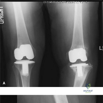
You are presented with a 68-year-old patient 5 years post-primary TKR, complaining of a 3-month history of increasing dull ache in the knee and "giving way" on stairs. Clinical exam reveals a neutral mechanical axis, no warmth, and no sinus tract. Radiographs are requested. What are you looking for, and what is your diagnostic priority?
Candidate: I would examine the radiographs for radiolucent lines at the cement-bone interface, specifically looking for lines >2mm in width or progressive changes compared to post-operative films. I would also check for component migration or subsidence. My priority is to rule out infection first, as it is a "can't miss" diagnosis, even if the clinical exam is non-specific.
Candidates often focus solely on the "loosening" aspect (mechanical) and forget to articulate that they must definitively rule out PJI despite the lack of systemic inflammatory signs. Another failure is not mentioning the need for *comparison* with previous radiographs—static lines may be benign, while progressive lines indicate pathology.
Start by classifying the workup into Ruling out Infection vs. Assessing Mechanical Failure. Mention the 2018 ICM criteria for PJI. Regarding the radiographs, categorize findings systematically: Bone-Cement Interface (Look for progressive >2mm radiolucencies in the zones of the Knee Society Roentgenographic Score), Implant position (Subsidence or tilting), and Osteolysis (Wear-debris related). Conclude by stating: "If radiographs are equivocal, I would proceed with inflammatory markers (ESR/CRP) and, if indicated, a diagnostic joint aspiration."
The joint aspiration returns a WBC count of 4,500 cells/µL with 85% neutrophils. What does this indicate, and how does this change your surgical management plan compared to a diagnosis of aseptic loosening?
Candidate: This indicates a Periprosthetic Joint Infection (PJI), given the threshold is typically >3,000 cells/µL and >80% neutrophils. My management shifts from a single-stage revision for aseptic loosening to a two-stage exchange arthroplasty as the gold standard for chronic PJI.
Failing to mention the "Two-Stage" vs "One-Stage" distinction. A poor candidate might just say "revision surgery." You must explain that two-stage involves an antibiotic spacer to allow for infection eradication, whereas aseptic loosening allows for a one-stage, definitive revision with reconstruction of bone stock.
State clearly that these values are diagnostic for PJI (ICM 2018 criteria). Explain the surgical divergence: 1. Infection: Requires a two-stage exchange. Stage 1: Radical debridement, synovectomy, removal of all components/cement, and implantation of an antibiotic-loaded articulating spacer. 2. Aseptic: Requires one-stage revision with immediate reconstruction of bone defects using augments/cones/stems to restore the joint line. Highlight that the goal of Stage 1 is not stability, but tissue health and antibiotic delivery, whereas the goal of aseptic revision is structural reconstruction.
During the first stage of your revision for infection, you remove the components and encounter significant bone loss on the tibial side, consistent with AORI Type 2B. How do you plan to manage this at the time of reimplantation?
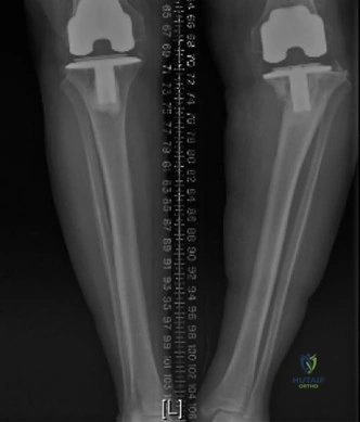
Candidate: An AORI Type 2B defect involves significant bone loss on one condyle, compromising the joint line. I would use modular metal augments, or possibly metaphyseal cones or sleeves, along with a long stem to bypass the defect and achieve stable fixation in the diaphysis.
Ignoring the "stem." A candidate who mentions augments but fails to mention that the stem is required to offload the stressed, deficient metaphyseal bone is essentially setting the patient up for secondary failure/subsidence.
Structure your response by the AORI logic: 1. Restore joint line: Address the metaphyseal void using metal augments (wedges) or porous tantalum/titanium cones/sleeves. 2. Stability: Use a long-stemmed component to achieve diaphyseal fixation, offloading the reconstructed metaphysis. 3. Constraint: Depending on the collateral ligament status following debridement, consider a Constrained Condylar Knee (CCK) or a Rotating Hinge Knee (RHK) to compensate for potential instability, ensuring you don't over-constrain if soft tissue balancing is adequate.
