العربية
Part of the Master Guide

100 High-Yield Orthopedic MCQs: Shoulder & Elbow | Mock Exam 251

Orthopedic Trauma Board Review MCQs (Set 2): Femoral & Tibial Fractures, Shoulder Dislocations

23 Apr 2026 57 min read 97 Views
Trauma 2006 MCQs - Part 2

Key Takeaway

This high-yield MCQ set (Set 2) is crucial for AAOS and ABOS board preparation. It features challenging questions on femoral shaft and periarticular fractures, including tibial plateau injuries. Additionally, test your knowledge on the diagnosis and management of various shoulder dislocations and associated neurovascular considerations.

Orthopedic Trauma Board Review MCQs (Set 2): Femoral & Tibial Fractures, Shoulder Dislocations

Comprehensive 100-Question Exam


00:00

Start Quiz

Question 1

Figures 14a and 14b show the initial radiographs of an 18-year-old man who fell while snowboarding. Figures 14c and 14d show the radiographs obtained following closed reduction. Examination reveals that the elbow is stable with range of motion. Management should now consist of


Explanation

The initial radiographs reveal a simple elbow dislocation without associated fractures. After successful closed reduction, the range of stability should be assessed. If the elbow is stable, nonsurgical management should consist of a short period of immobilization followed by range-of-motion exercises. Immobilization for more than 3 weeks results in significant elbow stiffness. Surgical repair is indicated for dislocations that are irreducible, have associated fractures, or where stability cannot be maintained with closed treatment. Cohen MS, Hastings H II: Acute elbow dislocations: Evaluation and management. J Am Acad Orthop Surg 1998;6:15-23.

Question 2

A 12-year-old boy sustains open comminuted midshaft tibial and fibular fractures while playing indoor soccer. The wound is grossly clean and measures 7 cm with some periosteal stripping. Antibiotics and tetanus toxoid are administered immediately in the emergency department. Following irrigation and debridement of the wound in the operating room, treatment should include


Explanation

Open fractures in children have similar rates of short-term complications such as compartment syndrome, vascular injury, and nerve injury when compared to adult fractures. Primary wound closure should be used for Gustillo and Anderson type 1 or uncomplicated type 2 fractures after surgical debridement. Skeletal stabilization may consist of external fixation, flexible nails, or casting with or without supplementary pin fixation. For an open comminuted midshaft fracture, external fixation is the treatment of choice. Reamed intramedullary nailing is contraindicated in children with an open physis. Plate fixation has a high complication rate in severe open fractures. Jones BG, Duncan RD: Open tibial fractures in children under 13 years of age-10 years experience. Injury 2003;34:776-780. Bartlett CS III, Weiner LS, Yang EC: Treatment of type II and type III open tibia fractures in children. J Orthop Trauma 1997;11:357-362. Robertson P, Karol LA, Rab GT: Open fractures of the tibia and femur in children. J Pediatr Orthop 1996;16:621-626.


Question 3

Which of the following is an advantage of unreamed nailing of the tibia compared to reamed nailing?


Explanation

The debate between reamed versus unreamed intramedullary nailing of the tibia continues. Although unreamed nailing was proposed for open fractures to minimize infection, its simplicity made it appealing for closed fractures. However, most studies to date show that the only advantage of unreamed nailing is less surgical time. All studies show higher nonunion rates with increased hardware failure and increased time to union for unreamed nailing. Even in open fractures graded up to Gustilo Grade IIIA, the reamed tibial nail performs better. Larsen LB, Madsen JE, Hoiness PR, et al: Should insertion of intramedullary nails for tibial fractures be with or without reaming? A prospective, randomized study with 3.8 years' follow-up. J Orthop Trauma 2004;18:144-149.


Question 4

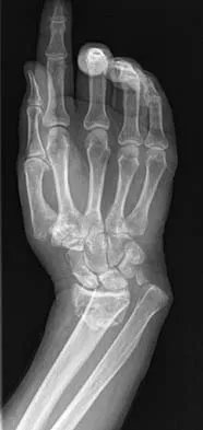
A 12-year-old boy sustained a both bone forearm fracture 10 weeks ago and underwent closed reduction and casting. Examination now reveals that the injury is healed, but he is unable to extend his little and ring fingers of the injured hand with his wrist extended. Full extension is possible with the wrist flexed. A radiograph and clinical photograph are shown in Figures 15a and 15b. The remainder of his hand and wrist examination and neurologic evaluation in the hand are normal. What is the most likely diagnosis?


Explanation

In this patient, examination reveals an inability to extend the fingers with the wrist extended, but full extension is possible with wrist flexion. These findings demonstrate isolated tenodesis of the flexor digitorum to the ring and little fingers. These findings are not consistent with compartment syndrome or nerve injury. Scarring or entrapment of tendons in forearm fractures can occur. Watson PA, Blair W: Entrapment of the index flexor digitorum profundus tendon after fracture of both forearm bones in a child. Iowa Orthop J 1999;19:127-128. Shaw BA, Murphy KM: Flexor tendon entrapment in ulnar shaft fractures. Clin Orthop 1996;330:181-184. Kolkman KA, van Niekerk JL, Rieu PN, et al: A complicated forearm greenstick fracture: Case report. J Trauma 1992;32:116-117.


Question 5

An otherwise healthy 35-year-old woman reports dorsal wrist pain and has trouble extending her thumb after sustaining a minimally displaced fracture of the distal radius 3 months ago. What is the next most appropriate step in management?


Explanation

Extensor pollicis longus tendon rupture can occur after a fracture of the distal radius, even a minimally displaced one. Poor vascularity of the tendon within the third dorsal compartment is the suspected etiology, not the displaced fracture fragments. Tendon transfer will suitably restore active extension of the thumb interphalangeal joint. Christophe K: Rupture of the extensor pollicis longus tendon following Colles fracture. J Bone Joint Surg Am 1953;35:1003-1005.


Question 6

Figure 16a shows the radiograph of a 34-year-old woman who sustained a basicervical fracture of the femoral neck. The fracture was treated with a compression screw and side plate. Seven months postoperatively, she continues to have significant hip pain and cannot bear full weight on her hip. A recent radiograph is shown in Figure 16b. Management should now consist of


Explanation

The patient sustained a high-angle femoral neck fracture. The follow-up clinical findings and radiograph show that she now has a nonunion with failed internal fixation. The joint appears preserved. In a healthy, young patient, arthroplasty of the femoral head, although possible, is not ideal. Excellent healing and function can be obtained in 70% to 80% of patients with femoral neck nonunion with a valgus intertrochanteric osteotomy. Marti RK, Schuller HM, Raaymakers EL: Intertrochanteric osteotomy for non-union of the femoral neck. J Bone Joint Surg Br 1989;71:782-787.


Question 7

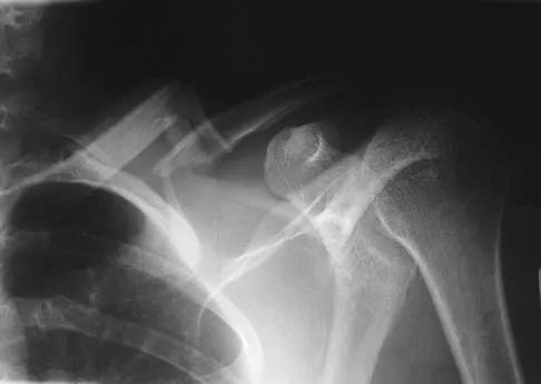
An 18-year-old man was in a motor vehicle accident and sustained a closed head injury, right displaced scapular body and glenoid fractures, a right proximal humeral fracture, fractures of ribs one through three, facial fractures, and bilateral pubic rami fractures with minimal displacement. He has a systolic blood pressure of 80/40 mm Hg despite fluid resuscitation. A radiograph is shown in Figure 17. Spiral CT does not identify any thoracic or abdominal injuries. What is the next most appropriate step in management?


Explanation

The patient has sustained high-energy upper extremity and chest injuries. He continues to remain hemodynamically unstable with no obvious thoracic or abdominal injury responsible for bleeding. The pelvic fracture is unlikely to be causing significant bleeding. A scapulothoracic dissociation and possible disruption of one of the great vessels of the upper extremity should be considered. Evaluation of peripheral pulses or blood pressure indices bilaterally in the upper extremities is a simple way to evaluate the need for further work-up. If there is any discrepancy or further concern, angiography of the involved extremity is necessary. Althausen PL, Lee MA, Finkemeier CG: Scapulothoracic dissociation: Diagnosis and treatment. Clin Orthop 2003;416:237-244.


Question 8

What is the major difference in outcome following open reduction and internal fixation (ORIF) of the tibial plafond at 2 to 5 days versus 10 to 20 days?


Explanation

Long-term outcomes following tibial plafond fractures treated with ORIF are satisfactory in most patients despite a high incidence of posttraumatic osteoarthritis. If ORIF is delayed until 10 to 20 days following injury, the major difference in outcomes is fewer complications associated with wound healing. Ankle strength, pain, range of motion, and the development of arthritis are equal regardless of the time until fixation. Sirkin M, Sanders R, DePasquale T, et al: A staged protocol for soft tissue management in the treatment of complex pilon fractures. J Orthop Trauma 1999;13:78-84.


Question 9

Figure 18a shows the initial lateral radiograph of a 6-year-old girl who sustained a fracture in a motor vehicle accident and was treated in a cast 1 year ago. She now has the valgus deformity seen in Figure 18b. Treatment should consist of


Explanation

Proximal tibial metaphyseal fractures may result in late genu valgum as a result of asymmetric growth of the proximal tibia. These patients are best treated with observation because the deformity is likely to remodel. Osteotomy is not indicated and potentially will lead to recurrence. Stapling of the medial tibial physis is appropriate in patients who have a severe and progressive deformity. Cozen L: Knock-knee deformity in children: Congenital and acquired. Clin Orthop 1990;258:191-203. Jackson DW, Cozen L: Genu valgum as a complication of proximal tibial metaphyseal fractures in children. J Bone Joint Surg Am 1971;53:1571-1578. Brammar TJ, Rooker GD: Remodeling of valgus deformity secondary to proximal metaphyseal fracture of the tibia. Injury 1998;29:558-560. Ogden JA, Ogden DA, Pugh L, et al: Tibia valga after proximal metaphyseal fractures in childhood: A normal biologic response. J Pediatr Orthop 1995;15:489-494.


Question 10

Figure 19 shows the radiograph of a 45-year-old woman who has a painful nonunion. Treatment should consist of


Explanation

The radiograph reveals a reverse obliquely subtrochanteric/intertrochanteric fracture. Open reduction and internal fixation should be accomplished with a 95-degree fixed angle device. An intramedullary nail with screw fixation into the head is another possible technique. Either method should correct the varus deformity. Exchange of a high-angled screw and plate device to a longer side plate and bone grafting does not afford any improvement in the mechanical stability. Hardware removal and retrograde intramedullary nailing is not indicated for this level of a proximal femoral injury. Placement of an implantable bone stimulator may change local biologic factors but would not enhance mechanical stability. The patient's femoral head is intact without signs of collapse; therefore, hardware removal, proximal femoral resection, and total hip arthroplasty are not warranted. Haidukewych GJ, Israel TA, Berry DJ: Reverse obliquity fractures of the intertrochanteric region of the femur. J Bone Joint Surg Am 2001;83:643-650.


Question 11

A 7-year-old boy has a swollen and deformed right arm after falling off his bicycle. Radiographs reveal a completely displaced posterolateral supracondylar humeral fracture. Examination reveals a warm, pink hand and forearm but absent pulses. What is the next most appropriate step in management?


Explanation

The incidence of vascular injury in supracondylar humeral fractures is directly related to the degree and direction of displacement. Significant posterior lateral displacement tends to result in brachial artery and median nerve injuries, and posterior medial displacement may lead to radial nerve injury. The brachial artery is always injured at the level of the fracture; therefore, angiography or MRA will not assist in locating the injury. The treatment of choice is surgical reduction and stabilization of the fracture, followed by reassessment of the vascular status. If the hand is pink and warm or pulses can be detected with doppler, it is reasonable to follow the extremity closely after surgery. If the arm becomes pulseless and white, immediate anterior exploration of the arm is indicated. The artery is often entrapped in the fracture and once extricated, will provide adequate blood flow. If the artery is injured, a primary repair or vein graft is needed. Shaw BA: The role of angiography in assessing vascular injuries associated with supracondylar humerus fractures remains controversial. J Pediatr Orthop 1998;18:273. Sabharwal S, Tredwell SJ, Beauchamp RD, et al: Management of pulseless pink hand in pediatric supracondylar fractures of humerus. J Pediatr Orthop 1997;17:303-310.


Question 12

What is the treatment of choice for the injury shown in Figures 20a through 20c?


Explanation

The radiographs show multiple carpometacarpal dislocations. Reduction is often obtainable but difficult to maintain. Internal fixation is required to maintain the reduction, preferably with Kirschner wires. Closed reduction and percutaneous pinning is preferred by some surgeons. Others recommend open reduction to remove irreconstructable osteochondral fragments from the individual joints and to ensure correct reduction of the carpometacarpal joints. Kirschner wires are removed at 6 to 8 weeks. Prokuski LJ, Eglseder WA Jr: Concurrent dorsal dislocations and fracture-dislocations of the index, long, ring, and small (second to fifth) carpometacarpal joints. J Orthop Trauma 2001;15:549-554.


Question 13

A 32-year-old man has intense right hand and wrist pain, a deformed wrist, and numbness in his fingers after falling off his motorcycle. This is an isolated injury. Examination reveals a swollen wrist, normal capillary refill to all fingers, and limited flexion of all fingers. Radiographs are shown in Figures 21a and 21b. Neurologic examination of the hand will most likely reveal


Explanation

The patient has a perilunate dislocation. A volar dislocation of the lunate is often associated with median nerve dysfunction. This injury to the wrist is often overlooked because of its benign clinical appearance and the presence of other injuries, as it is caused by high-energy mechanisms. Ruby LK, Cassidy C: Fractures and dislocations of the carpus, in Browner BD (ed): Skeletal Trauma, ed 3. Philadelphia, PA, WB Saunders, 2003, pp 1297-1300.


Question 14

A 55-year-old woman fell and sustained an elbow dislocation with a coronoid fracture and a radial head fracture. The elbow is reduced and splinted. What is the most common early complication?


Explanation

The patient has a dislocation of the elbow with displaced coronoid process and radial head fractures. The elbow is extremely unstable after this injury, and recurrent dislocation in a splint is the most common early complication. Skeletal stabilization of the fractures is required to restore stability of the joint. Characteristics of the fractures will determine the techniques required to restore stability. Ring D, Jupiter JB, Zilberfarb J: Posterior dislocation of the elbow with fractures of the radial head and coronoid. J Bone Joint Surg Am 2002;84:547-551.


Question 15

A 25-year-old man sustained the closed injury shown in Figures 22a and 22b. Examination reveals that this is an isolated injury, and he is hemodynamically stable. Treatment should consist of


Explanation

The treatment of choice for closed diaphyseal femoral fractures in adults is reamed intramedullary nailing with static interlocking. Reaming allows placement of a larger, stronger implant and offers better healing rates than unreamed nailing. Static interlocking ensures that there is no loss of reduction because of underappreciated fracture lines or comminution. Brumback RJ, Virkus WW: Intramedullary nailing of the femur: Reamed versus nonreamed. J Am Acad Orthop Surg 2000;8:83-90.


Question 16

Figure 23 shows the radiograph of an elderly man who fell on his right arm. What is the most important determinate of a good outcome following this injury?


Explanation

Minimally displaced fractures of the proximal humerus have a good outcome if physical therapy is initiated within 2 weeks of the injury. Results are not affected by age, open reduction and internal fixation, or involvement of the greater tuberosity. Immobilization for longer than 3 weeks will often result in stiffness. Koval KJ, Gallagher MA, Marsicano JG, et al: Functional outcome after minimally displaced fractures of the proximal part of the humerus. J Bone Joint Surg Am 1997;79:203-207.


Question 17

A 40 year-old-man was involved in a motor vehicle accident and sustained the pelvic injury seen in Figures 24a and 24b. Definitive management of the injury should consist of reduction by


Explanation

The radiograph reveals disruption of the symphysis pubis and a displaced left sacral fracture. A posterior injury with displacement of greater than 1 cm is unstable, and a sacral fracture is particularly unstable. Surgical stabilization is required for these unstable anterior and posterior injuries. External fixation provides little stability to an unstable posterior pelvic injury. Reduction and internal fixation of the symphysis pubis and sacral fracture will provide the most stable pelvis with the least resultant deformity and allow patient mobilization. Tile M: Management of pelvic ring injuries, in Tile M, Helfet DL, Kellam JF (eds): Fractures of the Pelvis and Acetabulum, ed 3. Philadelphia, PA, Lippincott Williams & Wilkins, 2003, pp 168-202.


Question 18

A 35-year-old patient sustained a bimalleolar ankle fracture. What is the most reliable method of predicting a tear of the interosseous membrane?


Explanation

The Weber and Lauge-Hansen fracture classifications suggest that the interosseous membrane (IOM) is torn with certain fracture patterns. In a recent study that evaluated ankle fractures with MRI, Nielson and associates identified 30 patients with IOM tears. Ten of the tears did not correspond with the level of the fibular fracture. The authors concluded that stability of the syndesmosis should not be based on the level of the fibular fracture alone but should also include an intraoperative stress test. Transsyndesmotic fixation should be considered for those fractures where the intraoperative stress test demonstrates instability. A widened medial clear space may occur with a deltoid injury and distal fibular fracture in the absence of a significant tear of the interosseous membrane.


Question 19

When performing a flexor tendon repair of a digit other than the thumb, what structures of the flexor tendon sheath should be preserved?


Explanation

The A2 and A4 pulleys are considered the most important parts of the pulley system. If these two structures are preserved, 80% of finger flexion can be maintained. If the pulley system is not left intact or is not reconstructed, "bow-stringing" of the flexor tendons occurs with loss of full flexion. The A2 pulley is over the proximal phalanx and the A4 pulley is over the middle phalanx. Doyle JR: Anatomy of the finger flexor tendon sheath and pulley system. J Hand Surg Am 1988;13:473-484.


Question 20

A distal radius fracture in an elderly man is strongly predictive for what subsequent injury?


Explanation

Fractures of the distal radius increase the relative risk of a subsequent hip fracture significantly more in men than in women. A previous spinal fracture has an equally important impact on the risk of a subsequent hip fracture in both genders.


Question 21

A 13-year-old girl injures her ankle playing soccer. Radiographs reveal a displaced Tillaux fracture. CT scans are shown in Figure 25. What is the most important consideration for appropriate management?


Explanation

Tillaux and triplane fractures occur in adolescents as the result of an external rotation injury of the ankle. As seen on the CT scan, the growth plate starts to close during adolescence; therefore, growth arrest resulting in limb-length discrepancy or angulation is less of a concern in this age group than achieving joint congruity. The joint should be surgically reduced if displacement is greater than 2 mm to minimize the chances of late arthrosis. Kay RM, Matthys GA: Pediatric ankle fractures: Evaluation and treatment. J Am Acad Orthop Surg 2001;9:268-278. Kling TF Jr: Operative treatment of ankle fractures in children. Orthop Clin North Am 1990;21:381-392.


Question 22

What measure of physiologic status best evaluates whether an injured patient is fully resuscitated and best predicts that perioperative complications will be minimized following definitive stabilization of long bone fractures?


Explanation

Serum lactate levels can be used to evaluate the effectiveness of the resuscitation of patients who have multiple injuries. Even after resuscitation, patients may have occult hypoperfusion as defined by a serum lactate level of greater than 2.5 mmol/L. The studies referenced indicate that these patients are at increased risk of perioperative complications such as organ failure or adult respiratory distress syndrome if definitive surgical fixation of the orthopaedic injuries is pursued prior to correction of the occult hypoperfusion. The other markers may be an indication of current physiology but have not been correlated with perioperative risks. Blow O, Magliore L, Claridge JA, et al: The golden hour and silver day: Detection and correction of occult hypoperfusion within 24 hours improves outcomes from major trauma. J Trauma 1999;47:964-977. Crowl A, Young JS, Kahler DM, et al: Occult hypoperfusion is associated with increased morbidity in patients undergoing early femur fracture fixation. J Trauma 2000;48:260-267.


Question 23

Based on the findings seen in the radiograph in Figure 26, emergent management should consist of


Explanation

The radiograph shows a volarly dislocated lunate. Initial emergent treatment of perilunate dislocations should consist of closed reduction and splinting, especially if the patient exhibits median nerve compression. Open reduction and pinning or ligament repair are necessary but are not emergent. A dorsal approach is sometimes required for ligament repair or bony visualization; however, this can be done in a more semi-elective manner. Isenberg J, Prokop A, Schellhammer F, et al: Palmar lunate dislocation. Unfallchirurg 2002;105:1133-1138.


Question 24

A 10-year-old girl has a midshaft both bone forearm fracture. After attempted closed reduction, alignment consists of bayonet apposition, 10 degrees of malrotation, and 8 degrees of volar angulation. Management should now consist of


Explanation

Acceptable alignment in both bone forearm fractures is related to age and location. In children younger than age 9 years, angulations of 15 degrees and malrotation of 45 degrees are acceptable. In children older than age 9 years, acceptable alignment is 10 degrees of angulation and 30 degrees of malrotation. Bayonet apposition is acceptable provided that the angular and rotational reductions are held within these guidelines. A long arm cast provides better control of deforming forces than a short arm cast. Do TT, Strub WM, Foad SL, et al: Reduction versus remodeling in pediatric distal forearm fractures: A preliminary cost analysis. J Pediatr Orthop B 2003;12:109-115. Flynn JM: Pediatric forearm fractures: Decision making, surgical techniques, and complications. Instr Course Lect 2002;51:355-360. Ring D, Waters PM, Hotchkiss RN, et al: Pediatric floating elbow. J Pediatr Orthop 2001;21:456-459.


Question 25

In the treatment of ankle fractures, the superficial peroneal nerve is most commonly injured by


Explanation

In the treatment of ankle fractures, the superficial peroneal nerve is most commonly injured by the use of a direct lateral approach to the ankle. The superficial peroneal nerve and its branches exit the fascial hiatus approximately 9 cm to 10 cm proximal to the tip of the distal fibula with a range of 4 cm to 13 cm, and their course is typically anterior to the midlateral plane of the fibula. However, small branches may course across the surgical plane directly laterally. A posterior-lateral approach diminishes the risk of injury to the superficial peroneal nerve and its branches; however, by moving farther posterior, the sural nerve and its branches may be at increased risk. Cast immobilization may injure the cutaneous nerves about the ankle; however, the risks are greater with surgical intervention. A medial or anterior-medial approach to the ankle will not injure the superficial peroneal nerve at the ankle level. Redfern DJ, Sauve PS, Sakellariou A: Investigation of incidence of superficial peroneal nerve injury following ankle fracture. Foot Ankle Int 2003;24:771-774.


Question 26

A 25-year-old man sustains a subtrochanteric femur fracture. During closed reduction for intramedullary nailing, the proximal fragment typically assumes which of the following positions?


Explanation

The proximal fragment in a subtrochanteric fracture is flexed by the iliopsoas, abducted by the gluteus medius and minimus, and externally rotated by the short external rotators.

Question 27

A 30-year-old male is involved in a high-speed MVC and sustains an ipsilateral femoral neck and shaft fracture. Which of the following is the most appropriate management strategy?


Explanation

In ipsilateral femoral neck and shaft fractures, anatomic reduction and fixation of the femoral neck should be prioritized to minimize the risk of avascular necrosis and nonunion. A common construct is provisional or definitive neck fixation followed by retrograde shaft nailing.

Question 28

A 65-year-old woman with a 10-year history of alendronate use presents with right thigh pain. Radiographs reveal localized lateral cortical thickening and a transverse radiolucent line in the proximal third of the femoral shaft. What is the most appropriate next step in management?


Explanation

Atypical femoral fractures are associated with prolonged bisphosphonate use. Patients with symptomatic incomplete fractures (thigh pain and lateral cortical radiolucency) should undergo prophylactic intramedullary nailing to prevent completion of the fracture.

Question 29

A 35-year-old man undergoes intramedullary nailing of a proximal third tibial shaft fracture. Postoperatively, the most common malalignment seen is:


Explanation

Intramedullary nailing of proximal third tibial fractures often results in an apex anterior (procurvatum) and valgus deformity. This is due to the pull of the patellar tendon and an anterior starting point.

Question 30

A 40-year-old man presents to the emergency department with severe shoulder pain and an inability to externally rotate his arm after suffering an electrical shock. An AP radiograph shows a 'lightbulb' sign. What is the most likely diagnosis?


Explanation

Posterior shoulder dislocations classically occur following seizures or electrical shocks due to the strong internal rotators overpowering the external rotators. The 'lightbulb' sign on an AP radiograph indicates the humerus is locked in internal rotation.

Question 31

A 65-year-old woman sustains a first-time traumatic anterior shoulder dislocation. After successful closed reduction, she continues to have profound weakness in active shoulder abduction and external rotation. The most likely cause is:


Explanation

In patients older than 40 years, an anterior shoulder dislocation is highly associated with massive rotator cuff tears. Persistent weakness in abduction and external rotation post-reduction warrants an MRI to evaluate for a cuff tear.

Question 32

A 28-year-old man sustains a closed midshaft tibial fracture. He develops severe leg pain out of proportion to the injury. Which of the following pressure measurements is most diagnostic for acute compartment syndrome requiring immediate fasciotomy?


Explanation

Acute compartment syndrome is a clinical diagnosis, but when utilizing intracompartmental pressures, a Delta P (diastolic blood pressure minus compartment pressure) of less than 30 mm Hg is an accepted threshold for performing a four-compartment fasciotomy.

Question 33

A 24-year-old male sustains a displaced, vertically oriented (Pauwels type III) femoral neck fracture. What is the most appropriate definitive management?


Explanation

In young patients with displaced, vertically oriented femoral neck fractures (Pauwels III), there are high shear forces. Open reduction and internal fixation using a fixed-angle construct (like a dynamic hip screw with a derotational screw) provides better biomechanical stability than parallel cannulated screws.

Question 34

A 45-year-old man sustains a distal femur fracture. CT scan reveals a coronal plane fracture of the lateral femoral condyle (Hoffa fracture). This specific fracture pattern is best treated with:


Explanation

A Hoffa fracture is a coronal shear fracture of the femoral condyle. It requires anatomic reduction and rigid fixation, typically with anterior-to-posterior or posterior-to-anterior lag screws placed perpendicular to the fracture line to allow early range of motion.

Question 35

A 35-year-old man presents with a locked posterior shoulder dislocation following a seizure. CT scan reveals an anteromedial humeral head defect (reverse Hill-Sachs lesion) involving 35% of the articular surface. Which of the following is the most appropriate surgical management?


Explanation

For locked posterior shoulder dislocations with an articular defect between 20% and 40%, a modified McLaughlin procedure (transferring the lesser tuberosity into the defect) is indicated. This restores rotational stability and prevents the defect from engaging the posterior glenoid rim.

Question 36

A 25-year-old polytrauma patient presents with a closed femoral shaft fracture, bilateral pulmonary contusions, and a Glasgow Coma Scale score of 7. His serum lactate is 4.5 mmol/L and base deficit is -8. What is the most appropriate initial management of the femur fracture?


Explanation

In an unstable or borderline polytrauma patient with severe chest and head injuries (elevated lactate and base deficit), Damage Control Orthopedics (DCO) is indicated. External fixation of the femur minimizes the 'second hit' phenomenon and systemic inflammatory response.

Question 37

When treating an extra-articular distal third tibial shaft fracture with an intramedullary nail, which of the following postoperative malalignments is most frequently observed?


Explanation

Intramedullary nailing of distal tibia fractures is notoriously complicated by valgus and procurvatum (apex anterior) deformities. This is due to the mismatch between the nail and the widening metaphysis, along with muscular deforming forces.

Question 38

A 24-year-old sustains an anterior shoulder dislocation. Closed reduction in the emergency department is unsuccessful despite adequate procedural sedation and muscle relaxation. Which structure is most likely interposing and preventing closed reduction?


Explanation

Irreducible anterior shoulder dislocations are relatively rare and are most commonly caused by interposition of the long head of the biceps tendon, the subscapularis tendon, or fracture fragments. Open reduction is required to clear the interposed tissue.

Question 39

A 30-year-old man sustains a closed high-energy tibial shaft fracture. Within 12 hours, he develops out-of-proportion pain and pain with passive toe flexion. Which compartment of the lower leg is most frequently involved in acute compartment syndrome following this injury?


Explanation

The anterior compartment of the leg is the most commonly affected compartment in acute compartment syndrome following a tibial shaft fracture. Diagnosis is confirmed clinically and with compartment pressure measurements.

Question 40

A 28-year-old sustains a displaced, vertically oriented (Pauwels III) femoral neck fracture. To maximize biomechanical stability and resist the high shear forces inherent to this fracture pattern, which fixation construct is preferred?


Explanation

In young patients with vertical (Pauwels III) femoral neck fractures, shear forces are high. A sliding hip screw coupled with a derotation screw provides superior biomechanical stability against vertical shear compared to multiple cancellous screws.

Question 41

A 40-year-old patient presents with an open type IIIB tibia fracture with a 6 cm soft tissue defect directly over the middle third of the tibia. Following adequate debridement, which flap is the most appropriate choice for local soft tissue coverage?


Explanation

The soleus muscle flap is the primary workhorse for soft tissue defects over the middle third of the tibia. The gastrocnemius flap is typically utilized for proximal third defects, while distal third defects often require free flaps.

Question 42

A 32-year-old sustains a high-energy femoral shaft fracture. Upon secondary survey, a non-displaced ipsilateral femoral neck fracture is discovered. Which of the following surgical strategies represents an optimal approach to manage both injuries?


Explanation

Using a retrograde intramedullary nail for the femoral shaft combined with cancellous screws for the femoral neck allows optimal, independent fixation of both fractures. Crucially, it prioritizes anatomic fixation of the neck and avoids displacement during shaft nailing.

Question 43

Following a traumatic anterior shoulder dislocation, a patient is unable to actively abduct the arm and has decreased sensation over the lateral aspect of the shoulder. Due to the most likely nerve injury, which of the following muscles will also exhibit weakness?


Explanation

The axillary nerve is frequently injured during anterior shoulder dislocations. It innervates both the deltoid and the teres minor muscles and provides sensation to the lateral shoulder (superior lateral cutaneous nerve of the arm).

Question 44

A 45-year-old sustains a coronal plane fracture of the lateral femoral condyle (Hoffa fracture). Which of the following statements regarding the surgical fixation of this injury is true?


Explanation

Biomechanical studies have demonstrated that posterior-to-anterior (PA) directed lag screws provide superior fixation and stability for Hoffa fractures compared to anterior-to-posterior (AP) screws. At least two screws are required to control rotation.

Question 45

When treating a proximal third tibial shaft fracture with an intramedullary nail using a standard infrapatellar approach, the most commonly encountered post-operative malalignment is:


Explanation

The classic deformity associated with standard intramedullary nailing of proximal third tibia fractures is apex anterior (procurvatum) and valgus. This is primarily driven by the pull of the patellar tendon and the anatomical widening of the metaphysis.

Question 46

A 55-year-old active woman sustains an anterior shoulder dislocation with an associated displaced greater tuberosity fracture. After successful closed reduction of the glenohumeral joint, radiographs show the greater tuberosity remains displaced 10 mm superiorly. What is the recommended treatment?


Explanation

Greater tuberosity fractures that remain displaced by more than 5 mm (and frequently >3 mm in active patients) after shoulder reduction require surgical fixation. Left untreated, they cause significant subacromial impingement and functional rotator cuff deficits.

Question 47

A 65-year-old woman on long-term alendronate therapy presents with an atraumatic subtrochanteric femur fracture. Which of the following radiographic features is considered a hallmark of a bisphosphonate-related atypical femur fracture?


Explanation

Atypical femur fractures associated with prolonged bisphosphonate use characteristically present as transverse or short oblique fracture lines originating at the lateral cortex. They are associated with localized lateral cortical thickening (beaking) and a lack of comminution.

Question 48

A 35-year-old man sustains a severe, closed, highly comminuted tibial pilon fracture with massive soft tissue swelling and impending fracture blisters. What is the most appropriate management strategy?


Explanation

For high-energy pilon fractures with significantly compromised soft tissues, the standard of care is a staged protocol. Initial spanning external fixation allows the soft tissue envelope to recover, followed by delayed definitive ORIF typically 10-21 days later.

Question 49

During arthroscopy for recurrent anterior shoulder instability, a lesion is noted where the anteroinferior labrum is avulsed but displaced medially along the glenoid neck with an intact periosteal sleeve. This pathoanatomic lesion is best described as an:


Explanation

An Anterior Labroligamentous Periosteal Sleeve Avulsion (ALPSA) lesion involves the anteroinferior labrum tearing and displacing medially. Unlike a classic Bankart lesion, the anterior periosteum remains intact, creating a sleeve that strips down the glenoid neck.

Question 50

According to the Winquist-Hansen classification of femoral shaft fractures, how is a Type III fracture defined?


Explanation

In the Winquist-Hansen classification, Type III fractures feature a large butterfly fragment with less than 50% cortical contact between the major proximal and distal fragments. These fractures are considered highly unstable regarding length and rotation.

Question 51

A 42-year-old pedestrian is struck by a motor vehicle and sustains an isolated medial tibial plateau fracture. According to the Schatzker classification system, what type of fracture is this?


Explanation

A Schatzker Type IV fracture is an isolated fracture of the medial tibial plateau. It is typically the result of a high-energy trauma and is highly associated with varus instability, as well as peroneal nerve and popliteal artery injuries.

Question 52

A 25-year-old man sustains a high-energy femoral shaft fracture. What is the most commonly missed associated ipsilateral injury, and what is the best imaging modality to rule it out during the initial trauma evaluation?


Explanation

Ipsilateral femoral neck fractures occur in 2-9% of femoral shaft fractures and are frequently missed initially. A dedicated CT or high-quality AP/lateral hip radiographs are mandatory to rule out this injury.

Question 53

A 28-year-old polytrauma patient sustains bilateral closed femoral shaft fractures. He has a GCS of 14, pulmonary contusions, and is hemodynamically stable after initial fluid resuscitation. Which of the following parameters is the most reliable indicator that the patient has been adequately resuscitated to safely undergo definitive early total care (intramedullary nailing) rather than damage control orthopedics?


Explanation

Serum lactate (< 2.5 mmol/L) and base deficit are the most reliable indicators of adequate tissue perfusion and resuscitation in trauma. Normalizing these parameters prior to early total care reduces the risk of "second hit" phenomena such as ARDS and systemic inflammatory response syndrome.

Question 54

A 25-year-old man undergoes reamed intramedullary nailing of a closed midshaft tibial fracture. Twelve hours postoperatively, he complains of severe, escalating leg pain that is not relieved by intravenous narcotics. On examination, the leg is tense, and passive plantar flexion of the great toe elicits excruciating pain. Which muscle compartment is most likely primarily involved?


Explanation

The anterior compartment of the leg contains the extensor hallucis longus (EHL). Passive stretch of the EHL by plantar flexing the great toe elicits severe pain, which is the classic sign of anterior compartment syndrome.

Question 55


A 40-year-old man presents after a tonic-clonic seizure. His right arm is locked in internal rotation and adduction. An initial anteroposterior (AP) radiograph demonstrates a symmetric "lightbulb" appearance of the humeral head without obvious fracture. Which of the following is the most appropriate next step in imaging to confirm the suspected diagnosis?


Explanation

The clinical presentation and "lightbulb" sign on AP radiograph strongly suggest a posterior shoulder dislocation. An axillary lateral (or Velpeau) radiograph is the most appropriate next step to definitively confirm posterior displacement of the humeral head.

Question 56

A 35-year-old male sustains a high-energy trauma resulting in a coronal plane fracture of the lateral femoral condyle (Hoffa fracture). When performing open reduction and internal fixation, which of the following lag screw configurations provides the most biomechanically stable construct?


Explanation

Biomechanical studies have demonstrated that posterior-to-anterior (PA) lag screw placement is significantly stronger for Hoffa fractures than anterior-to-posterior placement. This is due to the denser bone in the posterior condyle and the trajectory being more perpendicular to the fracture plane.

Question 57

A 45-year-old woman is struck by a car, sustaining a lateral tibial plateau fracture with both a split and central depression (Schatzker Type II). There is 8 mm of joint depression. Which of the following is the most appropriate surgical management?


Explanation

Schatzker Type II fractures require open reduction, elevation of the depressed articular fragment, filling of the metaphyseal void with bone graft or substitute, and support with a lateral buttress plate. This prevents late valgus collapse and post-traumatic arthritis.

Question 58

A 30-year-old construction worker sustains a Gustilo-Anderson Type IIIB open tibia fracture with a 10 x 5 cm anterior soft tissue defect requiring a free tissue transfer. Assuming the patient is hemodynamically stable and the wound is adequately debrided, what is the optimal timing for definitive soft tissue coverage to minimize infection rates?


Explanation

Early soft tissue coverage, ideally within 72 hours (or up to 5-7 days), is associated with the lowest rates of deep infection and flap failure in Type IIIB open tibia fractures. Delays beyond this window significantly increase complication rates.

Question 59

During the intramedullary nailing of a proximal third tibial shaft fracture using a standard infrapatellar approach, the surgeon notes a post-reduction malalignment. What is the most common deformity encountered during this specific procedure?


Explanation

The most common deformity when nailing proximal third tibia fractures is apex anterior (procurvatum) and valgus. This is caused by the unopposed pull of the patellar tendon and the anterior approach of the nail pushing the proximal fragment into extension.

Question 60

A 22-year-old athlete sustains a traumatic anterior shoulder dislocation. Following closed reduction in the emergency department, he reports numbness over the lateral aspect of his shoulder. Which muscle's function is most likely to be impaired due to the associated nerve injury?


Explanation

Numbness over the lateral shoulder (regimental badge area) indicates axillary nerve neuropraxia, the most common nerve injury in anterior shoulder dislocations. The axillary nerve innervates the deltoid and teres minor muscles.

Question 61

A 35-year-old male is involved in a high-speed motor vehicle collision and sustains an ipsilateral basicervical femoral neck fracture and a midshaft femur fracture. What is the standard priority and sequence of fixation for these injuries?


Explanation

In ipsilateral femoral neck and shaft fractures, the priority is precise anatomic reduction and stable fixation of the femoral neck to minimize the risk of avascular necrosis and nonunion. The shaft fracture is addressed subsequently.

Question 62

A 45-year-old male sustains a high-energy tibial pilon fracture. On presentation, the ankle is grossly swollen with hemorrhagic fracture blisters over the medial and lateral malleoli. What is the most appropriate initial management strategy?


Explanation

High-energy pilon fractures with severe soft tissue compromise are best managed with a staged protocol: initial spanning external fixation ("span and scan"). Definitive ORIF is delayed until soft tissue swelling resolves and the "wrinkle sign" appears, typically 10-21 days later.

Question 63

A 19-year-old college football player experiences recurrent anterior shoulder instability. An MRI arthrogram reveals an impaction fracture on the posterolateral aspect of the humeral head. What is the proper eponym for this osseous defect?


Explanation

A Hill-Sachs lesion is a posterolateral humeral head impaction fracture caused by the humeral head striking the anterior glenoid rim during an anterior shoulder dislocation. A Bankart lesion refers to the anterior inferior glenoid labral tear.

Question 64

A 65-year-old woman, who has been taking alendronate for 12 years, presents with a low-energy transverse subtrochanteric femur fracture. Radiographs show lateral cortical thickening and a medial spike. Following cephalomedullary nailing of the fracture, what is the most appropriate pharmacological recommendation?


Explanation

This is a classic atypical femur fracture associated with long-term bisphosphonate use, which severely suppresses bone remodeling. Bisphosphonates must be stopped, and teriparatide (recombinant PTH) is often started due to its anabolic effect to aid in fracture healing.

Question 65

A 28-year-old male presents with a "floating knee" injury (ipsilateral fractures of the femoral and tibial shafts) after a motorcycle collision. Which of the following factors is most predictive of a poor long-term functional outcome in this patient?


Explanation

The most significant prognostic factor for poor functional outcome in floating knee injuries is intra-articular involvement (Fraser Type II). Intra-articular fractures lead to much higher rates of arthrofibrosis, post-traumatic arthritis, and permanent knee stiffness.

Question 66

A 62-year-old man sustains an acute anterior shoulder dislocation after falling on an outstretched hand. The dislocation is successfully reduced. Three weeks later, he complains of persistent, profound weakness in active external rotation and abduction. Deltoid sensation is intact. What is the most likely underlying pathology?


Explanation

In patients older than 40-50 years, anterior shoulder dislocations are highly associated with concomitant rotator cuff tears (up to 30-80% incidence). Persistent weakness in external rotation and abduction post-reduction in this age group strongly suggests a massive cuff tear rather than isolated nerve injury.

Question 67

A 35-year-old pedestrian is struck by a truck, sustaining a severe crush injury to the right lower extremity with a mangled lower leg. When evaluating the patient for potential amputation versus limb salvage, which of the following is NOT a formal component of the Mangled Extremity Severity Score (MESS)?


Explanation

The components of the Mangled Extremity Severity Score (MESS) are skeletal/soft-tissue injury, limb ischemia, shock, and patient age. While severe nerve deficits heavily influence clinical decision-making, they are not a formal scoring criterion within the traditional MESS system.

Question 68

A 32-year-old man presents with a comminuted closed midshaft tibial fracture. Two hours after admission, he develops severe, unrelenting leg pain exacerbated by passive stretch of the hallux. The most reliable diagnostic parameter for acute compartment syndrome is a difference of less than 30 mmHg between:


Explanation

The delta P (diastolic blood pressure minus compartment pressure) is the most reliable threshold for diagnosing acute compartment syndrome. A delta P of less than 30 mmHg is an indication for immediate four-compartment fasciotomy to prevent irreversible muscle and nerve necrosis.

Question 69

A 22-year-old rugby player presents with recurrent anterior shoulder instability. A CT scan with 3D reconstruction reveals a glenoid bone loss of 28%. Which of the following is the most appropriate surgical treatment?


Explanation

In patients with significant anterior glenoid bone loss (typically greater than 20-25%), soft tissue stabilization alone has an unacceptably high failure rate. The Latarjet procedure, involving coracoid transfer to the anterior glenoid, restores the bony arc and provides a dynamic sling effect.

Question 70

A 28-year-old multiple trauma patient sustains bilateral femoral shaft fractures and a severe closed head injury. On arrival, his lactate is 6.5 mmol/L, pH is 7.1, and core temperature is 34.0°C. What is the most appropriate initial management of the femoral fractures?


Explanation

This patient is physiologically unstable (in extremis) with severe acidosis, hypothermia, and elevated lactate. Damage control orthopedics (DCO) using rapid temporary external fixation is indicated to minimize the physiologic "second hit" associated with prolonged intramedullary nailing.

Question 71

During intramedullary nailing of a proximal third tibial shaft fracture, the surgeon notes a persistent apex anterior and valgus deformity. Where should a blocking (Poller) screw be placed relative to the intended nail path to correct this deformity?


Explanation

Blocking screws are placed on the concave side of the deformity to narrow the metaphyseal corridor and force the nail to the center of the medullary canal. For an apex anterior (procurvatum) and valgus deformity, the blocking screw should be placed posterior and medial to the nail in the proximal segment.

Question 72

A 45-year-old man presents with severe shoulder pain and an inability to externally rotate his arm following a generalized tonic-clonic seizure. Radiographs demonstrate a posterior shoulder dislocation with a reverse Hill-Sachs lesion involving 25% of the articular surface. Which of the following is the most appropriate surgical management?


Explanation

Posterior shoulder dislocations are frequently accompanied by an anteromedial humeral head impaction fracture (reverse Hill-Sachs lesion). For defects involving 20% to 40% of the articular surface, transfer of the lesser tuberosity into the defect (modified McLaughlin procedure) prevents engagement and restores stability.

Question 73

A 35-year-old man sustains a subtrochanteric femur fracture. During closed reduction for intramedullary nailing, the proximal fragment is noted to be flexed, abducted, and externally rotated. Which muscle is primarily responsible for the flexion deformity of the proximal fragment?


Explanation

In subtrochanteric femur fractures, the proximal fragment is subjected to distinct deforming forces that complicate reduction. The iliopsoas flexes the fragment, the abductors (gluteus medius and minimus) abduct it, and the short external rotators externally rotate it.

Question 74

A 42-year-old pedestrian is struck by a car and sustains a pure centrally depressed fracture of the lateral tibial plateau with an intact lateral cortical rim (Schatzker type III). What is the optimal surgical approach and fixation strategy?


Explanation

A Schatzker type III fracture is a pure depression of the lateral tibial plateau, typically seen in softer, osteopenic bone. The optimal treatment involves a lateral approach with a cortical window to elevate the depressed articular fragment, followed by bone grafting and subchondral raft screw support.

Question 75

A 28-year-old man sustains a proximal third tibial shaft fracture. He undergoes intramedullary nailing via a standard infrapatellar approach. Which of the following deformities is most commonly seen postoperatively in this specific fracture pattern?


Explanation

Proximal third tibia fractures treated with infrapatellar intramedullary nails classically drift into procurvatum (apex anterior) and valgus. This malalignment is driven by the anterior pull of the patellar tendon and the wide metaphysis preventing tight cortical fit of the nail.

Question 76

A 22-year-old athlete presents with his first episode of an anterior shoulder dislocation following a rugby tackle. After closed reduction, what is the single most important prognostic factor for recurrent instability?


Explanation

The age of the patient at the time of the initial dislocation is the most significant prognostic factor for recurrence. Patients under 20 years old have recurrence rates exceeding 80%, whereas those over 40 have recurrence rates less than 15%.

Question 77

A 32-year-old man sustains a Pauwels type III (vertical) femoral neck fracture in a motor vehicle collision. Which of the following biomechanical constructs provides the most stable fixation for this specific fracture pattern?


Explanation

Pauwels type III fractures are highly vertically oriented (angle > 50 degrees), which subjects them to extreme shear forces. A sliding hip screw combined with an anti-rotation screw provides superior biomechanical stability against shear compared to multiple parallel cancellous screws.

Question 78

A 45-year-old man presents with severe shoulder pain following a generalized tonic-clonic seizure. Examination shows the arm is locked in internal rotation. Radiographs confirm a posterior shoulder dislocation with a 45% anteromedial humeral head impression defect (reverse Hill-Sachs lesion). What is the most appropriate surgical management?


Explanation

For posterior shoulder dislocations with an articular defect (reverse Hill-Sachs) larger than 40-45%, the joint is generally considered non-reconstructible with tendon or tuberosity transfers. Arthroplasty (hemiarthroplasty or total shoulder arthroplasty) is the indicated treatment.

Question 79

A 35-year-old construction worker sustains a Gustilo-Anderson Type IIIB open tibia fracture. Following initial thorough debridement and stabilization with an external fixator, what is the optimal timing for definitive soft-tissue coverage to minimize infection rates?


Explanation

Early soft-tissue coverage of Type IIIB open tibia fractures, ideally within 72 hours, significantly reduces the rate of deep infection and promotes fracture healing. Delays in coverage beyond 5 to 7 days are associated with markedly higher infection rates.

Question 80

A 78-year-old woman sustains a reverse obliquity intertrochanteric femur fracture. Which of the following best describes the biomechanical rationale for using a cephalomedullary nail rather than a sliding hip screw (SHS) for this fracture?


Explanation

In reverse obliquity fractures, the primary fracture line exits lateral to the shaft, rendering the lateral wall incompetent. A sliding hip screw allows the femoral shaft to medialize and the proximal fragment to slide laterally, commonly leading to construct failure.

Question 81

A 25-year-old male is brought in after a high-speed motorcycle crash. He has bilateral closed midshaft femur fractures and a pulmonary contusion. After initial fluid resuscitation, his serum lactate is 4.2 mmol/L. What is the most appropriate initial orthopedic management of the femur fractures?


Explanation

This patient is an "unstable" or "borderline" polytrauma patient, indicated by pulmonary injury and inadequately cleared lactate (> 2.5 mmol/L). Damage Control Orthopedics (DCO) using temporary external fixation is indicated to prevent the systemic "second hit" associated with intramedullary nailing.

Question 82

A 40-year-old male sustains a high-energy Schatzker VI tibial plateau fracture. He presents with severe, unrelenting leg pain out of proportion to the injury, pain on passive stretch of the toes, and tense compartments. What is the most critical diagnostic step prior to surgical intervention?


Explanation

The clinical presentation is classic and unequivocal for acute compartment syndrome. When the clinical diagnosis is clear based on physical exam, time should not be wasted on measuring compartment pressures; emergent four-compartment fasciotomy is indicated.

Question 83

A 65-year-old woman taking alendronate for 10 years presents with a low-energy transverse subtrochanteric femur fracture. Radiographs show cortical thickening of the lateral cortex. What is the most appropriate management of the contralateral, asymptomatic femur if radiographs show lateral cortical beaking?


Explanation

Patients with bisphosphonate-related atypical femur fractures often have bilateral involvement. If the asymptomatic side demonstrates radiographic changes such as lateral cortical "beaking" or a stress line, prophylactic intramedullary nailing is indicated to prevent completion of the fracture.

Question 84

A 45-year-old smoker is 9 months out from a reamed intramedullary nailing of a closed midshaft tibia fracture. He reports continued pain with weight-bearing. Radiographs show an oligotrophic nonunion with a broken distal locking screw. What is the most successful surgical intervention?


Explanation

Exchange intramedullary nailing with reaming to a larger diameter is the treatment of choice for an oligotrophic or hypertrophic nonunion of the tibial shaft. It provides both biological stimulation through reaming and increased mechanical stability.

Question 85

An 80-year-old woman sustains an anterior shoulder dislocation after a fall. The dislocation is successfully reduced in the emergency department. Three weeks later, she complains of profound weakness with active shoulder elevation and external rotation, though passive motion is preserved. Plain radiographs are normal. What is the most likely diagnosis?


Explanation

In elderly patients, anterior shoulder dislocations are highly associated with acute rotator cuff tears, with incidences exceeding 80% in patients older than 60. Profound weakness after reduction in this age group should raise high suspicion for a massive cuff tear, which must be evaluated with an MRI.

Question 86

A 30-year-old driver presents after a dashboard injury with a swollen knee. Radiographs reveal a coronal plane fracture of the lateral femoral condyle (Hoffa fracture). What is the most appropriate fixation strategy for this specific fracture pattern?


Explanation

A Hoffa fracture is a coronal plane fracture of the femoral condyle. Biomechanically, anterior-to-posterior (AP) directed lag screws placed perpendicular to the fracture plane provide the best compression and stability for this intra-articular fracture.

Question 87

A 50-year-old man falls from a height, sustaining a highly comminuted, displaced intra-articular distal tibia fracture (OTA/AO 43-C3) with severe soft tissue swelling and fracture blisters. What is the most appropriate initial step in the operative sequence?


Explanation

High-energy pilon fractures with severe soft tissue compromise require a staged approach. Initial management involves a spanning external fixator across the ankle joint to restore length and alignment while allowing soft tissue swelling to subside before definitive fixation.

None

Clinic OS
Medically Verified Content by
Prof. Clinic OS
Consultant Orthopedic & Spine Surgeon
Chapter Index