العربية
Part of the Master Guide

100 High-Yield Orthopedic MCQs: Shoulder & Elbow | Mock Exam 251

AAOS Orthopedic MCQs (Set 3): Shoulder, Elbow & Wrist Trauma | 2005 Board Review

23 Apr 2026 63 min read 96 Views
Upper Extremity 2005 MCQs - Part 3

Key Takeaway

This high-yield question set for the AAOS/ABOS exams focuses on common upper extremity pathologies. Questions cover the diagnosis, classification, and management of shoulder girdle injuries, elbow fractures, and specific wrist conditions. It is an essential resource for candidates preparing for orthopedic board certifications and residency exams.

AAOS Orthopedic MCQs (Set 3): Shoulder, Elbow & Wrist Trauma | 2005 Board Review

Comprehensive 100-Question Exam


00:00

Start Quiz

Question 1

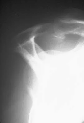
A 42-year-old woman with a long-standing history of rheumatoid arthritis undergoes total shoulder arthroplasty for persistent pain that has failed to respond to nonsurgical management. Intraoperative radiographs reveal an oblique, minimally displaced fracture of the greater tuberosity. Based on these findings, what is the best course of action?


Explanation

The risk of intraoperative fracture in osteoporotic bone in patients with rheumatoid arthritis is significant. Fractures most often occur during humeral head dislocation and positioning for canal reaming. If the fracture occurs at the greater tuberosity, cerclage suture fixation of the tuberosity fracture with autogenous cancellous bone graft from the resected humeral head is the treatment of choice. Wright TW, Cofield RH: Humeral fractures after shoulder arthroplasty. J Bone Joint Surg Am 1995;77:1340-1346. Norris TR (ed): Orthopaedic Knowledge Update: Shoulder and Elbow. Rosemont, IL, American Academy of Orthopaedic Surgeons, 1997, pp 215-225.

Question 2

A 13-year-old gymnast has had recurrent right elbow pain for the past year. She denies any history of trauma. Rest and anti-inflammatory drugs have failed to provide relief. Examination reveals no localized tenderness and only slight loss of both flexion and extension (10 degrees). What is the most likely diagnosis?


Explanation

Osteochondritis of the capitellum is characterized by pain, swelling, and limited motion. Catching, clicking, and giving way also can occur. It commonly affects athletes who participate in competitive sports with high stresses, such as pitching or gymnastics. Krijnen MR, Lim L, Willems WJ: Arthoscopic treatment of osteochondritis dissecans of the capitellum: Report of 5 female athletes. Arthroscopy 2003;19:210-214.

Question 3

The incidence of ipsilateral phrenic nerve blockade after an interscalene block approaches


Explanation

The most common side effect of an interscalene block is ipsilateral phrenic nerve blockade. The phrenic nerve arises chiefly from the fourth cervical ramus (with contributions from the third and fifth) and is the sole motor supply to the diaphragm. Phrenic nerve palsy usually is well tolerated in healthy patients but should be avoided in patients with limited pulmonary function (severe restrictive or obstructive lung disease, myasthenia gravis, or contralateral hemidiaphragmatic dysfunction). The incidence of ipsilateral phrenic nerve blockade afer interscalene block approaches 100%. Long T, Wass C, Burkle C: Perioperative interscalene blockade: An overview of its history and current clinical use. J Clin Anesthesia 2002;14;546-556.

Question 4

What is the most consistent finding regarding glenohumeral kinematics in patients with symptomatic tears of the rotator cuff?


Explanation

Normal glenohumeral kinematics are represented by ball-and-socket modeling when the rotator cuff is intact. This is true for motion that involves more than 30 degrees of abduction. In patients with shoulder pain and symptomatic rotator cuff tears, superior translation occurs with abduction beyond 30 degrees. This is quite evident in massive tears but is seen consistently to a lesser degree with smaller tears. Yamaguchi K, Sher JS, Anderson WK, et al: Glenohumeral motion in patients with rotator cuff tears: A comparison of asymptomatic and symptomatic shoulders. J Shoulder Elbow Surg 2000;9:6-11.

Question 5

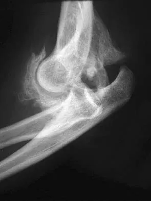
A 28-year-old man sustained numerous injuries in an accident including a dislocation of the elbow and a severe closed head injury that resulted in unconsciousness. The elbow was reduced in the emergency department. After 1 month of rehabilitation, the patient reports pain and stiffness. A radiograph is shown in Figure 23. Management should now consist of


Explanation

In a young individual with a chronic dislocation of the elbow and heterotopic bone formation, the treatment of choice is open reduction, heterotopic bone excision, anterior and posterior capsular releases, and a dynamic hinged fixator to begin protected early postoperative range of motion. It is important to understand that the fixator protects the reconstruction and allows early range of motion, but it does not maintain the reduction and should not be expected to do so. Pin fixation across the elbow delays early motion and is not recommended. Total elbow arthroplasty is not indicated, and ulnohumeral arthroplasty is for a primary arthritic condition. Garland DE, Hanscom DA, Keenan MA, et al: Resection of heterotopic ossification in the adult with head trauma. J Bone Joint Surg Am 1985;67:1261-1269.


Question 6

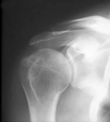
A 52-year-old man has had right shoulder pain in the deltoid region that increases at night for the past 2 months. He denies any history of trauma. Examination reveals mild tenderness over the greater tuberosity, and the Neer and Hawkins impingement signs are positive. AP and outlet lateral radiographs are shown in Figures 24a and 24b. Initial management should consist of


Explanation

The patient has the findings of classic subacromial impingement. Initial management should consist of stretching exercises directed at the posterior capsule and a program of rotator cuff and deltoid strengthening exercises performed below the horizontal in a "safe" plane. The judicious use of subacromial cortisone injections (one or two) may be helpful. Anterior acromioplasty is reserved for patients who have failed to respond to nonsurgical management. Morrison DS, Frogameni AD, Woodworth P: Non-operative treatment of subacromial impingement syndrome. J Bone Joint Surg Am 1997;79:732-737. Neer CS: Impingement lesions. Clin Orthop 1983;173:70-77.


Question 7

A 70-year-old woman is brought to the emergency department with a two-part greater tuberosity fracture with an anterior subcoracoid dislocation. One day after successful closed reduction, examination reveals marked swelling of the involved arm, forearm, and hand, as well as large amounts of "weeping" serous fluid but no obvious lacerations. The fingers are warm and pink, and the pulses are normal distally with good refill. Edema is present. There is no pain with passive and active motion of the elbow, wrist, and fingers. What is the next most appropriate step in management?


Explanation

Although not as common as arterial injury, venous thrombosis secondary to trauma of the subclavian or axillary vein can be problematic; therefore, venous duplex ultrasound scanning is the diagnostic study of choice. Arteriography may not show venous thrombosis in the venous run-off phase. The clinical history does not fit the usual presentation of a compartment syndrome or complex regional pain syndrome.

Question 8

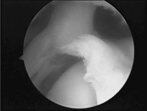
A baseball pitcher has intractable posterior and superior shoulder pain. The arthroscopic view seen in Figure 25 shows no Bankart or Hill-Sachs lesion and a negative drive-through sign. There are no signs of ligamentous laxity, but active compression and anterior slide tests are positive. Treatment should consist of


Explanation

According to Morgan and associates, a type II SLAP lesion can create or is associated with a superior instability pattern. They suggest that this can exist without a co-existing anteroinferior instability pattern. They reported that repair of the SLAP lesion alone resulted in satisfactory outcomes in 90% of patients and a return to throwing in more than 90% of pitchers. The arthroscopic findings in this patient do not support a diagnosis of anteroinferior laxity or instability; therefore, thermal capsular shift or capsular placation is not necessary. Morgan CD, Burkhart SS, Palmeri M, et al: Type II SLAP lesions: Three subtypes and their relationships to superior instability and rotator cuff tears. Arthroscopy 1998;14:553-565. Mileski RA, Snyder RJ: Superior labral lesions in the shoulder: Pathoanatomy and surgical management. J Am Acad Orthop Surg 1998;6:121-131.


Question 9

With increasing abduction in the scapular plane, maintaining neutral rotation, contact area, and contact pressure per unit area between the humeral head and glenoid follows what pattern if the total load across the joint is held constant?


Explanation

The glenohumeral joint becomes more congruent at higher levels of abduction. As a consequence, contact area increases. As the load is spread more evenly across the joint, contact pressure per unit area decreases as long as the total load across the joint is held constant. Warner JJP, Bowen MK, Deng XH, et al: Articular contact patterns of the normal glenohumeral joint. J Shoulder Elbow Surg 1998;7:381-388.

Question 10

A 21-year-old patient has had pain and a marked decrease in active and passive shoulder motion after having had a seizure 2 months ago as the result of alcohol abuse. Current AP and axillary radiographs and a CT scan are shown in Figures 26a through 26c. Management should consist of


Explanation

Open reduction and subscapularis and lesser tuberosity transfer into the defect is the treatment of choice in young individuals who have defects that involve between 20% to 45% of the head. Disimpaction and bone grafting is an option in injuries that are less than 3 weeks old. Closed reduction 2 to 3 months after injury usually is unsuccessful and increases the risk of fracture or neurovascular injury. Total shoulder arthroplasty is reserved for defects of greater than 50% or with associated glenoid surface damage. Hemiarthroplasty should be avoided in young individuals unless 50% or more of the head is involved. Gerber C: Chronic locked anterior and posterior dislocations, in Warner JJ, Iannotti JP, Gerber C (eds): Complex and Revision Problems in Shoulder Surgery. Philadelphia, PA, Lippincott-Raven, 1997, pp 99-113.


Question 11

Which of the following ligaments are the primary static restraints to inferior translation of the arm when the shoulder is in 0 degrees of abduction and neutral rotation?


Explanation

Biomechanical ligament sectioning studies have implicated both the superior glenohumeral and coracohumeral ligaments as restraints to inferior translation when the shoulder is in 0 degrees of abduction and neutral rotation. Although there is controversy over the significance of each ligament, both are involved to some degree. The middle glenohumeral ligament is more important in the midranges of abduction, and the inferior ligament is more important at 90 degrees of abduction. The coracoacromial and coracoclavicular ligaments play no role in glenohumeral restraint. Warner JJ, Deng XH, Warren RF, et al: Static capsuloligamentous restraints to superior-inferior translation of the glenohumeral joint. Am J Sports Med 1992;20:675-685.

Question 12

A 44-year-old man who sustained an elbow dislocation 3 months ago now reports pain and restricted elbow motion. Radiographs are shown in Figures 27a and 27b. Management should consist of


Explanation

The treatment of choice for an ankylosed chronically dislocated elbow is surgical reduction. Open reduction with application of an external fixator provides excellent results for this complex problem. Radial head arthroplasty is indicated for a radial head fracture that cannot be reconstructed. Attempts at closed reduction will be unsuccessful and should not be attempted in a stiff elbow. In chronic dislocations, direct reinsertion of injured ligaments is not feasible because of soft-tissue contracture. Jupiter J, Ring D: Treatment of unreduced elbow dislocation with hinged external fixation. J Bone Joint Surg Am 2002;84:1630-1635.


Question 13

A 67-year-old man who underwent humeral head arthroplasty for a four-part fracture 6 months ago reports that he is still unable to actively elevate his arm. Rehabilitation after surgery consisted of a sling with passive range-of-motion exercises for 2 weeks and then progressed to active-assisted and strengthening exercises at 3 weeks. Radiographs are shown in Figures 28a and 28b. What is the primary cause of his inability to elevate the arm?


Explanation

The radiographs show nonunion of both the greater and lesser tuberosities. Tuberosity pull-off and nonunion remain among the most common causes of failed humeral head arthroplasty for fracture. Strict attention to securing the tuberosities to each other and to the shaft, and autogenous bone grafting from the excised humeral head will decrease the incidence of pull-off and improve healing rates. Active-assisted range-of-motion and strengthening exercises should be delayed until tuberosity healing is noted radiographically, usually at 6 to 8 weeks postoperatively. Hartsock LA, Estes WJ, Murray CA, et al: Shoulder hemiarthroplasty for proximal humeral fractures. Orthop Clin North Am 1998;29:467-475. Hughes M, Neer CS: Glenohumeral joint replacement and postoperative rehabilitation. Phys Ther 1975;55:850-858.


Question 14

Initial postoperative management after repair of an acute rotator cuff tear includes


Explanation

In the immediate postoperative period following repair of an acute rotator cuff tear, passive forward elevation and external rotation should be performed within the safe zone determined at surgery. Early active range of motion (prior to tendon healing), internal rotation behind the back, and resistive exercises increase the risk of rupture of the repair. Iannotti JP: Full-thickness rotator cuff tear: Factors affecting surgical outcome. J Am Acad Orthop Surg 1994;2:87-95.

Question 15

A 34-year-old woman reports constant midlateral arm pain after sustaining minimal trauma to the shoulder. Radiographs and a biopsy specimen are shown in Figures 29a and 29b. What is the most likely diagnosis?


Explanation

Eighty percent of giant cell tumors occur in patients older than age 20 years, with the peak incidence in the third decade of life. Most of these tumors are eccentrically located and epiphyseal in location. They are lytic in nature as in this patient. Although named for the hallmarked multinucleated giant cells seen in the lesion, the basic cell type is the spindle-shaped stromal cell. Chondroblastoma is highly cellular and contains large multinucleated giant cells with intercellular chondroid material, some of which is calcified. Chondromyxoid fibroma has chondroid tissue separated by strands of more cellular tissue with occasional multinucleated giant cells. Desmoplastic fibroma is characterized by poorly cellular fibrous tissue, and lymphoma is highly cellular with characteristic round cells. Campanacci M, Baldini N, Boriani S, et al: Giant cell tumor of bone. J Bone Joint Surg Am 1987;69:106-114.


Question 16

A 25-year-old professional baseball pitcher reports a 4-month history of gradually increasing medial elbow pain that occurs during the late cocking and acceleration phases of throwing. The pain occasionally refers distally along the ulnar aspect of the forearm. He denies any weakness; however, he notes occasional paresthesias. A nerve conduction velocity study demonstrates increased latency across the cubital tunnel. Management consisting of 6 weeks of rest and rehabilitation fails to provide relief as the symptoms returned when he resumed throwing. What is the best course of action?


Explanation

In the thrower's elbow, ulnar neuritis is felt to result from both chronic compression and traction on the nerve that occurs during the throwing motion. Occasionally, subluxation of the nerve also can lead to symptoms. If nonsurgical management fails to provide relief, transposition of the nerve to an anterior subcutaneous location is the surgical procedure of choice. The nerve is held in its new position by one or two fascial slings created from the fascia of the common flexor origin. Schickendantz MS: Diagnosis and treatment of elbow disorders in the overhead athlete. Hand Clin 2002;18:65-75.

Question 17

What artery provides the only direct vascularizaton to both the intraneural and extraneural blood supply of the ulnar nerve just proximal to the cubital tunnel?


Explanation

The superior ulnar collateral, inferior ulnar collateral, and posterior ulnar recurrent arteries provide consistent vascular supply to the ulnar nerve. This supply is segmental in nature. No identifiable direct anastomosis is seen between the superior ulnar collateral and the posterior ulnar recurrent arteries. The inferior ulnar collateral artery provides the only direct vascularization to the nerve and is located in the region just proximal to the cubital tunnel. The segmental nature of the blood supply to the ulnar nerve underscores the importance of its preservation during transposition.

Question 18

A 56-year-old man underwent right total shoulder arthroplasty 2 months ago. Recently while reaching with his shoulder in a flexed and adducted position, he noted shoulder pain and afterwards he could not externally rotate his arm. An axillary radiograph is shown in Figure 30. What is the most likely cause of this problem?


Explanation

Anteversion of the humeral component may result in anterior instability of the component. Posterior instability after total shoulder arthroplasty is usually the result of some combination of the following factors: untreated anterior soft-tissue contractures, excessive posterior capsular laxity, and excessive retroversion of the humeral and/or glenoid components. Cofield RH, Edgerton BC: Total shoulder arthroplasty: Complications and revision surgery. Instr Course Lect 1990;39:449-462.


Question 19

A 70-year-old man seen in the emergency department has had left shoulder pain and a fever of 101.5 degrees F (38.6 degrees C) for the past 3 days. He denies any history of trauma. Examination reveals tenderness anterosuperiorly and at the posterior glenohumeral joint line. He has very limited range of motion (passive and active). Laboratory studies show a WBC count of 12,000/mm3 and an erythrocyte sedimentation rate of 48 mm/h. Initial management should consist of


Explanation

It appears that the patient has septic arthritis of the glenohumeral joint; therefore, initial management should consist of aspiration of the glenohumeral joint and subacromial space separately, followed by Gram stain and culture of the fluid. Based on the findings, broad-spectrum IV antibiotics should be started. If the diagnosis of septic arthritis is confirmed, then arthroscopic or open surgical drainage usually is indicated. Sawyer JR, Esterhai JL Jr: Shoulder infections, in Warner JJ, Iannotti JP, Gerber C (eds): Complex and Revision Problems in Shoulder Surgery. Philadelphia, PA, Lippincott-Raven, 1997.

Question 20

A 40-year-old man who is an avid weight lifter has had chronic pain in the proximal anterior shoulder for the past year. He denies any history of trauma. Examination reveals tenderness at the intertubercular groove, a positive speed test, and a positive Neer impingement sign. Nonsurgical management has failed to provide relief, and he is now considering surgery. Arthroscopic findings in the glenohumeral joint are shown in Figure 31. Based on these findings, treatment should consist of


Explanation

The arthroscopic image shows a tear through more than 50% of the biceps tendon; therefore, treatment should consist of tenodesis or tenotomy of the tendon. However, because this patient is relatively young and active, the treatment of choice is tenodesis of the biceps tendon. Sethi N, Wright R, Yamaguchi K: Disorders of the long head of the biceps tendon. J Shoulder Elbow Surg 1999;8:644-654. Eakin CL, Faber KJ, Hawkins RJ, et al: Biceps tendon disorders in athletes. J Am Acad Orthop Surg 1999;7:300-310.


Question 21

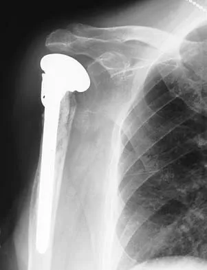
A 59-year-old man reports moderate shoulder pain and very restricted range of motion after undergoing humeral arthroplasty for osteoarthritis 1 year ago. An AP radiograph is shown in Figure 32. Management should now consist of


Explanation

The radiograph reveals that an insufficient amount of the proximal humerus was excised in the index procedure, resulting in malalignment of the humeral component, overstuffing of the glenohumeral joint, and glenoid arthritis. It is unlikely that physical therapy or soft-tissue releases alone will be adequate. Revision of the humeral component, recutting of the proximal humerus to allow a more anatomic alignment of the humeral component, appropriate soft-tissue releases, and glenoid arthroplasty will offer the best chance of improvement in this difficult situation. Neer CS II, Kirby RM: Revision of humeral head and total shoulder arthroplasties. Clin Orthop 1982;170:189-195.


Question 22

A 70-year-old woman has a preoperative anterior interscalene block prior to undergoing a total shoulder arthroplasty. After seating her in the beach chair position, she becomes acutely hypotensive. What is the most likely cause for the hypotension?


Explanation

The beach chair position may cause sudden hypotension and bradycardia as a result of the Bezold-Jarisch reflex. This reflex occurs when venous pooling and increased sympathetic tone induce a low-volume, hypercontractile ventricle, resulting in activation of the parasympathetic nervous system and sympathetic withdrawal. The reported incidence of this phenomenon associated with the sitting position is between 13% to 24%. Left untreated, the result may be cardiac arrest. Pneumothorax or central nervous system toxicity after interscalene block is rare and has an incidence of less than 0.2%. Laryngeal nerve block associated with interscalene nerve block can occur but usually results in hoarseness secondary to ipsilateral vocal cord palsy. Long T, Wass C, Burkle C: Perioperative interscalene blockade: An overview of its history and current clinical use. J Clin Anesthesia 2002;14:546-556.

Question 23

What structure is considered the single most important soft-tissue restraint to anterior-posterior stability of the sternoclavicular joint?


Explanation

In a cadaver ligament sectioning study, the posterior capsular ligament was considered the most important structure for anterior-posterior stability of the sternoclavicular joint. The anterior capsular ligament also helps prevent anterior displacement but not to the same degree as the posterior ligament. The interclavicular ligament provides little support for anteroposterior translation. Spencer EE, Kuhn JE, Huston LJ, et al: Ligamentous restraints to anterior and posterior translation of the sternoclavicular joint. J Shoulder Elbow Surg 2002;11:43-47.

Question 24

A 35-year-old man has atraumatic painless limited elbow motion. Radiographs are shown in Figures 33a and 33b. What is the most likely diagnosis?


Explanation

Based on the radiographic findings, the patient has melorheostosis, a rare, benign connective tissue disorder that is characterized by a cortical thickening of bone. It produces a "dripping candle wax" appearance with dense hyperostosis that flows along the cortex. Ectopic bone formation is a consideration but is associated with injuries or burns. Bone infarcts produce intraosseous sclerosis typically affecting the distal femur with the "smoke up chimney" appearance. Infection is always a consideration but typically does not have the linear osteitis seen in melorheostosis. Juxacortical chondroma is a benign cartilage growth that arises from the capsule and may involve the underlying cortical bone but rarely the medullary canal. Campbell CJ, Papademetriou T, Bonfiglio M: Melorheostosis: A report of the clinical, roentgenographic, and pathological findings in fourteen cases. J Bone Joint Surg Am 1968;50:1281-1304.


Question 25

A 64-year-old man who underwent total shoulder arthroplasty 4 weeks ago is making satisfactory progress in physical therapy, but his therapist notes limitations in external rotation to neutral. A stretching program is started, and the patient suddenly gains 90 degrees of external rotation but now reports increased pain and weakness. What is the best course of action?


Explanation

Nearly all approaches to shoulder arthroplasty require detachment of the subscapularis tendon from the humerus and subsequent repair. Healing of this tenotomy is one of the limiting factors in postoperative recovery. Failure of the tenotomy repair must be recognized and treated early with repeat repair or pectoralis muscle transfer for optimal results. Failure of the subscapularis is diagnosed clinically as excessive external rotation and weakness, especially in the lift-off or belly press position. Muscle testing can be difficult in the postoperative period and may not be possible to assess in those positions. Although MRI might be useful to confirm the diagnosis, studies may be limited by artifact. CT or electromyography would not be diagnostic. Wirth MA, Rockwood CA Jr: Complications of total shoulder-replacement arthroplasty. J Bone Joint Surg Am 1996;78:603-616.

Question 26

A 24-year-old man falls on an outstretched hand and sustains a proximal pole scaphoid fracture. Which of the following best describes the blood supply to the proximal pole of the scaphoid and its clinical implication?


Explanation

The scaphoid's blood supply enters distally from the dorsal carpal branch of the radial artery and flows retrograde. Proximal pole fractures disrupt this intraosseous supply, leading to high rates of avascular necrosis and nonunion.

Question 27

A 35-year-old woman falls from a ladder and sustains a 'terrible triad' injury of the elbow. Which of the following describes the standard sequence of surgical reconstruction to restore elbow stability?


Explanation

The standard surgical protocol for a terrible triad injury involves fixing the coronoid first to restore the anterior buttress, followed by fixing or replacing the radial head, and finally repairing the lateral collateral ligament (LCL) complex.

Question 28

A 40-year-old man sustains a closed spiral fracture of the middle third of the humerus. On initial exam, neurological function is intact. Immediately following a closed reduction and splinting, he loses active wrist and finger extension and sensation in the first dorsal web space. What is the most appropriate next step in management?


Explanation

A secondary radial nerve palsy that develops immediately after closed manipulation or reduction of a humeral shaft fracture is an absolute indication for surgical exploration, as the nerve may be entrapped in the fracture site.

Question 29

A 78-year-old woman with severe osteoporosis presents with a 4-part proximal humerus fracture after a mechanical fall. The humeral head is entirely devoid of soft tissue attachments, and the tuberosities are comminuted. What is the preferred surgical treatment to maximize functional outcome and pain relief in this patient?


Explanation

In elderly patients with poor bone quality and a 4-part proximal humerus fracture, reverse total shoulder arthroplasty (RTSA) provides more predictable functional outcomes and pain relief compared to ORIF or hemiarthroplasty, as it does not rely on tuberosity healing for overhead function.

Question 30

A 30-year-old man presents with a highly comminuted, un-reconstructable radial head fracture and severe ipsilateral wrist pain following a fall. Examination reveals distal radioulnar joint (DRUJ) instability. If the radial head is excised and not replaced, which of the following complications is most likely to occur?


Explanation

This is an Essex-Lopresti injury, involving a radial head fracture, interosseous membrane disruption, and DRUJ instability. Excising the radial head without replacing it with a prosthesis will lead to proximal migration of the radius and severe ulnar impaction syndrome.

Question 31

A 25-year-old man sustains a traumatic injury to his right forearm. Radiographs demonstrate a fracture of the distal third of the radial shaft with associated dorsal displacement of the distal ulna. Which of the following is the most appropriate definitive management?


Explanation

A Galeazzi fracture (distal third radius fracture with DRUJ disruption) is highly unstable and requires ORIF of the radius in adults. The DRUJ should then be assessed and, if unstable, stabilized via pinning or immobilization in supination.

Question 32

Which of the following is considered an absolute indication for operative fixation of an acute midshaft clavicle fracture?


Explanation

While severe displacement, complete lack of bony contact, and a floating shoulder are relative indications for surgery, an open clavicle fracture is an absolute indication for immediate operative debridement and fixation.

Question 33

A 32-year-old male presents with persistent wrist pain following a hyperextension injury 6 weeks ago. Radiographs show a widened scapholunate interval of 4 mm and a "cortical ring sign" of the scaphoid. What does the cortical ring sign indicate?


Explanation

The cortical ring sign is seen on an AP radiograph in scapholunate dissociation. It represents the axially aligned scaphoid viewed end-on due to abnormal volar flexion and rotary subluxation of the scaphoid.

Question 34

A 22-year-old gymnast falls on an outstretched arm and sustains a Type I (Hahn-Steinthal) capitellum fracture. Which of the following best describes this fracture pattern?


Explanation

A Type I (Hahn-Steinthal) capitellum fracture is a large coronal shear fracture containing a significant amount of subchondral bone. A Type II (Kocher-Lorenz) fracture involves articular cartilage with minimal attached subchondral bone.

Question 35

According to the Bado classification, which of the following characterizes a Type I Monteggia lesion?


Explanation

A Bado Type I Monteggia fracture is characterized by a fracture of the proximal or middle third of the ulna with an anterior dislocation of the radial head. It is the most common type.

Question 36

A 28-year-old man presents to the emergency department after a motorcycle accident. His wrist is grossly deformed, and he reports numbness in his thumb, index, and middle fingers. Radiographs reveal a volar dislocation of the lunate with the capitate lying dorsally. Which nerve is most commonly injured in this scenario?


Explanation

Lunate and perilunate dislocations frequently cause acute carpal tunnel syndrome due to the displaced lunate compressing the median nerve within the carpal tunnel, requiring prompt reduction and possible carpal tunnel release.

Question 37

A 45-year-old male bodybuilder feels a sudden "pop" in his anterior elbow while performing heavy bicep curls. He presents with weakness in forearm supination and elbow flexion. During an anatomic repair of the distal biceps tendon using a single-incision anterior approach, which nerve is at greatest risk of iatrogenic injury?


Explanation

The lateral antebrachial cutaneous nerve (LABCN) is the most commonly injured nerve during a single-incision anterior approach for distal biceps repair due to its superficial location near the cephalic vein and biceps tendon.

Question 38

A 35-year-old woman undergoes tension-band wiring for a transverse, non-comminuted olecranon fracture. What is the most common complication associated with this specific surgical technique?


Explanation

Symptomatic hardware prominence is the most common complication of tension-band wiring for olecranon fractures, occurring in up to 80% of patients and often requiring secondary surgery for hardware removal.

Question 39

A 35-year-old man falls on an outstretched hand and sustains the injury shown in

. The injury includes a posterior elbow dislocation, radial head fracture, and coronoid fracture. During operative management, what is the most appropriate sequence of repair?


Explanation

The standard inside-out approach for a terrible triad injury involves repairing the coronoid first, followed by the radial head (fixation or replacement), and finally the lateral collateral ligament (LCL) complex.

Question 40

A 28-year-old man presents with chronic wrist pain after an untreated injury 5 years ago. Radiographs shown in

demonstrate a scaphoid nonunion advanced collapse (SNAC). Which of the following articulations is typically preserved the longest in the natural history of a SNAC wrist?


Explanation

In both SLAC and SNAC wrists, the radiolunate articulation is classically spared until late in the disease process due to the concentric shape of the fossa and the intact short radiolunate ligament.

Question 41

When evaluating a displaced 4-part proximal humerus fracture for the risk of avascular necrosis, which of the following provides the primary arterial supply to the humeral head?


Explanation

Recent perfusion studies demonstrate that the posterior circumflex humeral artery provides the majority (up to 64%) of the blood supply to the humeral head, challenging older concepts that emphasized the anterior circumflex artery.

Question 42

Which of the following is considered an absolute indication for operative fixation of an acute midshaft clavicle fracture?


Explanation

Absolute indications for ORIF of a clavicle fracture include open fractures, neurovascular compromise, and severe skin tenting with impending necrosis. Displacement and shortening are considered relative indications.

Question 43

A 55-year-old woman is treated in a cast for a minimally displaced distal radius fracture. Six weeks later, she notes a sudden inability to extend her thumb at the interphalangeal joint. What is the most likely etiology of this complication?


Explanation

Extensor pollicis longus (EPL) ruptures typically occur after minimally displaced distal radius fractures. They result from local ischemia due to increased pressure within the intact third extensor compartment, followed by mechanical attrition.

Question 44

A 24-year-old professional baseball player complains of hypothenar wrist pain and paresthesias in his small finger after a forceful swing. A carpal tunnel view radiograph is shown in

. He is diagnosed with a hook of hamate fracture. What is the most definitive surgical treatment to allow earliest return to professional sports?


Explanation

Excision of the hook of the hamate fragment is the treatment of choice for athletes with symptomatic nonunions or acute fractures who require rapid return to play, yielding high satisfaction and low complication rates.

Question 45

After closed reduction of a simple posterior elbow dislocation, the elbow is stable in pronation but subluxates when extended in supination. This finding indicates insufficiency of which of the following structures?


Explanation

The LUCL is the primary stabilizer against posterolateral rotatory instability (PLRI). Pronation tightens the medial soft tissues and flexor-pronator mass, stabilizing the elbow when the lateral side is incompetent.

Question 46

A 22-year-old motorcyclist is brought in after a high-speed collision. He has an entirely flail, pulseless upper extremity. Radiographs shown in

demonstrate significant lateral displacement of the scapula. What is the most critical immediate step in management?


Explanation

Scapulothoracic dissociation is associated with a high incidence of devastating vascular injuries and complete brachial plexus avulsions. Immediate vascular imaging and management are life-saving priorities.

Question 47

A 30-year-old man falls onto his extended wrist. Lateral radiographs shown in

reveal a dorsal perilunate dislocation. Which of the following nerve injuries is most commonly associated with this specific trauma?


Explanation

Median nerve neuropathy (acute carpal tunnel syndrome) is the most common neurologic complication associated with lunate and perilunate dislocations due to volar displacement of the lunate into the carpal tunnel.

Question 48

A 45-year-old woman presents with elbow pain after a fall. Imaging shown in

reveals a fracture of the capitellum involving the lateral trochlear ridge. Which surgical approach is most appropriate for direct visualization and fixation of this specific fracture pattern?


Explanation

The extended lateral approach provides excellent exposure of the anterior capitellum and lateral trochlea. It is required for complex coronal shear fractures that extend medially into the trochlea.

Question 49

A 62-year-old woman undergoes volar locked plating for a distal radius fracture. Postoperatively, she develops flexor pollicis longus (FPL) tenosynovitis. What technical error is the most likely cause of this complication?


Explanation

Volar plates placed too distally (beyond the watershed line of the distal radius) can impinge on the flexor tendons, most commonly the FPL, leading to tenosynovitis or attrition rupture.

Question 50

A 19-year-old college football player sustains an acute anterior shoulder dislocation. MRI confirms an anterior labral tear (Bankart lesion). If he is treated nonoperatively with a sling, what is his approximate risk of recurrent instability?


Explanation

The risk of recurrent shoulder instability following an acute first-time anterior dislocation in a young athlete (under 20 years old) treated nonoperatively is extremely high, generally reported between 70% and 90%.

Question 51

During surgical management of a 'terrible triad' injury of the elbow, after fixing the coronoid and replacing the radial head, the elbow remains unstable in extension. What is the next most appropriate step?


Explanation

The standard surgical algorithm for terrible triad injuries involves fixing the coronoid, addressing the radial head, and repairing the lateral ulnar collateral ligament (LUCL). The medial collateral ligament is typically only addressed or an external fixator placed if instability persists after LUCL repair.

Question 52

A 22-year-old man falls on an outstretched hand and presents with anatomic snuffbox tenderness. Radiographs are negative, but MRI confirms a nondisplaced fracture of the proximal pole of the scaphoid. The blood supply placing this region at high risk for avascular necrosis originates primarily from which vessel?


Explanation

The proximal pole of the scaphoid relies on retrograde blood supply from the dorsal carpal branch of the radial artery. Because vessels enter the scaphoid distally and flow proximally, proximal pole fractures are highly susceptible to avascular necrosis.

Question 53

A 65-year-old woman is evaluated 6 months after volar locked plating of a distal radius fracture. She now presents with a new-onset inability to actively flex the interphalangeal joint of her thumb. What is the most likely etiology?


Explanation

Hardware placed prominent or volar to the watershed line of the distal radius can lead to attritional rupture of the flexor pollicis longus (FPL) tendon. The clinical presentation is a loss of active thumb interphalangeal joint flexion.

Question 54

A 55-year-old woman presents with a severely displaced 4-part proximal humerus fracture after a fall. Examination reveals profound weakness in active arm abduction and numbness over the lateral aspect of the shoulder. Which nerve is most likely injured?


Explanation

The axillary nerve is the most commonly injured nerve in proximal humerus fractures and anterior shoulder dislocations. Injury presents as deltoid weakness and sensory deficits over the lateral shoulder (regimental badge area).

Question 55

A 25-year-old cyclist sustains a completely displaced midshaft clavicle fracture with 2.5 cm of shortening. Compared to nonoperative management, what is the primary advantage of open reduction and internal fixation (ORIF) for this specific injury pattern?


Explanation

For completely displaced and significantly shortened (>2 cm) midshaft clavicle fractures, operative fixation significantly decreases the risk of nonunion and symptomatic malunion compared to nonoperative sling immobilization.

Question 56

A 7-year-old boy falls from the monkey bars. Radiographs demonstrate a fracture of the ulnar shaft with an anterior dislocation of the radial head (Bado Type I Monteggia variant). What is the preferred initial management?


Explanation

In pediatric Bado Type I Monteggia fractures, closed reduction of the ulnar fracture typically allows spontaneous reduction of the radial head. The most stable position to maintain reduction is in a long arm cast with the elbow flexed to roughly 100 degrees and the forearm supinated.

Question 57

A 30-year-old man is involved in a motorcycle crash. His wrist is grossly swollen, and he has numbness in the median nerve distribution. A lateral wrist radiograph demonstrates that the lunate maintains its normal articulation with the distal radius, but the capitate is displaced dorsally. What is the diagnosis?


Explanation

In a dorsal perilunate dislocation, the lunate maintains its alignment within the radiolunate fossa, but the capitate and the distal carpal row are displaced dorsally. Prompt closed reduction is required to relieve median nerve compression prior to definitive surgical management.

Question 58

A 40-year-old man sustains a severe traction injury to his right arm. Examination reveals massive swelling around the shoulder girdle and absent radial and ulnar pulses. Radiographs show extreme lateral displacement of the scapula. What is the most critical next step in management?


Explanation

Scapulothoracic dissociation involves a high-energy disruption of the scapulothoracic articulation and is associated with a very high rate of severe subclavian or axillary artery injury. Immediate vascular assessment via CT angiography or formal angiogram is critical to prevent limb loss.

Question 59

An 82-year-old community-dwelling woman with severe osteoporosis presents with a highly comminuted, intra-articular distal humerus fracture. She is a low-demand patient but requires her arms for basic transfers. What is the most appropriate surgical intervention?


Explanation

Total elbow arthroplasty (TEA) is the treatment of choice for comminuted, intra-articular distal humerus fractures in low-demand, elderly patients with poor bone quality. TEA provides predictable pain relief and allows for immediate weight-bearing for transfers.

Question 60

A 35-year-old man presents to the ER after a grand mal seizure. His shoulder is locked in internal rotation. An AP radiograph shows a 'lightbulb sign.' What is the most common associated bony defect in this injury pattern?


Explanation

Posterior shoulder dislocations, classically presenting after seizures or electrical shock with a 'lightbulb' appearance on AP radiographs, are frequently associated with an impaction fracture of the anteromedial humeral head, known as a reverse Hill-Sachs lesion.

Question 61

A 28-year-old man suffers a severe fall on an outstretched hand, resulting in a comminuted radial head fracture, diffuse forearm pain, and distal radioulnar joint (DRUJ) tenderness. If the radial head is surgically excised without replacement, what is the most likely complication?


Explanation

This presentation describes an Essex-Lopresti injury, which involves a radial head fracture, tear of the interosseous membrane, and DRUJ disruption. Excision of the radial head without prosthetic replacement leads to catastrophic proximal radial migration and ulnocarpal impaction.

Question 62

A 40-year-old woman falls onto her outstretched hand and presents with lateral elbow pain. A lateral elbow radiograph demonstrates a 'double arc' sign. What specific type of injury does this radiographic finding represent?


Explanation

The 'double arc' sign on a lateral elbow radiograph represents the displaced subchondral bone of the capitellum and the lateral ridge of the trochlea. This is pathognomonic for a Type IV capitellum fracture (McKee modification), which indicates extension into the trochlea.

Question 63

A 50-year-old man with a chronic, untreated scapholunate ligament tear presents with progressive wrist pain. Radiographs show joint space narrowing exclusively at the radioscaphoid articulation. As Scapholunate Advanced Collapse (SLAC) progresses to Stage III, which articulation is typically affected next?


Explanation

Scapholunate Advanced Collapse (SLAC) arthritis progresses in a predictable pattern: Stage I involves the radial styloid, Stage II involves the entire radioscaphoid fossa, and Stage III progresses to the capitolunate joint. The radiolunate joint is classically spared.

Question 64

A 5-year-old boy presents with a completely displaced extension-type III supracondylar humerus fracture. His hand is pale and pulseless. Following closed reduction and percutaneous pinning in the OR, the hand immediately becomes pink and well-perfused, but the radial pulse remains unpalpable. What is the most appropriate next step?


Explanation

In a pediatric supracondylar humerus fracture presenting with a 'pink, pulseless' hand after successful fracture reduction and stabilization, collateral circulation is typically adequate. The standard of care is observation and close clinical monitoring rather than surgical exploration.

Question 65

A 45-year-old weightlifter feels a sudden 'pop' in his anterior elbow during a heavy deadlift. Examination reveals a positive hook test. He undergoes a single-incision anterior approach for distal biceps tendon repair. What is the most common complication associated with this specific surgical approach?


Explanation

The single-incision anterior approach for distal biceps tendon repair retracts structures laterally, carrying a high risk of injury to the lateral antebrachial cutaneous nerve. This typically results in transient neuropraxia and numbness over the lateral forearm.

Question 66

A 32-year-old man sustains a Galeazzi fracture. After achieving anatomic open reduction and internal fixation of the radius, intraoperative assessment reveals the distal radioulnar joint (DRUJ) remains unstable in supination. What is the most appropriate next step in management?


Explanation

In a Galeazzi fracture-dislocation, if the DRUJ remains unstable despite anatomic radius fixation, the joint should be transfixed with K-wires in the position of maximum stability, which is typically full supination.

Question 67

During open reduction and internal fixation of a midshaft clavicle fracture, the surgeon inadvertently plunges the drill bit deep to the clavicle and the subclavius muscle. Which neurovascular structure is at greatest risk of direct injury in this immediate anatomic region?


Explanation

The subclavian vein is the most superficial and anterior neurovascular structure located directly posterior to the subclavius muscle and the middle third of the clavicle, making it highly vulnerable to plunging drills or excessively long screws.

Question 68

A 35-year-old woman undergoes ORIF for a Mason Type II radial head fracture with a mechanical block. To avoid hardware impingement on the proximal radioulnar joint during forearm rotation, plates must be placed within the radial head 'safe zone.' Which clinical landmarks define this safe zone?


Explanation

The non-articulating 'safe zone' of the radial head is a 90- to 110-degree lateral arc. Intraoperatively, it can be reliably identified by keeping the forearm in neutral rotation and using the proximal projections of the radial styloid and Lister's tubercle as boundaries.

Question 69

A 28-year-old manual laborer presents with chronic wrist pain 5 years after an untreated scaphoid fracture. Radiographs demonstrate a scaphoid nonunion with advanced radioscaphoid and capitolunate osteoarthritis, while the radiolunate joint is preserved. What is the most appropriate surgical treatment?


Explanation

This patient has Scaphoid Nonunion Advanced Collapse (SNAC) stage III, which involves the capitolunate joint. Proximal row carpectomy is contraindicated due to capitate head arthritis, making four-corner fusion the treatment of choice.

Question 70

A 35-year-old man sustains a 'terrible triad' injury of the elbow. Intraoperatively, the coronoid fracture is fixed, the comminuted radial head is replaced, and the lateral ulnar collateral ligament (LUCL) is repaired. During range of motion testing, the elbow remains persistently unstable and subluxates in 30 degrees of extension. What is the next most appropriate step?


Explanation

The surgical algorithm for terrible triad injuries is: fix the coronoid, fix/replace the radial head, and repair the LCL complex. If the elbow remains unstable after these steps, the MCL should be repaired or a hinged external fixator applied.

Question 71

A 65-year-old woman sustains a 4-part proximal humerus fracture and undergoes shoulder hemiarthroplasty. Which of the following technical factors is most critical for restoring forward elevation and predicting a good long-term functional outcome?


Explanation

Healing of the tuberosities in anatomic or near-anatomic position is the single most important prognostic factor for functional return (particularly forward elevation) following hemiarthroplasty for a proximal humerus fracture.

Question 72

A 28-year-old motorcycle rider presents with severe wrist pain after a crash. Radiographs reveal a volar dislocation of the lunate. The rest of the carpus remains aligned with the radius. What is the most commonly associated nerve injury with this specific pattern?


Explanation

Volar lunate dislocations classically protrude into the carpal tunnel, placing direct pressure on the median nerve. This frequently causes acute carpal tunnel syndrome, which requires urgent reduction.

Question 73

A 40-year-old woman falls on an outstretched hand and sustains a comminuted, unfixable Mason type III radial head fracture combined with distal radioulnar joint instability (Essex-Lopresti lesion).

What is the most appropriate management of the radial head?


Explanation

In an Essex-Lopresti injury, the interosseous membrane is disrupted, leading to longitudinal radioulnar instability. Radial head excision is absolutely contraindicated; radial head arthroplasty is required to restore longitudinal stability.

Question 74

A 32-year-old man undergoes superior plating for a completely displaced midshaft clavicle fracture. Postoperatively, he notes a localized area of numbness over the anterosuperior aspect of his chest wall, directly inferior to the surgical incision. Which nerve was most likely injured during the exposure?


Explanation

The supraclavicular nerves cross directly over the clavicle. Incisions for clavicle ORIF frequently traumatize or divide these branches, leading to a common complication of postoperative numbness just inferior to the incision.

Question 75

An 82-year-old woman with advanced osteoporosis presents with a severely comminuted intra-articular distal humerus fracture (AO/OTA type 13-C3). She lives independently but uses a walker. Which surgical treatment provides the most reliable early return to function while minimizing the risk of reoperation for fixation failure?


Explanation

Total elbow arthroplasty is the preferred treatment for elderly, lower-demand patients with osteoporotic, highly comminuted intra-articular distal humerus fractures. It allows immediate postoperative mobilization and avoids the high complication rates of ORIF in poor bone.

Question 76

A 55-year-old woman undergoes volar locked plating for a displaced distal radius fracture. Six months later, she presents with a sudden, painless inability to actively flex the interphalangeal joint of her thumb.

What is the most likely cause of this complication?


Explanation

Flexor pollicis longus (FPL) rupture is a well-documented complication of volar plating for distal radius fractures. It is typically caused by plate placement distal to the watershed line, resulting in attrition of the FPL tendon over the prominent hardware.

Question 77

A 19-year-old football player presents with shortness of breath, dysphagia, and a choking sensation after a direct blow to the medial aspect of his shoulder. Plain radiographs of the shoulder and clavicle are inconclusive. What is the most appropriate next diagnostic step?


Explanation

The patient's symptoms (dysphagia, shortness of breath) strongly suggest a posterior sternoclavicular dislocation, which can compress the trachea and great vessels. A CT scan is the gold standard imaging modality to definitively diagnose and evaluate the direction of sternoclavicular dislocations.

Question 78

A 7-year-old boy falls from the monkey bars and sustains a Bado Type I Monteggia fracture equivalent. What is the classic anatomic deformity seen in a typical Bado Type I injury?


Explanation

Bado Type I Monteggia fractures are the most common variant, characterized by an anterior dislocation of the radial head and a diaphyseal fracture of the ulna with anterior angulation.

Question 79

During open reduction and internal fixation of a Galeazzi fracture-dislocation in a 30-year-old man, the distal radius is anatomically plated. Intraoperative evaluation reveals that the distal radioulnar joint (DRUJ) remains grossly unstable in supination. What is the most appropriate next step in management?


Explanation

In a Galeazzi fracture, if the DRUJ remains unstable after anatomic stabilization of the radius, operative intervention of the DRUJ is required. This consists of repairing the TFCC or pinning the DRUJ in the position of maximum stability (usually supination).

Question 80

A 45-year-old woman falls on her outstretched hand and sustains a coronal shear fracture of the distal humerus. The fracture fragment includes the capitellum and a significant medial extension that encompasses most of the trochlea. What is the correct classification for this fracture pattern?


Explanation

A Type IV capitellum fracture (McKee modification) describes a coronal shear fracture that involves the capitellum and extends medially to include a significant portion of the trochlea. This typically requires an extensile exposure for fixation.

Question 81

A 34-year-old man presents with a high-energy multitrauma including a displaced scapular neck fracture. Which of the following radiographic parameters is an accepted indication for operative fixation of the scapula?


Explanation

A glenopolar angle of less than 20 to 22 degrees causes severe medialization and distalization of the glenoid, leading to poor functional outcomes and rotator cuff dysfunction. This is a primary indication for ORIF of a scapular neck/body fracture.

Question 82

According to the O'Driscoll classification of coronoid fractures, an isolated fracture of the anteromedial facet of the coronoid is pathognomonic for which specific instability pattern of the elbow?


Explanation

Anteromedial facet fractures of the coronoid result from varus posteromedial rotatory forces. This injury pattern typically involves disruption of the lateral collateral ligament complex and can lead to rapid arthrosis if the facet is not stabilized.

Question 83

A 50-year-old man sustains a highly comminuted, high-energy distal radius fracture. Two hours later in the emergency department, he complains of severe, progressively worsening pain and numbness in his thumb, index, and middle fingers. Passive extension of his fingers causes excruciating pain out of proportion to the injury. What is the most urgent intervention?


Explanation

The patient is exhibiting signs of acute carpal tunnel syndrome with impending volar compartment syndrome (pain with passive stretch). This is an orthopedic emergency requiring immediate gross reduction and, if symptoms persist, emergent carpal tunnel release and forearm fasciotomies.

Question 84

A 45-year-old woman sustains a nondisplaced distal radius fracture treated in a short arm cast. Six weeks later, after cast removal, she suddenly loses the ability to actively extend the interphalangeal joint of her thumb. What is the most likely diagnosis?


Explanation

Extensor pollicis longus (EPL) ruptures frequently occur following nondisplaced distal radius fractures due to mechanical attrition or local ischemia at Lister's tubercle. Treatment typically involves an extensor indicis proprius (EIP) to EPL tendon transfer.

Question 85

A 35-year-old man falls from a height and sustains a 'terrible triad' injury to his elbow. When proceeding with operative management, what is the most accepted sequence of surgical repair to restore elbow stability?


Explanation

The classic sequence for repairing a terrible triad injury proceeds from deep to superficial. The coronoid is addressed first, followed by the radial head, and finally the lateral collateral ligament complex.

Question 86

Which of the following factors most significantly increases the risk of avascular necrosis and nonunion following a scaphoid fracture?


Explanation

The blood supply to the scaphoid enters at the dorsal ridge distally and flows in a retrograde fashion. Consequently, proximal pole fractures are at the highest risk for ischemia, avascular necrosis, and nonunion.

Question 87

A 40-year-old woman presents with elbow pain after a fall on an outstretched hand. The lateral radiograph reveals a 'double arc' sign. Which of the following is the most likely diagnosis?


Explanation

The 'double arc' sign on a lateral elbow radiograph represents the subchondral bone of the capitellum and the lateral trochlear ridge. It is pathognomonic for a Type IV (McKee modification) capitellum fracture that extends medially into the trochlea.

Question 88

A 30-year-old manual laborer sustains a comminuted radial head fracture, wrist pain, and distal radioulnar joint (DRUJ) instability. If the radial head is excised and not replaced, what is the most likely long-term complication?


Explanation

This presentation describes an Essex-Lopresti injury, which involves a radial head fracture, interosseous membrane tear, and DRUJ disruption. Excision of the radial head without replacement leads to proximal radial migration and severe ulnocarpal impaction.

Question 89

Which of the following scenarios is considered an absolute indication for operative internal fixation of a Galeazzi fracture?


Explanation

A Galeazzi fracture consists of a distal radius shaft fracture with disruption of the distal radioulnar joint. In adults, these injuries are highly unstable and require absolute operative internal fixation of the radius.

Question 90

According to Hertel's criteria, which of the following radiographic findings is the strongest predictor of humeral head ischemia in the setting of a proximal humerus fracture?


Explanation

Hertel established that a short calcar segment (<8 mm), a disrupted medial hinge, and complex fracture patterns are the most reliable predictors of ischemia to the humeral head.

Question 91

A 6-year-old child sustains an extension-type Monteggia fracture-dislocation. Which of the following neurological deficits is most commonly associated with this injury?


Explanation

The posterior interosseous nerve (PIN) is most commonly injured in Monteggia fractures due to stretching of the nerve by the anteriorly dislocated radial head. This presents as weakness in extending the metacarpophalangeal joints and thumb.

Question 92

A 22-year-old cyclist sustains a midshaft clavicle fracture. Which of the following is a widely accepted relative indication for open reduction and internal fixation?


Explanation

Significant shortening (>2 cm) and 100% displacement are strong relative indications for operative fixation of clavicle fractures. Nonoperative management in these cases is associated with higher rates of nonunion and decreased shoulder strength.

Question 93

In the setting of an acute perilunate dislocation, what neurological complication is most frequently encountered on initial presentation?


Explanation

Perilunate dislocations frequently cause acute carpal tunnel syndrome due to the lunate rotating into the carpal canal and compressing the median nerve. Emergent reduction and potential carpal tunnel release are required.

Question 94

A 5-year-old boy falls from monkey bars and sustains a significantly displaced extension-type supracondylar humerus fracture with posteromedial displacement. Which nerve is at the greatest risk of injury?


Explanation

In extension-type supracondylar fractures, posteromedial displacement typically puts the radial nerve at risk as it stretches over the anteriorly displaced proximal fragment. Posterolateral displacement places the anterior interosseous nerve at risk.

Question 95

During surgical exposure for a comminuted intra-articular distal humerus fracture via an olecranon osteotomy, what is the ideal configuration of the osteotomy to maximize stability upon repair?


Explanation

A chevron-shaped osteotomy with the apex pointing distally provides optimal interdigitation and maximized rotational stability when repaired with a tension band or plate construct.

Question 96

A 30-year-old man presents to the emergency department following a high-speed motor vehicle collision. Radiographs reveal an isolated, minimally displaced fracture of the scapular body. What is the most appropriate initial management?


Explanation

The vast majority of extra-articular scapular body fractures heal well with nonoperative management. A sling for comfort followed by early range of motion is the standard of care for minimally displaced fractures.

Question 97

A 19-year-old rugby player presents with severe medial chest pain, shortness of breath, and dysphagia after being tackled. Examination reveals a depression at the medial end of the clavicle. What is the most crucial next step in management?


Explanation

This patient has a posterior sternoclavicular joint dislocation, a true emergency due to potential compression of the trachea, esophagus, or great vessels. A CT scan confirms the diagnosis, and reduction requires general anesthesia with cardiothoracic standby.

Question 98

Following a closed reduction of a simple posterolateral elbow dislocation in a 40-year-old male, the elbow is noted to be stable in pronation and flexion but subluxates in full extension. What is the best rehabilitation protocol?


Explanation

Early active range of motion with an extension block prevents stiffness while protecting the healing collateral ligaments. Active muscle contraction also provides dynamic stability to the joint during rehabilitation.

Question 99

A 28-year-old active manual laborer complains of ulnar-sided wrist pain 6 months after a nonoperatively treated distal radius fracture. Radiographs show a shortened, dorsally angulated distal radius with DRUJ incongruity. What is the most appropriate definitive surgical intervention?


Explanation

In a young, active patient with an extra-articular distal radius malunion causing secondary DRUJ dysfunction, a corrective osteotomy of the radius restores native anatomy, mechanics, and DRUJ congruity. Procedures like the Darrach are reserved for low-demand or elderly patients.

Question 100

A 28-year-old man falls onto his shoulder and is diagnosed with a Type V acromioclavicular (AC) joint injury. Which of the following best describes the anatomic disruption characteristic of this injury type?


Explanation

A Type V AC joint injury features disruption of both the AC and CC ligaments along with the deltotrapezial fascia, resulting in severe superior displacement of the clavicle by greater than 100% to 300% relative to the acromion.

None

Clinic OS
Medically Verified Content by
Prof. Clinic OS
Consultant Orthopedic & Spine Surgeon
Chapter Index