العربية
Part of the Master Guide

100 High-Yield Orthopedic MCQs: Shoulder & Elbow | Mock Exam 251

AAOS & ABOS Ortho MCQs (Set 5): Upper Extremity Trauma & Rotator Cuff | 2005 Board Prep

27 Apr 2026 51 min read 93 Views
Upper Extremity 2005 MCQs - Part 5

Key Takeaway

This high-yield Set 5 question set for AAOS, ABOS, and OITE board review focuses on key upper extremity orthopedics. Questions cover the diagnosis and management of rotator cuff tears, common elbow fractures, wrist trauma, and peripheral nerve conditions. Essential for orthopedic exam preparation.

AAOS & ABOS Ortho MCQs (Set 5): Upper Extremity Trauma & Rotator Cuff | 2005 Board Prep

Comprehensive 100-Question Exam


00:00

Start Quiz

Question 1

A 45-year-old woman who recently underwent biopsy of a lymph node in the right posterior cervical triangle now finds it difficult to hold objects overhead and has diffuse aching in the right shoulder region. What is the most likely diagnosis?


Explanation

The trapezius is innervated by the spinal accessory nerve. The nerve is superficial in the area of the posterior cervical triangle and is prone to injury during dissection. Paralysis of the trapezius causes loss of scapular stability when forward flexion or abduction of the shoulder is attempted. Vastamaki M, Solonen KA: Accessory nerve injury. Acta Orthop Scand 1984;55:296-299.

Question 2

The posterior cord of the brachial plexus terminates into what two main branches?


Explanation

The posterior cord of the brachial plexus terminates into the radial and axillary nerves. The lateral cord terminates in branches to the musculocutaneous and the lateral root of the median nerve. The medial cord terminates in branches to the ulnar and medial roots of the median nerve.

Question 3

Atraumatic neuropathy of the suprascapular nerve usually occurs at what anatomic location?


Explanation

The suprascapular nerve passes through the suprascapular notch and the spinoglenoid notch before innervating the infraspinatus muscle. At both locations, the suprascapular nerve is prone to nerve compression, which often results from a ganglion cyst. The other anatomic locations are not associated with suprascapular nerve impingement. Romeo AA, Rotenberg DD, Bach BR: Suprascapular neuropathy. J Am Acad Orthop Surg 1999;7:358-367.

Question 4

A 22-year-old patient underwent successful reduction of a posterolateral elbow dislocation. Management should now consist of


Explanation

The elbow usually is stable after reduction in most elbow dislocations. Ross and associates reported that supervised motion begun immediately after reduction was effective in uncomplicated dislocations. The elbow will become stiff if immobilization is applied for an extended period of time. Immediate open treatment is not indicated for a simple elbow dislocation. Ross G, McDevitt ER, Chronister R, et al: Treatment of simple elbow dislocation using an immediate motion protocol. Am J Sports Med 1999;27:308-311.

Question 5

A 56-year-old woman who underwent axillary node dissection 4 months ago now reports shoulder pain, weakness of forward elevation, and obvious winging of the scapula. What structure has been injured?


Explanation

The long thoracic nerve, which innervates the serratus anterior, is prone to injury because of its superficial location along the chest wall. The long thoracic nerve is derived from the roots of C5, C6, and C7. The spinal accessory nerve innervates the trapezius, and the thoracodorsal nerve innervates the latissimus dorsi. The posterior cord of the brachial plexus provides the axillary and the radial nerves. Hollinshead WH: Anatomy for Surgeons: The Back and Limbs, ed 3. Philadelphia, PA, Harper and Row, 1982, pp 259-340.

Question 6

The lateral arm flap is based on what arterial supply?


Explanation

The lateral arm flap is based on the posterior radial collateral artery, a branch of the profunda brachial artery. Katsaros J, Tan E, Zoltie N: The use of the lateral arm flap in upper limb surgery. J Hand Surg 1991;16:598-604.

Question 7

A 32-year-old man has a closed oblique displaced fracture at the junction of the lower and middle third of the humeral shaft and a complete radial nerve palsy. Closed reduction is performed and is felt to be acceptable. Management of the radial nerve palsy should consist of


Explanation

In patients who have radial nerve dysfunction associated with a closed humeral fracture, nerve function usually will return to normal without surgical exploration. If clinical findings or electromyographic studies show no improvement at 3 months, surgical exploration and repair can be performed. Tendon transfers are performed if nerve repair is deemed unsuccessful. Pollock FH, Drake D, Bovill EG, et al: Treatment of radial neuropathy associated with fractures of the humerus. J Bone Joint Surg Am 1981;63:239-243.

Question 8

A 19-year-old man sustains a low-velocity gunshot wound to the forearm. What factor most strongly correlates with the development of compartment syndrome after this injury?


Explanation

In a multivariate analysis, the strongest factor for the development of compartment syndrome is fracture of the proximal third of the forearm. However, compartment syndrome can still occur without a fracture. Therefore, these patients should be followed with a high level of suspicion for the development of compartment syndrome. Moed BR, Fakhouri AJ: Compartment syndrome after low-velocity gunshot wounds to the forearm. J Orthop Trauma 1991;5:134-137.

Question 9

A 30-year-old farmer undergoes replantation of an above-the-elbow amputation. What form of management is most important following this surgery?


Explanation

After major limb replantation, the occurrence of ischemic rhabdomyonecrosis can result in lactic acidosis and myoglobulinemia. These complications can be limited by rapid repair of the arterial supply, potentially using a shunt before skeletal stability. Repair of the venous system should be performed after repair of the artery. High volume fluid replacement will maintain a diuresis, thus limiting the complications from myoglobulinemia. Wood MB: Replantations about the elbow, in Morrey BF (ed): The Elbow and Its Disorders. Philadelphia, PA, WB Saunders, 1985, pp 472-480.

Question 10

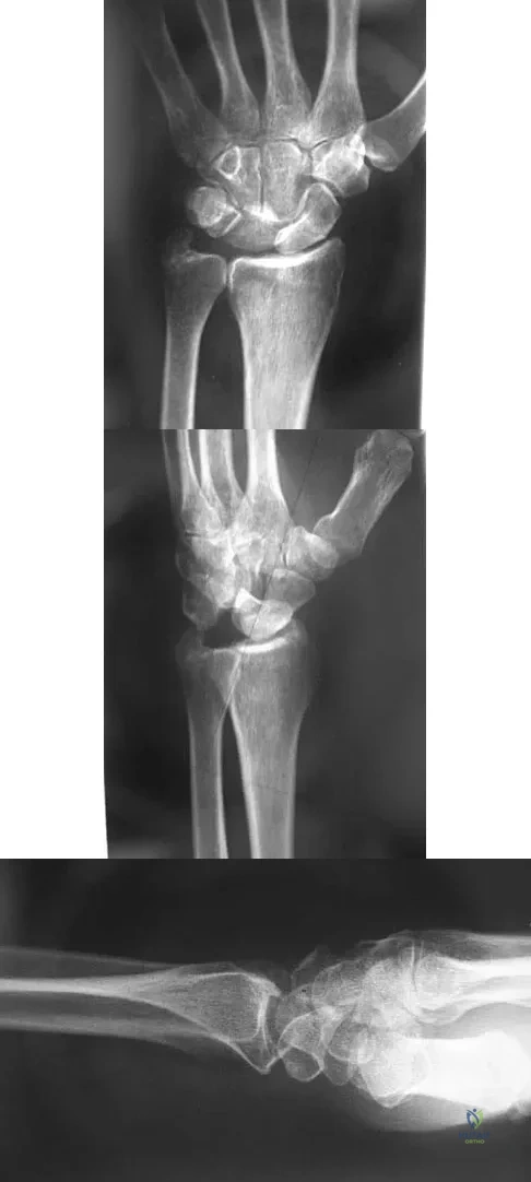
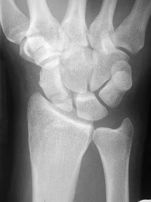
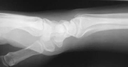
Figures 44a through 44c show the radiographs of an 18-year-old female soccer player who fell on her outstretched hand 1 day ago. She denies any history of wrist pain. Examination reveals tenderness at the anatomic snuffbox. Management should consist of
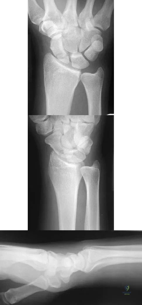


Explanation

The treatment of choice for proximal pole scaphoid fractures is open reduction and internal fixation with a differential pitch screw via a dorsal approach. Healing rates of 100% have been reported for these acute fractures. Casting results in slow healing, with recommendations including 16 weeks or more in a cast. Vascularized bone grafts are not indicated for acute fractures. Rettig ME, Raskin KB: Retrograde compression screw fixation of acute proximal pole scaphoid fractures. J Hand Surg 1999;24:1206-1210.

Question 11

An excessively large radial styloidectomy poses a risk for wrist instability. What ligament is at greatest risk for injury?


Explanation

The radioscaphocapitate ligament is the most radial of the extrinsic volar ligaments of the wrist. It has a mean attachment to the radius 4 mm from the tip of the radial styloid. Nakamura T, Cooney WP III, Lui WH, et al: Radial styloidectomy: A biomechanical study on the stability of the wrist joint. J Hand Surg Am 2001;26:85-93.

Question 12

What joint always remains uninvolved in all stages of scapholunate advanced collapse (SLAC) deformity of the wrist?


Explanation

The development of arthritis in SLAC wrist follows a consistent pattern. Beginning at the radial styloid to the scaphoid articulation, it progresses through the entire radioscaphoid joint and the midcarpal joint. In all stages, the radiolunate joint is spared, which is the basis for a scaphoid excision and four-corner fusion performed as a motion-sparing procedure for treatment of this condition. Wyrick JD: Proximal row carpectomy and intercarpal arthrodesis for the management of arthritis. J Am Acad Orthop Surg 2003;11:277-281. Watson HK, Ballett FL: The SLAC wrist: Scapholunate advanced collapse pattern of degenerative arthritis. J Hand Surg Am 1984;9:358-365.

Question 13

Free flap coverage for severe trauma to the upper extremity has the fewest complications when performed within what time period after injury?


Explanation

Flap necrosis and infection rates are lowest if free flap coverage is performed within 72 hours of injury. Delays beyond 72 hours are associated with a higher rate of complications. Godina M: Early microsurgical reconstruction of complex trauma of the extremities. Plast Reconstr Surg 1986;78:285-292.

Question 14

A 54-year-old woman with idiopathic carpal tunnel syndrome undergoes open carpal tunnel release with a flexor tenosynovectomy. The pathology from the tenosynovium is likely to show


Explanation

The tenosynovium excised at the time of a carpal tunnel release for idiopathic carpal tunnel syndrome rarely shows signs of acute or chronic inflammation. Fibrosis, edema, and vascular sclerosis are the most common histologic findings. A tenosynovectomy with a carpal tunnel release usually is not necessary in the treatment of idiopathic carpal tunnel syndrome. Shum C, Parisien M, Strauch RJ, et al: The role of flexor tenosynovectomy in the operative treatment of carpal tunnel syndrome. J Bone Joint Surg Am 2002;84:221-225. Fuchs PC, Nathan PA, Myers LD: Synovial histology in carpal tunnel syndrome. J Hand Surg Am 1991;16:753-758.

Question 15

Examination of a 10-year-old girl with a hypoplastic breast and atrophic pectoralis major may also reveal which of the following findings?


Explanation

Poland's syndrome has four main features: 1) short digits as the result of absence or shortening of the middle phalanx; 2) syndactyly of the short digits usually consisting of a simple, complete type; 3) hypoplasia of the hand and forearm; and 4) absence of the sternocostal head of the pectoralis major on the same side. Wilson MR, Louis DS, Stevenson TR: Poland's syndrome: Variable expression and associated anomalies. J Hand Surg 1988;13:880-882.

Question 16

Figures 45a and 45b show the radiographs of a 40-year-old woman with rheumatoid arthritis who is unable to straighten her ring and little fingers. Examination reveals that the fingers can be passively corrected, but she is unable to actively maintain the fingers in extension. Management should consist of
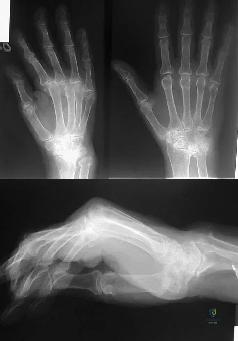


Explanation

The patient has extensor tendon ruptures at the level of the wrist that are the result of synovitis at the distal radioulnar joint (Vaughn-Jackson syndrome). Extensor indius proprius transfer appropriately matches strength and excursion of the ruptured extensor digiti quinti and extensor digitorum communis tendons. An extensor tenosynovectomy with distal radioulnar joint resection decreases the synovitis, which if left untreated may cause additional tendon ruptures. Radial head resection is used for posterior interosseous nerve compression secondary to radial head synovitis, and in this patient only two fingers are involved, which rules out this diagnosis. Dynamic splinting is not indicated for ruptured tendons. Metacarpophalangeal arthroplasties and imbrication of the sagittal bands are used for metacarpophalangeal arthritis and extensor tendon subluxation. If this was the problem, the patient should be able to maintain the fingers in extension after they are passively extended. Total wrist arthrodesis prevents the tenodesis effect, thus limiting effective tendon excursion and making the proposed transfer less effective. Feldon P, Terrono AL, Nalebuff EA, et al: Rheumatoid arthritis and other connective tissue diseases: Tendon ruptures, in Green DP, Hotchkiss RN, Pederson WC (eds): Green's Operative Hand Surgery, ed 4. New York, NY, Churchill Livingstone, 1999, pp 1669-1684. Moore JR, Weiland AJ, Valdata L: Tendon ruptures in the rheumatoid hand: Analysis of treatment and functional results in 60 patients. J Hand Surg Am 1987;12:9-14.

Question 17

Figures 46a through 46e show the radiographs of a 22-year-old man who injured his wrist in a motorcycle accident. He has no other injuries. What is the best course of action?
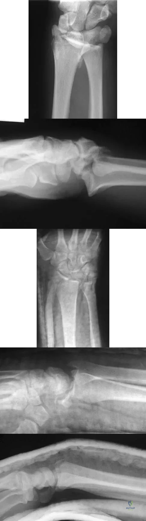


Explanation

The patient has a fracture-dislocation of the radiocarpal joint. Attached to the large radial styloid fragment are the extrinsic wrist ligaments to the carpus. This injury should be treated with open reduction and internal fixation of the styloid fracture. Radiolunate fusion or extrinsic ligament repair is suggested when the extrinsic ligaments are ruptured, resulting in ulnar translocation of the carpus. Dumontier C, Meyer ZU, Reckendorf G, et al: Radiocarpal dislocations: Classification and proposal for treatment: A review of twenty-seven cases. J Bone Joint Surg Am 2001;83:212.

Question 18

A 36-year-old nurse has had redness, pain, and small vesicles on the pulp of her middle finger for the past 3 days. Management should consist of


Explanation

Small vesicles on the fingers of a health care worker suggest a herpetic infection, and the management of choice is observation. Incision and drainage may result in a bacterial infection. Marsupialization is used in the treatment of a chronic paronychia. Calcium gluconate is used for hydrofluoric acid burns, and copper sulfate is used for white phosphorus burns. Fowler JR: Viral Infections. Hand Clin 1989;5:613-627.

Question 19

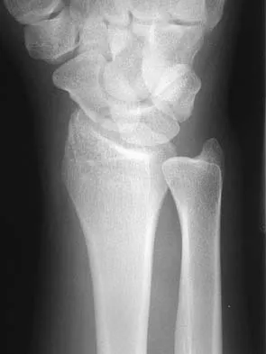
A 35-year-old man has numbness and tingling in the index, middle, and ring fingers. History reveals that he also has had vague wrist pain and stiffness since being injured in a motorcycle accident 1 year ago. Radiographs are shown in Figures 47a through 47c. Management should consist of


Explanation

The patient has a chronic unrecognized volar lunate dislocation. Median nerve compression is the result of the lunate displaced into the carpal tunnel. The diagnosis can be made by radiographs; MRI is not necessary. A volar approach allows median nerve decompression with excision of the lunate, whereas a dorsal approach facilitates excision of the scaphoid and triquetrum. Rettig ME, Raskin KB: Long-term assessment of proximal row carpectomy for chronic perilunate dislocations. J Hand Surg Am 1999;24:1231-1236.

Question 20

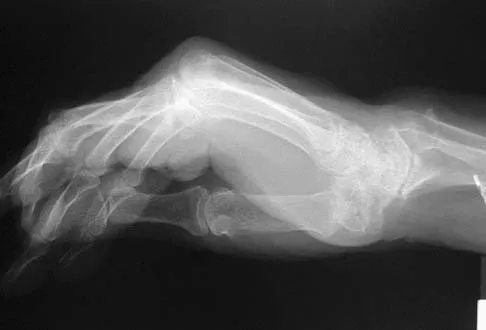
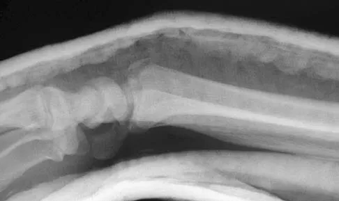
A 42-year-old woman has persistent thumb pain that she notes is worse with opening jars and turning her car key. Opponens splinting provides some relief, but she is poorly tolerant of the splint. Finkelstein's test is negative, and a carpometacarpal grind test is positive. The radiographs shown in Figures 48a and 48b reveal minimal degenerative changes at the first carpometacarpal joint. What is the best course of action?
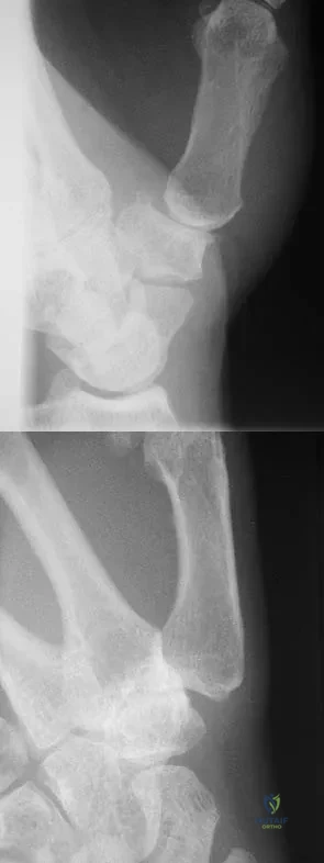


Explanation

The woman has early basilar thumb arthritis. An extension osteotomy will redirect the force to the dorsal, more uninvolved portion of the first carpometacarpal joint and has been reported to alleviate pain in these patients. Arthrodesis is usually reserved for young, typically male laborers. Thermal shrinkage and denervation are considered experimental at this time. Interposition arthroplasty is typically used for more advanced stages of arthritis. Tomaino MM: Treatment of Eaton stage I trapeziometacarpal disease with thumb metacarpal extension osteotomy. J Hand Surg Am 2000;25:1100-1106. Pellegrini VD Jr, Parentis M, Judkins A, et al: Extension metacarpal osteotomy in the treatment of trapeziometacarpal osteoarthritis: A biomechanical study. J Hand Surg Am 1996;21:16-23.

Question 21

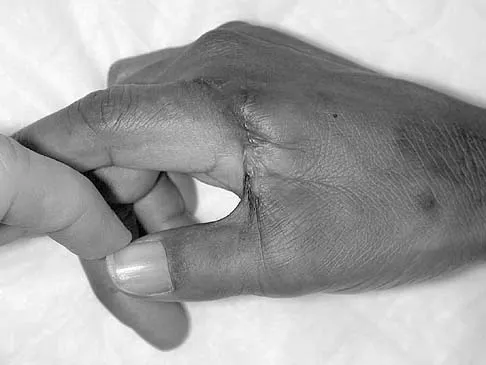
A 45-year-old man sustains a low-velocity gunshot wound to the base of the right thumb. The open wound is allowed to heal by secondary intention, resulting in a contracture of the first web space. Clinical photographs are shown in Figures 49a through 49c. Treatment should now consist of
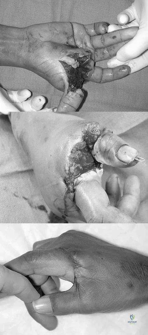


Explanation

The contracture is too large for a Z-plasty, which allows a 75% increase in length. Excision of the scar with placement of a skin graft is prone to contracture. A posterior interosseous fasciocutaneous flap will provide enough well-vascularized tissue and is well suited to reach the first dorsal web space. Buchler U, Frey HP: Retrograde posterior interosseous flap. J Hand Surg Am 1991;16:283-292.

Question 22

The vessel seen in the clinical photographs shown in Figures 50a and 50b (1,2 intercompartmental supraretinacular artery) is being dissected to be used as a source of vascularized bone graft for a patient who is scheduled to undergo internal fixation of a scaphoid nonunion. This vessel is a branch of what artery?
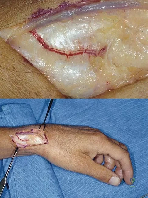


Explanation

The 1,2 intercompartmental supraretinacular artery is a branch of the radial artery. The vessel provides a reliable source of vascularized bone graft with an adequate pedicle length for use in scaphoid nonunions. Sheetz KK, Bishop AT, Berger RA: The arterial blood supply of the distal radius and ulna and its potential use in vascularized pedicled bone grafts. J Hand Surg 1995;20:902-914.

Question 23

The flap shown in the clinical photograph seen in Figure 51 is based on what arterial supply?

Upper Extremity 2005 Practice Questions: Set 5 (Solved) - Figure 21


Explanation

The groin flap is based on the superficial circumflex iliac artery, an axial flap that has been a mainstay of providing soft-tissue coverage of the upper extremity. Flaps as large as 35 cm in length and 15 cm in width have been reported. An advantage of the flap is that when used as a pedicle flap, the donor site can be closed directly. A disadvantage of the flap is that it can be quite bulky and can have a thick layer of subcutaneous fat. The superficial circumflex iliac artery travels lateral and superficial to the fascia and below and parallel to the inguinal ligament. It is helpful to elevate the fascia at the medial border of the sartorius muscle to include the deep and superficial branches of the artery for improved flap survival. McGregor IA, Jackson IT: The groin flap. Br J Plast Surg 1972;25:3-9.

Question 24

A 63-year-old woman who sustained a distal radial fracture 2 months ago now reports that she is unable to achieve active extension of the thumb at the interphalangeal joint. What type of trauma may lead to this clinical finding?


Explanation

Nondisplaced distal radial fractures have a higher rate of spontaneous rupture of the extensor pollicis longus tendon. The extensor mechanism is felt to impinge on the tendon following a nondisplaced fracture and causes either a mechanical attrition of the tendon or a local area of ischemia in the tendon. Helal B, Chen SC, Iwegbu G: Rupture of the extensor pollicis longus tendon in undisplaced Colles' type of fracture. Hand 1982;14:41-47.

Question 25

What radiographic view will best reveal degeneration of the pisotriquetral joint in a patient who is being evaluated for pisotriquetral arthrosis?


Explanation

The pisotriquetral joint is best seen on a lateral view in 30 degrees of supination. The carpal tunnel view provides visualization of the joint but to a lesser extent. The other views do not provide clear and accurate visualization. Paley D, McMurty RY, Cruickshank B: Pathologic conditions of the pisiform and pisotriquetral joint. J Hand Surg Am 1987;12:110-119.

Question 26

In the treatment of a 4-part proximal humerus fracture with a shoulder hemiarthroplasty in an elderly patient, what is the most critical factor for achieving a good functional outcome?


Explanation

Anatomic reduction and secure healing of the tuberosities around the prosthesis is the most critical factor determining a successful functional outcome in proximal humerus hemiarthroplasty. Failure of tuberosity healing leads to poor active elevation and poor clinical results.

Question 27

A 25-year-old weightlifter feels a sudden pop in his anterior axilla while bench pressing. He has ecchymosis and loss of the anterior axillary fold. If surgical repair is chosen, to which anatomical structure should the tendon be reattached?


Explanation

The pectoralis major tendon normally inserts onto the lateral lip of the bicipital groove of the humerus. Surgical repair of a rupture involves reattaching the tendon to this native footprint.

Question 28

A 35-year-old man falls onto his shoulder. Radiographs show a 100% to 300% superior displacement of the clavicle relative to the acromion. Which ligaments are disrupted in this classic Type V acromioclavicular injury?


Explanation

A Type V AC joint separation is characterized by severe superior displacement of the distal clavicle. This requires complete disruption of both the AC and CC ligaments, as well as significant tearing of the deltotrapezial fascia.

Question 29

An anterior single-incision approach to distal biceps tendon repair places which of the following nerves at greatest risk of iatrogenic injury?


Explanation

The lateral antebrachial cutaneous (LABC) nerve is the most commonly injured nerve during the single-incision anterior approach to the distal biceps. The posterior interosseous nerve (PIN) is classically at higher risk during a two-incision approach.

Question 30

A 42-year-old male presents with isolated weakness of the infraspinatus muscle. MRI shows a ganglion cyst compressing a nerve. At what anatomical location is the compression most likely occurring?


Explanation

Compression of the suprascapular nerve at the spinoglenoid notch results in isolated infraspinatus weakness, as the branch to the supraspinatus has already been given off. Compression at the suprascapular notch would affect both the supraspinatus and infraspinatus.

Question 31

A 28-year-old overhead athlete is diagnosed with a Type II SLAP tear. During diagnostic arthroscopy, which of the following defines a Type II tear?


Explanation

A Type II SLAP lesion involves pathological detachment of the superior labrum and the origin of the long head of the biceps from the glenoid. It is the most common type of SLAP tear requiring surgical repair in overhead athletes.

Question 32

A 65-year-old man with chronic massive rotator cuff tear presents with pseudo-paralysis of the shoulder and severe glenohumeral arthritis. Which of the following structures must remain intact for a reverse total shoulder arthroplasty to be successful?


Explanation

A functional deltoid muscle is an absolute prerequisite for a reverse total shoulder arthroplasty. It provides the primary motor function to elevate the arm once the center of rotation is medialized and distalized.

Question 33

A 50-year-old man complains of inability to internally rotate his arm behind his back following a fall. Examination shows increased passive external rotation compared to the contralateral side. The lift-off and belly-press tests are positive. Which tendon is ruptured?


Explanation

The subscapularis is the primary internal rotator of the shoulder. A tear results in increased passive external rotation and profound weakness in active internal rotation, demonstrated by positive lift-off and belly-press tests.

Question 34

A patient with a mid-shaft humeral fracture treated non-operatively in a functional brace develops a new-onset radial nerve palsy 3 weeks post-injury. What is the most appropriate next step in management?


Explanation

While primary radial nerve palsies with closed humeral fractures are typically observed, a secondary (delayed) radial nerve palsy developing during non-operative management (e.g., bracing or manipulation) is a strong indication for surgical exploration.

Question 35

During the standard deltopectoral approach to the proximal humerus, the cephalic vein is identified. To minimize bleeding and preserve its main venous drainage, the cephalic vein should ideally be retracted in which direction?


Explanation

The cephalic vein marks the deltopectoral interval. It is typically retracted laterally with the deltoid muscle to preserve its main venous tributaries from the deltoid, thereby minimizing bleeding.

Question 36

A 22-year-old man presents to the ER after a seizure. His arm is locked in internal rotation. An axillary radiograph reveals a posterior shoulder dislocation with a 30% reverse Hill-Sachs lesion. What is the most appropriate surgical management?


Explanation

A reverse Hill-Sachs lesion involving 20-40% of the articular surface is typically managed with a McLaughlin procedure (or modified McLaughlin). This transfers the lesser tuberosity and/or subscapularis tendon into the anterior humeral defect to prevent re-engagement.

Question 37

A 30-year-old patient falls from a height, sustaining a displaced transverse fracture of the olecranon. The fracture is treated with tension band wiring. What is the most common complication of this procedure?


Explanation

The most common complication of tension band wiring for olecranon fractures is symptomatic hardware prominence. This frequently necessitates a second surgery for hardware removal after the fracture has united.

Question 38

A patient sustains an anterior shoulder dislocation that is reduced in the ER. Post-reduction, there is a large area of numbness over the lateral aspect of the shoulder, and the patient cannot contract the deltoid muscle. Which nerve is most likely injured?


Explanation

The axillary nerve is the most commonly injured nerve during an anterior shoulder dislocation. It provides motor innervation to the deltoid and teres minor, and sensory innervation to the lateral shoulder (the badge area).

Question 39

A 55-year-old female presents with severe shoulder pain. Radiographs reveal superior migration of the humeral head. Which physical exam finding would most reliably indicate a massive, irreparable posterior-superior rotator cuff tear involving the teres minor?


Explanation

A positive Hornblower's sign (inability to maintain external rotation in 90 degrees of abduction) indicates severe weakness or absence of the teres minor. This is highly suggestive of a massive, irreparable posterior-superior rotator cuff tear.

Question 40

A 35-year-old male suffers a high-energy dashboard injury, sustaining a posterior sternoclavicular dislocation. What is the most immediate life-threatening complication associated with this specific injury?


Explanation

Posterior sternoclavicular dislocations can compress or injure critical mediastinal structures. Tracheal compression causing rapid airway compromise is the most immediate, life-threatening complication, requiring urgent reduction.

Question 41

In a patient with a 'terrible triad' injury of the elbow (elbow dislocation, radial head fracture, coronoid fracture), what is the most appropriate surgical sequence to restore elbow stability?


Explanation

The standard surgical sequence for a terrible triad injury works deep to superficial. This involves fixation of the coronoid first, followed by radial head repair or replacement, and finally repair of the lateral collateral ligament (LCL) complex.

Question 42

A patient falls on an outstretched hand and complains of lateral elbow pain. Radiographs show a comminuted radial head fracture. There is also marked tenderness over the distal radioulnar joint (DRUJ). If the radial head is excised without replacement, what complication is most likely to occur?


Explanation

The patient has an Essex-Lopresti lesion, consisting of a radial head fracture, interosseous membrane disruption, and DRUJ injury. Excision of the radial head without prosthetic replacement in this setting will lead to profound proximal radial migration and chronic wrist pain.

Question 43

A 72-year-old man presents with chronic right shoulder pain, an inability to actively elevate his arm past 45 degrees, and a preserved passive range of motion. Radiographs demonstrate superior migration of the humeral head with articulation against the acromion. What is the most appropriate surgical management to restore active elevation in this patient?


Explanation

Reverse total shoulder arthroplasty is indicated for patients with rotator cuff tear arthropathy and pseudoparalysis. It shifts the center of rotation medially and distally, tensioning the deltoid and maximizing its mechanical advantage for shoulder elevation.

Question 44

Which of the following is the most reliable radiographic predictor of humeral head ischemia (avascular necrosis) following a proximal humerus fracture?


Explanation

Hertel identified specific radiographic risk factors for humeral head ischemia. A disrupted medial hinge (>2 mm displacement) and a short metaphyseal head extension (<8 mm) are the strongest predictors of avascular necrosis due to compromise of the ascending branch of the anterior humeral circumflex artery.

Question 45

A 35-year-old male cyclist sustains a completely displaced midshaft clavicle fracture with 2.5 cm of shortening. According to prospective randomized trials, what is the primary benefit of operative fixation compared to nonoperative management for this specific injury?


Explanation

In totally displaced, shortened (>2 cm) midshaft clavicle fractures, nonoperative management has historically been associated with a significantly higher rate of nonunion and symptomatic malunion. Operative fixation in these specific cases lowers these risks and often leads to earlier functional recovery.

Question 46

A 28-year-old competitive weightlifter feels a tearing sensation in his anterior chest while bench pressing. Examination reveals loss of the anterior axillary fold and weakness in internal rotation. Where is the most common anatomic location for this specific tendon rupture?


Explanation

Pectoralis major ruptures almost exclusively occur during eccentric loading activities like bench pressing. The most common site of failure is an avulsion of the sternal head tendon from its insertion on the proximal humerus.

Question 47

A 45-year-old woman falls on an outstretched hand and sustains a comminuted radial head fracture, wrist pain, and instability of the distal radioulnar joint (DRUJ). Radial head excision is contraindicated in this setting primarily due to the risk of:


Explanation

This presentation is consistent with an Essex-Lopresti fracture-dislocation, which includes a longitudinal tear of the interosseous membrane. Excising the radial head removes the primary restraint to proximal radial migration, leading to severe ulnocarpal impaction and wrist pain.

Question 48

A 22-year-old football player has recurrent anterior shoulder instability. A 3D CT scan reveals 27% bone loss of the anteroinferior glenoid. What is the most appropriate surgical intervention to minimize the risk of recurrent dislocation?


Explanation

Soft-tissue Bankart repairs have unacceptably high failure rates when glenoid bone loss exceeds 20-25%. A bony augmentation procedure, such as the Latarjet (coracoid transfer), is required to restore the articular arc and provide a sling effect via the conjoint tendon.

Question 49

A 30-year-old man sustains a closed, spiral fracture of the distal third of the humeral shaft. Upon presentation in the emergency department, his radial nerve function is completely intact. Following closed reduction and splinting, he immediately exhibits a dense wrist drop and loss of finger extension. What is the most appropriate next step in management?


Explanation

A secondary radial nerve palsy that develops immediately after a closed reduction attempt of a humeral shaft fracture strongly suggests nerve entrapment within the fracture site. This is a classic, absolute indication for urgent surgical exploration and internal fixation.

Question 50

When evaluating massive rotator cuff tears on MRI, the Goutallier classification system is frequently used. What specific pathologic feature does this system grade to determine the prognosis of a rotator cuff repair?


Explanation

The Goutallier classification evaluates the severity of fatty infiltration within the rotator cuff muscle bellies, originally described on CT and adapted for MRI. High grades (greater than 50% fat) correlate with a poor prognosis for tendon healing and functional recovery following repair.

Question 51

A 35-year-old mechanic presents with vague posterior shoulder pain. MRI demonstrates isolated muscle edema and early atrophy isolated to the teres minor muscle. Which anatomic space is most likely compromised?


Explanation

Isolated atrophy of the teres minor is a hallmark of quadrilateral space syndrome. This condition involves compression of the axillary nerve and posterior humeral circumflex artery within the quadrilateral space.

Question 52

Which of the following defines the specific pattern of injury known as the 'terrible triad' of the elbow?


Explanation

The terrible triad of the elbow consists of an elbow dislocation, a radial head fracture, and a coronoid fracture. It is nearly always accompanied by a complete avulsion or tear of the lateral ulnar collateral ligament (LUCL) complex.

Question 53

A 7-year-old boy falls on an outstretched arm and sustains a Monteggia fracture-dislocation. According to the Bado classification, what represents a Type I injury?


Explanation

The Bado classification categorizes Monteggia fractures based on the direction of radial head dislocation. Type I, the most common type in children, involves an anterior dislocation of the radial head with an associated ulnar diaphyseal fracture.

Question 54

A 45-year-old laborer undergoes surgical repair of a distal biceps tendon rupture using a two-incision technique. Compared to a single anterior incision technique, the two-incision approach is associated with a higher risk of which specific complication?


Explanation

The two-incision technique for distal biceps repair was developed to minimize the risk to the radial nerve (PIN) and lateral antebrachial cutaneous nerve seen in single-incision approaches. However, it carries a higher risk of heterotopic ossification and potentially debilitating radioulnar synostosis.

Question 55

A 55-year-old woman presents with a highly comminuted, intra-articular fracture of the distal radius. Radiographs show a distinct volar marginal fragment that has subluxated palmarly with the carpus. When plating this specific fracture pattern, what is the most critical biomechanical principle for stabilization?


Explanation

A volar shear fracture of the distal radius (Volar Barton's fracture) represents an inherently unstable injury due to carpal subluxation. It requires an open reduction and volar buttress plating to mechanically counteract the proximal and palmar shearing forces.

Question 56

A 50-year-old man presents with chronic shoulder weakness. Clinical examination demonstrates a positive lift-off test and an asymmetric increase in passive external rotation compared to the contralateral side. Which rotator cuff tendon is predominantly injured?


Explanation

The subscapularis is the primary internal rotator of the shoulder. A complete tear results in weakness during internal rotation (positive lift-off or belly-press test) and an unresisted increased passive external rotation due to the loss of the anterior capsular restraint.

Question 57

A 17-year-old rugby player sustains high-energy trauma to the anterior chest. Clinical exam shows a prominence over the medial clavicle with dyspnea. Radiographs appear to show a posterior sternoclavicular dislocation. In a patient of this age, what is the most likely true underlying pathology?


Explanation

The medial clavicular physis is the last growth plate in the body to fuse, often remaining open until age 25. Injuries mimicking a sternoclavicular dislocation in adolescents and young adults are typically Salter-Harris physeal fractures.

Question 58

A 65-year-old female sustains a 4-part proximal humerus fracture. Which of the following radiographic and clinical findings is most predictive of humeral head ischemia and subsequent avascular necrosis?


Explanation

According to Hertel's criteria, a short calcar length (metaphyseal extension < 8 mm) and a disrupted medial hinge are highly predictive of humeral head ischemia following proximal humerus fractures.

Question 59

A 45-year-old heavy laborer presents with a massive, irreparable posterosuperior rotator cuff tear. He has intact subscapularis function, active forward elevation to 140 degrees, and no glenohumeral arthritis. What is the most appropriate surgical management?


Explanation

Latissimus dorsi transfer is indicated for massive, irreparable posterosuperior cuff tears in younger, active patients with an intact subscapularis and preserved forward elevation.

Question 60

A 30-year-old man sustains a closed distal-third spiral humeral shaft fracture (Holstein-Lewis pattern). Clinical examination reveals an immediate, complete radial nerve palsy. What is the most appropriate initial management?


Explanation

Closed humeral shaft fractures with primary radial nerve palsies are initially managed non-operatively with bracing. Over 80% of these palsies spontaneously recover.

Question 61

During surgical reconstruction of a 'terrible triad' injury of the elbow, which of the following is the generally recommended sequence of repair to best restore stability?


Explanation

The standard surgical protocol for a terrible triad injury proceeds from deep to superficial: fixation of the coronoid, followed by radial head repair or replacement, and finally lateral collateral ligament (LCL) repair.

Question 62

A 55-year-old man complains of weakness and pain in his shoulder following a fall. Examination demonstrates a positive 'belly-press' test and a positive 'lift-off' test. Which of the following structures is most likely injured?


Explanation

The belly-press and lift-off tests isolate and specifically evaluate the integrity and strength of the subscapularis tendon.

Question 63

A 35-year-old patient presents with a locked posterior shoulder dislocation after a seizure. CT reveals an anteromedial humeral head impression fracture (reverse Hill-Sachs lesion) involving 45% of the articular surface. What is the most appropriate surgical treatment?


Explanation

Reverse Hill-Sachs defects >40% typically require structural allograft reconstruction or arthroplasty to prevent recurrent instability. Lesser tuberosity transfer is generally reserved for defects between 20% and 40%.

Question 64

A 40-year-old bodybuilder feels a 'pop' in his antecubital fossa during a heavy deadlift, accompanied by bruising and a positive hook test. If a single-incision anterior approach is used for repair, which nerve is at the highest risk of iatrogenic injury?


Explanation

The lateral antebrachial cutaneous nerve is the most frequently injured structure during a single-incision anterior approach for distal biceps tendon repair due to its superficial course.

Question 65

The proximal pole of the scaphoid is highly susceptible to avascular necrosis following a fracture due to its unique retrograde blood supply. Which of the following arteries provides the primary vascularity to the proximal pole?


Explanation

The dorsal carpal branch of the radial artery enters the scaphoid at the dorsal ridge and supplies the proximal 80% of the bone via a precarious retrograde flow.

Question 66

A 30-year-old man sustains a Bado Type I Monteggia fracture. What is the most critical step in achieving and maintaining the anatomic reduction of the radial head?


Explanation

In Monteggia fractures, the primary requirement for reducing and stabilizing the dislocated radial head is achieving anatomic length and alignment of the ulna through rigid internal fixation.

Question 67

Which of the following clinical scenarios is an absolute indication for operative fixation of an acute midshaft clavicle fracture?


Explanation

Absolute indications for open reduction and internal fixation of a clavicle fracture include open fractures, neurovascular compromise, and skin tenting causing impending necrosis.

Question 68

A 24-year-old professional baseball pitcher complains of a 'dead arm' and pain during the late cocking phase of throwing. MR arthrography confirms an isolated Type II SLAP lesion. After 6 months of failed conservative therapy, what is the most appropriate surgical intervention?


Explanation

In young, elite overhead throwing athletes, symptomatic Type II SLAP tears that fail non-operative management are typically treated with arthroscopic repair. Tenodesis is generally favored in older, non-throwing populations.

Question 69

A 28-year-old man presents with acute median neuropathy after falling onto an extended wrist. Radiographs show volar displacement of the lunate (perilunate dislocation). According to the Mayfield classification, what is the initial ligament to fail in this progressive instability pattern?


Explanation

Mayfield described a progressive sequence of perilunate instability that strictly begins with the disruption of the scapholunate ligament (Stage I), progressing to capitate, triquetral, and finally lunate dislocation.

Question 70

When stabilizing a dorsally comminuted distal radius fracture with a volar locking plate, what is the primary biomechanical advantage provided by the distal locking screws?


Explanation

Volar locking plates utilize threaded screw heads that lock into the plate, creating a fixed-angle construct. This acts as a subchondral raft to support the joint surface and prevent late dorsal collapse without needing bicortical purchase.

Question 71

A 30-year-old competitive weightlifter sustains an acute pectoralis major tear while bench pressing. Examination reveals loss of the anterior axillary fold. Where do these ruptures most commonly occur?


Explanation

Pectoralis major ruptures almost exclusively occur at the distal musculotendinous junction or the tendon insertion onto the proximal humerus during maximal eccentric contraction.

Question 72

A 72-year-old female presents with severe right shoulder pain, pseudoparalysis, and radiographic evidence of superior humeral head migration with acetabularization of the acromion. What is the most reliable definitive surgical treatment to restore active elevation?


Explanation

Reverse total shoulder arthroplasty is the gold standard for rotator cuff tear arthropathy with pseudoparalysis. It medializes and distalizes the center of rotation, maximizing the moment arm of the deltoid to restore forward elevation.

Question 73

A 25-year-old cyclist falls directly onto his shoulder. Radiographs demonstrate superior displacement of the clavicle relative to the acromion by approximately 150%. Which ligaments are fully disrupted in this Type III acromioclavicular (AC) injury?


Explanation

A Type III AC joint separation involves complete disruption of both the acromioclavicular ligaments and the coracoclavicular (conoid and trapezoid) ligaments.

Question 74

A 45-year-old man sustains a diaphyseal fracture of the distal third of the radius with associated clinical disruption of the distal radioulnar joint (DRUJ). What is the standard of care for this Galeazzi fracture?


Explanation

Galeazzi fractures in adults are highly unstable and necessitate open reduction and rigid internal fixation (plating) of the radius. Anatomic radial restoration is required to secondarily stabilize the DRUJ.

Question 75

When surgically treating an adult intercondylar distal humerus fracture (AO Type 13-C), what is the optimal plate configuration required for stable internal fixation to allow early range of motion?


Explanation

Dual plating (either parallel or orthogonal) is the standard of care for bicolumnar distal humerus fractures in adults, providing adequate biomechanical stability to permit early elbow mobilization.

Question 76

A 20-year-old football player sustains an anterior shoulder dislocation. MRI reveals an avulsion of the anterior labroligamentous complex with an attached avulsed fragment of glenoid bone. What is the specific eponym for this lesion?


Explanation

A Bony Bankart lesion describes a detachment of the anteroinferior labrum that includes an osseous fracture of the anterior glenoid rim.

Question 77

A 72-year-old man presents with severe right shoulder pain and an inability to actively elevate his arm above 60 degrees. Radiographs demonstrate a massive, retracted rotator cuff tear with an acromiohumeral interval of 2 mm and advanced glenohumeral arthritis. What is the most reliable definitive surgical treatment for this patient?


Explanation

Reverse total shoulder arthroplasty is the treatment of choice for rotator cuff tear arthropathy with pseudoparalysis. It medializes and distalizes the center of rotation, relying on the deltoid to overcome the deficient rotator cuff.

Question 78

A 45-year-old man feels a 'pop' in his antecubital fossa while lifting a heavy box and experiences weakness in forearm supination. He undergoes a distal biceps tendon repair using a traditional two-incision technique. Compared to a single anterior incision approach, this technique carries a historically higher risk of which of the following complications?


Explanation

The two-incision technique for distal biceps repair carries a higher risk of heterotopic ossification and radioulnar synostosis. Conversely, the single-incision approach has a higher rate of lateral antebrachial cutaneous nerve (LABCN) neuropraxia.

Question 79

A 35-year-old woman falls on an outstretched hand and sustains a 'terrible triad' injury of the elbow. During the standard surgical protocol for this injury, which of the following structures is typically addressed or repaired last?


Explanation

The standard surgical algorithm for a terrible triad injury proceeds from deep to superficial and anterior to posterior: coronoid fixation, radial head fixation/replacement, followed by LCL repair. The MCL is typically only addressed if the elbow remains unstable after the lateral and anterior structures are stabilized.

Question 80

A 22-year-old collegiate baseball pitcher reports deep posterior shoulder pain during the late cocking phase of throwing. Arthroscopic evaluation reveals undersurface fraying of the rotator cuff. Where is this lesion most likely located?


Explanation

Internal impingement typically occurs in overhead athletes during maximal abduction and external rotation. The articular surface of the posterior supraspinatus and anterior infraspinatus abuts the posterosuperior glenoid labrum, causing fraying.

Question 81

A 30-year-old man sustains a closed, completely displaced spiral fracture of the distal third of the humerus (Holstein-Lewis fracture). In the emergency department, his radial nerve function is intact. Following a closed reduction and application of a coaptation splint, he loses the ability to extend his wrist and fingers. What is the most appropriate next step in management?


Explanation

A secondary radial nerve palsy that develops after a closed reduction attempt of a humeral shaft fracture is an absolute indication for surgical exploration. This clinical presentation suggests the nerve has become entrapped within the fracture fragments.

Question 82

A 55-year-old woman was treated in a cast for a non-displaced distal radius fracture 6 weeks ago. She now presents with a sudden, painless inability to actively extend her thumb interphalangeal joint. What is the most appropriate surgical treatment?


Explanation

EPL rupture is a known complication of non-displaced distal radius fractures due to ischemia or attrition at Lister's tubercle. Because the tendon ends are typically retracted and degenerated, primary repair is rarely possible, making EIP to EPL transfer the gold standard.

Question 83

A 40-year-old man sustains a highly comminuted radial head fracture from a high-energy fall. An isolated radial head excision is performed. Three months later, he develops progressive proximal migration of the radius and severe ulnar-sided wrist pain. Which associated injury was most likely missed at the initial presentation?


Explanation

An Essex-Lopresti lesion involves a radial head fracture combined with a longitudinal tear of the interosseous membrane and disruption of the distal radioulnar joint (DRUJ). Excising the radial head in this setting removes the remaining restraint to proximal radial migration, leading to ulnar impaction.

Question 84

A 28-year-old bodybuilder complains of vague shoulder weakness. Physical examination demonstrates prominent medial winging of the scapula when the patient performs a wall push-up. Which nerve is most likely injured?


Explanation

Medial winging of the scapula is caused by paralysis or weakness of the serratus anterior muscle. The serratus anterior is innervated by the long thoracic nerve, and this deficit is classically highlighted during a wall push-up.

Question 85

A 25-year-old cyclist sustains a closed, midshaft clavicle fracture after a fall over the handlebars. Which of the following radiographic findings is considered the strongest indication for operative fixation to prevent symptomatic nonunion?


Explanation

Displacement greater than 100% and shortening greater than 2 cm are strong indications for ORIF of a midshaft clavicle fracture. Non-operative management of fractures with these characteristics carries a significantly higher risk of nonunion and symptomatic malunion.

Question 86

During the physical examination of a patient with suspected rotator cuff pathology, which of the following tests is considered the most sensitive and specific for detecting an isolated tear of the upper border of the subscapularis tendon?


Explanation

The bear-hug test is highly sensitive for evaluating the upper portion of the subscapularis tendon. In contrast, the lift-off test primarily isolates the lower portion of the subscapularis muscle.

Question 87

A 30-year-old professional volleyball player presents with painless weakness in external rotation of the right shoulder. Physical examination reveals isolated atrophy in the infraspinatus fossa, while supraspinatus strength and bulk are perfectly normal. At which anatomic location is nerve compression most likely occurring?


Explanation

Compression of the suprascapular nerve at the spinoglenoid notch selectively denervates the infraspinatus muscle, causing isolated external rotation weakness. Compression more proximally at the suprascapular notch affects both the supraspinatus and infraspinatus.

Question 88

A 24-year-old man suffers an anterior shoulder dislocation. Prior to reduction, he has decreased sensation over the lateral aspect of his deltoid. Due to the involved nerve, which muscle's function must be most closely monitored for associated weakness?


Explanation

The axillary nerve provides sensation to the lateral shoulder via the superior lateral cutaneous nerve of the arm and motor innervation to the deltoid and teres minor. It is the most frequently injured nerve in anterior shoulder dislocations.

Question 89

A 32-year-old construction worker sustains a Galeazzi fracture-dislocation after being struck by a heavy beam. Which of the following accurately defines this specific injury pattern?


Explanation

A Galeazzi fracture-dislocation is characterized by a fracture of the distal third of the radial shaft accompanied by a disruption of the distal radioulnar joint (DRUJ). In adults, this unstable injury requires operative fixation of the radius.

Question 90

A 29-year-old man is struck on the forearm with a blunt object, resulting in an isolated, non-displaced midshaft ulnar fracture ('nightstick' fracture). Angulation is less than 5 degrees. What is the most appropriate treatment for this injury?


Explanation

Isolated, non-displaced or minimally displaced ulnar shaft fractures (<50% displacement, <10 degrees angulation) are highly stable. They are best treated non-operatively with a functional brace and early range of motion to prevent stiffness and promote healing.

None

Clinic OS
Medically Verified Content by
Prof. Clinic OS
Consultant Orthopedic & Spine Surgeon
Chapter Index