العربية
Part of the Master Guide

100 High-Yield Orthopedic MCQs: Shoulder & Elbow | Mock Exam 251

Shoulder Orthopedic MCQs (Set 1): Rotator Cuff & Instability | AAOS & ABOS Board Review

23 Apr 2026 63 min read 107 Views
Shoulder 2000 MCQs - Part 1

Key Takeaway

This high-yield question set (Set 1) for AAOS, ABOS, and OITE exams focuses on comprehensive shoulder pathology. It covers rotator cuff tears, glenohumeral instability, impingement syndrome, and arthroscopic techniques, providing essential practice for board certification.

Shoulder Orthopedic MCQs (Set 1): Rotator Cuff & Instability | AAOS & ABOS Board Review

Comprehensive 100-Question Exam


00:00

Start Quiz

Question 1

Which of the following statements best describes why the ulnar nerve is most prone to neuropathy at the elbow?


Explanation

The ulnar nerve is more prone to neuropathy than the radial or median nerves for many reasons. It has the greatest longitudinal excursion required to accommodate elbow range of motion, subjecting it to potential traction forces. The dimensions of the entrance of the cubital tunnel change with elbow motion, potentially causing compression in flexion. For these two reasons, the ulnar nerve is subjected to both compression and traction during elbow motion. Although it passes between two muscle heads as it enters the forearm, so do the median and radial nerves. Finally, the vascular supply is adequate because of the anastamoses between the superior ulnar collateral artery, the posterior ulnar recurrent artery, and the inferior ulnar collateral artery. Norris TR (ed): Orthopaedic Knowledge Update: Shoulder and Elbow. Rosemont, IL, American Academy of Orthopaedic Surgeons, 1997, pp 369-378. Prevel CD, Matloub HS, Ye Z, Sanger JR, Yousif NJ: The extrinsic blood supply of the ulnar nerve at the elbow: An anatomic study. J Hand Surg Am 1993;18:433-438.

Question 2

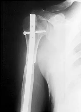
Figure 1 shows the radiograph of a 71-year-old man who has had increasing pain and weakness in his shoulder for the past 3 years. Nonsurgical management has failed to provide relief. Examination shows 130 degrees of active forward flexion and intact external rotation strength. During surgery, a 1- x 1-cm rotator cuff tear involving the supraspinatus is encountered. Treatment should include


Explanation

Given the size of the rotator cuff tear, it is likely to be repaired; therefore, the treatment of choice is a total shoulder replacement with rotator cuff repair. Severe rotator cuff insufficiency can lead to early glenoid failure because of superior instability, and glenoid resurfacing should be avoided in those instances. Boyd AD Jr, Thomas WH, Scott RD, Sledge CB, Thornhill TS: Total shoulder arthroplasty versus hemiarthroplasty: Indications for glenoid resurfacing. J Arthroplasty 1990;5:329-336.


Question 3

Which of the following is considered the cause of Milwaukee shoulder, a joint disease similar to rotator cuff arthropathy?


Explanation

Neer and associates focused on mechanical and nutritional factors as the etiology of rotator cuff arthropathy. McCarty and associates, in describing a similar syndrome known as Milwaukee shoulder, focused on an inflammatory cause in proposing the pathogenic role of hydroxyapatite, a basic calcium phosphate. Neer CS II, Craig EV, Fukuda H: Cuff-tear arthropathy. J Bone Joint Surg Am 1983;65:1232-1244.


Question 4

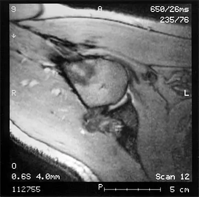
The MRI scan of the shoulder shown in Figure 2 was performed with the arm in abduction and external rotation. The image reveals what condition?


Explanation

Internal impingement of the shoulder is now a well-recognized cause of shoulder pain in the throwing athlete. First described by Walch and associates, it involves contact of the rotator cuff and labrum in the maximally externally rotated and abducted shoulder, such as in the late cocking phase of the throwing motion. Schickendantz and associates have shown this contact to be physiologic in most patients and becoming pathologic with repetitive overhead activity. Schickendantz MS, Ho CP, Keppler L, Shaw BD: MR imaging of the thrower's shoulder: Internal impingement, latissimus dorsi/subscapularis strains, and related injuries. Magn Reson Imaging Clin N Am 1999;7:39-49. Walch G, Boileau P, Noel E, et al: Impingement of the deep surface of the supraspinatus tendon on the posterosuperior glenoid rim: An arthroscopic study. J Shoulder Elbow Surg 1992;1:238-245.


Question 5

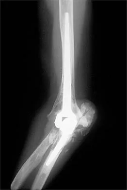
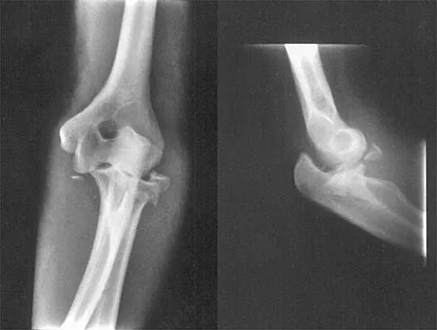
Figure 3 shows the radiographs of a 32-year-old man who fell 12 feet onto his outstretched arm and sustained a fracture-dislocation of the elbow. Initial management consisted of closed reduction of the dislocation. Surgical treatment should now include repair or reduction and fixation of the


Explanation

The radiographs show fractures of the coronoid and radial head. The medial collateral ligament has been avulsed from the ulnar insertion, and there is a valgus opening on the medial side. The lateral collateral ligament is always disrupted in elbow dislocations and fracture-dislocations that occur secondary to falls. This is known as the terrible triad injury (dislocation and fractures of the coronoid and radial head); it has a very poor prognosis because of its propensity for recurrent or persistent instability and late arthritis. The principle in treating this injury is to repair all of the injured parts or protect them with a hinged external fixator until they heal. Norris TR (ed): Orthopaedic Knowledge Update: Shoulder and Elbow. Rosemont, IL, American Academy of Orthopaedic Surgeons, 1997, pp 345-354. Kasser JR (ed): Orthopaedic Knowledge Update 5. Rosemont, IL, American Academy of Orthopaedic Surgeons, 1996, pp 283-294.


Question 6

It is important to avoid which of the following exercises in the immediate postoperative period after humeral head replacement for an acute four-part fracture?


Explanation

It is critical to withhold active range of motion of the shoulder within the first 6 weeks after arthroplasty for acute fracture to prevent tuberosity avulsion. When radiographic and clinical findings show that the tuberosities are healed, active motion may be instituted, usually at 6 to 8 weeks. Immediate passive range-of-motion exercises, including external rotation with a stick, pendulum, and passive elevation, should begin within the limits of the repair on the day of surgery to prevent stiffness. Hartstock LA, Estes WJ, Murray CA, et al: Shoulder hemiarthroplasty for proximal humerus fractures. Orthop Clin North Am 1998;29:467-475.


Question 7

A 38-year-old man has winging of the ipsilateral scapula after undergoing a transaxillary resection of the first rib 3 weeks ago. What is the most likely cause of this finding?


Explanation

During transaxillary resection of the first rib, the long thoracic nerve is at risk as it passes either through or posterior to the middle scalene muscle. Injury to this nerve may occur as the result of overly aggressive retraction of the middle scalene during the procedure. Leffert RD: Thoracic outlet syndrome. J Am Acad Orthop Surg 1994;2:317-325.


Question 8

A 73-year-old man who underwent repair of the left rotator cuff 6 years ago reports good pain relief but notes residual weakness of the left shoulder, especially with overhead tasks. He denies having pain at night and has minimal discomfort with activities of daily living but is dissatisfied with his shoulder strength. Radiographs show an acromiohumeral interval of 2 mm. Appropriate management should consist of


Explanation

An exercise program to strengthen the deltoid and remaining rotator cuff will most likely offer the best results. Revision rotator cuff surgery yields better results in decreasing pain than improving strength and function, and this patient has only minimal pain. Tendon transfers, involving the use of the latissimus dorsi or teres major, have been used when the rotator cuff is deemed irreparable but are not indicated in elderly patients with minimal symptoms. Bigliani LU, Cordasco FA, McIlveen SJ, Musso ES: Operative treatment of failed repairs of the rotator cuff. J Bone Joint Surg Am 1992;74:1505-1515. DeOrio JK, Cofield RH: Results of a second attempt at surgical repair of a failed initial rotator-cuff repair. J Bone Joint Surg Am 1984;66:563-567.


Question 9

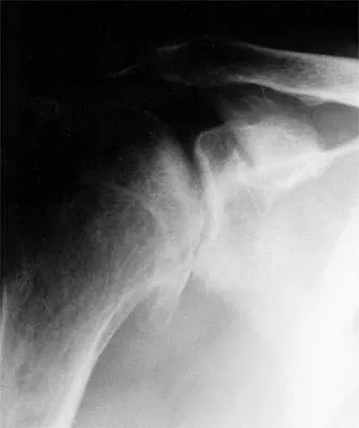
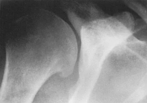
A 45-year-old woman has had progressive right shoulder pain for the past 6 months. She notes that the pain disrupts her sleep, she has pain at rest that requires the use of narcotic analgesics, and she has limited use of her left shoulder for most activities of daily living. History reveals the use of corticosteroids for systemic lupus erythematosus. Examination shows diminished range of motion. Radiographs of the right shoulder are shown in Figures 4a and 4b. Treatment should consist of


Explanation

Humeral arthroplasty provides excellent pain relief and function for stage IV osteonecrosis with humeral collapse. In late disease with glenoid involvement (stage V), total shoulder arthroplasty is preferred. Some authors have reported satisfactory results with core decompression of the humeral head for early stages of osteonecrosis, but results for stage IV osteonecrosis are less satisfactory when compared with those for humeral arthroplasty. Cruess RL: Steroid-induced avascular necrosis of the head of the humerus: Natural history and management. J Bone Joint Surg Br 1976;58:313-317. LePorte DM, Mont MA, Mohan V, Pierre-Jacques H, Jones LC, Hungerford DS: Osteonecrosis of the humeral head treated by core decompression. Clin Orthop 1998;355:254-260.


Question 10

The relocation test is most reliable for diagnosing anterior subluxation of the glenohumeral joint when


Explanation

The relocation test is most accurate when true apprehension is produced with the arm in combined abduction and external rotation and then relieved when posterior pressure is placed on the humeral head. Pain with this test is a less specific response and may occur with other shoulder disorders such as impingement.


Question 11

A 16-year-old high school pitcher notes acute pain on the medial side of his elbow during a pitch. Examination that day reveals medial elbow tenderness, pain with valgus stress, mild swelling, and loss of extension. Plain radiographs show closed physes and no fracture. Which of the following diagnostic studies will best reveal his injury?


Explanation

The history and findings are consistent with a diagnosis of a sprain of the medial collateral ligament (MCL) of the elbow; therefore, contrast-enhanced MRI is considered the most sensitive and specific study for accurately showing this injury. Arthroscopic visualization of the MCL is limited to the most anterior portion of the anterior bundle only; complete inspection of the MCL using the arthroscope is not possible. CT without the addition of contrast is of no value in this situation. Use of a technetium Tc 99m bone scan is limited to aiding in the diagnosis of occult fracture, a highly unlikely injury in this patient. There are no clinical indications for electromyography. Timmerman LA, Andrews JR: Undersurface tear of the ulnar collateral ligament in baseball players: A newly recognized lesion. Am J Sports Med 1994;22:33-36. Timmerman LA, Schwartz ML, Andrews JR: Preoperative evaluation of the ulnar collateral ligament by magnetic resonance imaging and computed tomography arthrography: Evaluation of 25 baseball players with surgical confirmation. Am J Sports Med 1994;22:26-32.


Question 12

Figures 5a and 5b show the radiographs of a 45-year-old patient. What is the most likely diagnosis?


Explanation

Glenoid dysplasia is an uncommon anomaly that usually has a benign course but may result in shoulder pain, arthritis, or multidirectional instability. Shoulder pain and instability often improve with shoulder strengthening exercises. Wirth MA, Lyons FR, Rockwood CA Jr: Hypoplasia of the glenoid: A review of sixteen patients. J Bone Joint Surg Am 1993;75:1175-1184.


Question 13

A 14-year-old boy sustains a twisting injury to his right shoulder and recalls feeling a snap during a wrestling match. Examination shows hesitancy to raise the arm away from the side, diffuse tenderness and swelling of the upper arm, and no evidence of neurovascular compromise. Figures 6a and 6b show an AP radiograph and MRI scan. What is the most likely diagnosis?


Explanation

While difficult to appreciate on the AP radiograph of the shoulder, the increased physeal signal demonstrated on the axial MRI scan is consistent with a nondisplaced growth plate fracture. A comparison radiograph of the left shoulder also could be considered and the injured shoulder evaluated for physeal widening. Proximal humeral fractures in children are somewhat unusual, representing less than 1% of all fractures seen in children and only 3% to 6% of all epiphyseal fractures. Physeal injuries are classified according to the Salter-Harris classification scheme. Salter-Harris type I fractures represent approximately 25% of physeal injuries to the proximal humerus in adolescents. The proximal humeral physis is responsible for 80% of the longitudinal growth of the humerus; therefore, there is tremendous potential for remodeling of fractures in this region. Management for nondisplaced Salter-Harris type I fractures is limited to a short period of immobilization followed by a gradual return to activities as clinical symptoms resolve. Curtis RJ, Rockwood CA Jr: Fractures and dislocations of the shoulder in children, in Rockwood CA Jr, Matsen FA III (eds): The Shoulder. Philadelphia, PA, WB Saunders, 1990, pp 991-1007.


Question 14

Figure 7 shows the radiograph of an otherwise healthy 65-year-old man who injured his right dominant shoulder while skiing 18 months ago. He did not seek treatment at the time of the injury. He now reports intermittent soreness when playing golf but has no other limitations. Examination reveals full range of motion and no tenderness, but he has slight pain with a crossed arm adduction stress test. He is neurologically intact. Initial management should consist of


Explanation

The radiograph shows a displaced type II distal clavicle fracture with nonunion. Because the patient's symptoms are minimal, the injury can be treated like a grade III acromioclavicular separation. Present management should consist of ice, anti-inflammatory drugs, activity modification, and perhaps physical therapy. If nonsurgical management fails to provide relief, the surgical options are varied with no uniformity in the literature regarding surgical treatment of this injury. Beaty JH (ed): Orthopaedic Knowledge Update 6. Rosemont, IL, American Academy of Orthopaedic Surgeons, 1999, pp 271-286.


Question 15

Figure 8 shows the AP radiograph of a 33-year-old woman who sustained a midshaft clavicle fracture from a motorcycle accident 15 months ago. She continues to have significant pain with activities of daily living. Management should consist of


Explanation

The patient has a symptomatic painful atrophic midclavicular nonunion, and the treatment of choice is rigid internal fixation with a dynamic compression plate and autogenous bone grafting. A tension band effect is desired and achieved by placing the plate superiorly. Excellent success rates of 90% to 100% have been reported using this technique. Intramedullary screw fixation without bone grafting has a decreased success rate. Partial claviculectomy is not a preferred option. Jupiter JB, Leffert RD: Non-union of the clavicle: Associated complications and surgical management. J Bone Joint Surg Am 1987;69:753-760.


Question 16

A 62-year-old patient with rheumatoid arthritis has had pain and instability of the elbow following total elbow replacement 2 years ago. A complete work-up, including aspiration and cultures, is negative. Figures 9a and 9b show the AP and lateral radiographs. Treatment should consist of


Explanation

The patient has aseptic loosening of the original semiconstrained prosthesis and significant proximal ulnar bone destruction; therefore, the treatment of choice is revision arthroplasty using a semiconstrained design. Although orthotic stabilization could be used, it will not provide long-term pain relief. Resection arthroplasty after removal of the components may lead to painful instability. Elbow arthrodesis would be difficult with the bone stock loss and is not considered the best option. Two main contraindications to the use of an unconstrained prosthesis are significant bone loss and previous use of a hinged or semiconstrained prosthesis. An ulnar allograft could be combined with the use of a semiconstrained long-stemmed ulnar prosthesis as a treatment modification. Ewald FC, Simmons ED Jr, Sullivan JA, et al: Capitellocondylar total elbow replacement in rheumatoid arthritis: Long-term results. J Bone Joint Surg Am 1993;75:498-507.


Question 17

A 21-year-old football player reports increasing pain and a deformity involving his chest after colliding with another player during a scrimmage. Imaging studies confirm an anterior sternoclavicular dislocation. Management should consist of


Explanation

For the patient with an anterior sternoclavicular dislocation, the most appropriate initial treatment should be symptomatic. Surgical options are usually contraindicated because the incidence of intraoperative and postoperative complications is high. A deformity from an anterior sternoclavicular dislocation is usually well tolerated. Return to play is allowed when symptoms resolve. Rockwood CA Jr: Disorders of the sternoclavicular joint, in Rockwood CA Jr, Matsen FA III (eds): The Shoulder. Philadelphia, PA, WB Saunders, 1998, vol 1, pp 477-525.


Question 18

During total shoulder replacement for rheumatoid arthritis, fracture of the humeral shaft occurs. An intraoperative radiograph shows a displaced short oblique fracture at the tip of the prosthesis. At this point, the surgeon should


Explanation

The risk of intraoperative fracture in osteopenic rheumatoid bone is significant. Fractures may occur with dislocation of the head and canal reaming, especially while extending and externally rotating the shoulder. If the fracture occurs at the distal tip of the prosthesis, the use of a long-stemmed prosthesis to bypass the fracture site and supplementation with wire cables has been reported with good results. Wright TW, Cofield RH: Humeral fractures after shoulder arthroplasty. J Bone Joint Surg Am 1995;77:1340-1346. Boyd AD Jr, Thornhill TS, Barnes CL: Fractures adjacent to humeral protheses. J Bone Joint Surg Am 1992;74:1498-1504.


Question 19

What is the most common contracture deformity of the spastic shoulder secondary to a cerebrovascular accident?


Explanation

The resultant spasticity and weakness (paresis) following a cerebrovascular accident leads to muscle imbalance that commonly results in contracture of the shoulder in adduction, internal rotation, and varying degrees of forward flexion. In addition, the elbow is usually flexed and the forearm pronated. Braun RM, Botte MJ: Treatment of shoulder deformity in acquired spasticity. Clin Orthop 1999;368:54-65.


Question 20

A 21-year-old collegiate pitcher has had pain in his dominant shoulder for the past 3 months despite management consisting of rest, rehabilitation, and an analysis of throwing mechanics. An arthroscopic photograph from the posterior portal is shown in Figure 10. The biceps anchor to the bone was not detached to probing. Treatment of the lesion to the left of the cannula should consist of arthroscopic


Explanation

The lesion is a variation of a type I superior labrum anterior and posterior lesion; therefore, appropriate treatment is simple debridement. Biceps tenodesis or release is not indicated because the biceps tendon and anchor are intact. There is no indication for labral repair or capsulorraphy. Beaty JH (ed): Orthopaedic Knowledge Update 6. Rosemont, IL, American Academy of Orthopaedic Surgeons, 1999, pp 261-270.


Question 21

After humeral head replacement for four-part fractures, what is the most commonly reported difficulty?


Explanation

Results show that patients who underwent humeral head replacement for fracture almost routinely report pain relief, but functional reports vary. The most commonly reported difficulty is the use of weight in the overhead position with wide variation in active elevation. Factors found to affect active elevation include age, humeral offset, greater tuberosity positioning, and four-part (as compared with three-part) fractures. Goldman RT, Koval KJ, Cuomo F, Gallagher MA, Zuckerman JD: Functional outcome after humeral head replacement for acute three- and fourth-part proximal humeral fractures. J Shoulder Elbow Surg 1995;4:81-86.


Question 22

Figures 11a and 11b show the AP and lateral radiographs of a 32-year-old patient on hemodialysis who has increasing elbow pain and a visibly growing mass over the extensor surface. Figure 11c shows the photomicrograph of the biopsy specimen. What is the most likely diagnosis?


Explanation

The radiographic findings are classic for tumoral calcinosis; they are not consistent with myositis ossificans, fungal granuloma, or hemochromatosis. The condition typically appears as large aggregations of dense calcified lobules confined to the surrounding soft tissues. Hyperphosphatemia is a fundamental factor in many patients with this condition. Tumoral calcinosis also occurs in the setting of chronic renal failure when mineral homeostasis is not controlled. The histologic appearance is essentially a foreign body granuloma reaction. Multilocular cysts with purplish amorphous material are surrounded by thick connective tissue capsules. The fibrous walls contain numerous foreign body giant cells. Surgical excision is indicated if the tumor causes discomfort or interferes with function. Sisson HA, Murray RO, Kemp HBS (eds): Orthopaedic Diagnosis: Clinical, Radiological and Pathological Coordinates. New York, NY, Springer-Verlag, 1984.


Question 23

A 52-year-old man who was a former high school pitcher now reports loss of elbow flexion and extension with pain at the extremes of motion. Nonsurgical management has failed to provide relief. Examination reveals movement from 50 degrees to 110 degrees and is painful only at the limits of motion. A radiograph is shown in Figure 12. Treatment should consist of


Explanation

Based on the history, examination, and radiograph, the patient has typical degenerative arthritis of the elbow. This condition is found almost exclusively in men, and there is almost universally a history of repetitive heavy use or overuse of the elbow. Patients report pain at terminal extension and usually have a flexion contracture. Radiographs reveal osteophytes on the coronoid and olecranon and in the coronoid and olecranon fossae. The osteophytes are often associated with loose bodies that sometimes are attached to the soft tissues. Treatment should consist of removal of all loose bodies and impinging osteophytes using open technique or by arthroscopy. The capsular contractures should be released at the same time. Kasser JR (ed): Orthopaedic Knowledge Update 5. Rosemont, IL, American Academy of Orthopaedic Surgeons, 1996, pp 283-294. Morrey BF: Primary degenerative arthritis of the elbow: Treatment by ulnohumeral arthroplasty. J Bone Joint Surg Br 1992;74:409-413. Redden JF, Stanley D: Arthroscopic fenestration of the olecranon fossa in the treatment of osteoarthritis of the elbow. Arthroscopy 1993;9:14-16.


Question 24

A 79-year-old woman with polyarticular rheumatoid arthritis has had progressively increasing right shoulder pain for the past year, and nonsurgical management has failed to provide relief. Her neurologic examination is entirely normal, but she is unable to elevate her arm against gravity. An AP radiograph is shown in Figure 13. Treatment should consist of


Explanation

Because the patient has end-stage rheumatoid arthritis with glenoid and rotator cuff deficiency, humeral arthroplasty is the treatment of choice. When a patient has an intact rotator cuff and there is sufficient glenoid bone stock to implant a glenoid component, total shoulder arthroplasty is the preferred method because it appears to provide more predictable pain relief. Glenohumeral arthrodesis is generally avoided when there is a functional deltoid or rotator cuff. Open synovectomy is appropriate in early rheumatoid disease before articular changes are present. Anterior acromioplasty with coracoacromial ligament resection is avoided in patients with rheumatoid arthritis because this procedure compromises the coracoacromial arch and may result in anterosuperior instability. Neer CS II, Watson KC, Stanton FJ: Recent experience in total shoulder replacement. J Bone Joint Surg Am 1982;64:319-337. Neer CS II: Glenohumeral arthroplasty, in Neer CS II (ed): Shoulder Reconstruction. Philadelphia, PA, WB Saunders, 1990, pp 143-271. Pollock RG, Deliz ED, McIlveen ST, et al: Prosthetic replacement in rotator cuff deficient shoulders. J Shoulder Elbow Surg 1992;1:173-186.


Question 25

A 22-year-old woman has had progressive upper extremity weakness for the past several years. History reveals no pain in her neck or shoulders. Examination reveals scapular winging of both shoulders and weakness in external rotation. She can abduct to only 120 degrees bilaterally, and there is mild supraspinatus weakness. She is otherwise neurologically intact with normal sensation and reflexes; however, she has difficulty whistling. A clinical photograph is shown in Figure 14. What is the most likely diagnosis?


Explanation

Progressive weakness is a common sign with a large differential diagnosis. Nerve, muscle, and joint problems should be excluded when a patient has diffuse weakness and atrophy. Fascioscapulohumeral dystrophy is a rare disease characterized by facial muscle weakness and proximal shoulder muscle weakness. The weakness is usually bilateral, and scapular winging is common. If the scapular winging becomes pronounced, elevation of the shoulder can be affected. In severe cases, scapulothoracic fusion or pectoralis muscle transfer to the scapula may be indicated. Duchenne muscular dystrophy is typically severe and progressive. The other diagnoses are not compatible with the history or the physical findings. Shapiro F, Specht L: The diagnosis and orthopaedic treatment of inherited muscular diseases of childhood. J Bone Joint Surg Am 1993;75:439-454.


Question 26

A 22-year-old collegiate rugby player presents with his fourth anterior shoulder dislocation this season. A 3D CT scan of the shoulder reveals 26% anterior glenoid bone loss. Which of the following is the most appropriate surgical management to minimize his risk of recurrent instability?


Explanation

Anterior glenoid bone loss exceeding 20-25% is a critical threshold where isolated soft-tissue stabilization (Bankart repair) carries a high failure rate. The Latarjet procedure (coracoid transfer) restores the bony arc and provides a sling effect, making it the treatment of choice.

Question 27

A 68-year-old man presents with severe shoulder pain and an inability to actively elevate his arm past 45 degrees, though passive motion is full. MRI reveals a massive, retracted, and fatty-infiltrated tear of the supraspinatus and infraspinatus. The subscapularis and teres minor are intact. Radiographs show no significant glenohumeral osteoarthritis. What is the most reliable surgical option to restore active elevation?


Explanation

Reverse total shoulder arthroplasty is indicated for patients with massive, irreparable rotator cuff tears associated with pseudoparalysis, especially in older patients. It restores elevation by increasing the deltoid moment arm, regardless of the presence of advanced arthritis.

Question 28

A 35-year-old man presents to the emergency department with a locked posterior shoulder dislocation following a seizure. Following closed reduction, a CT scan reveals an anteromedial humeral head impaction fracture (reverse Hill-Sachs lesion) involving 30% of the articular surface. Which of the following is the most appropriate surgical treatment?


Explanation

For a reverse Hill-Sachs lesion involving 20-40% of the articular surface, transferring the subscapularis tendon or lesser tuberosity into the defect (McLaughlin or modified McLaughlin procedure) stabilizes the joint and prevents recurrent engagement.

Question 29

A 55-year-old woman is diagnosed with a massive, chronically retracted rotator cuff tear involving the supraspinatus and infraspinatus. She demonstrates profound weakness in external rotation. Electromyography (EMG) reveals denervation of the infraspinatus. What is the most likely mechanism of nerve injury in this setting?


Explanation

Massive, medially retracted tears of the supraspinatus and infraspinatus can tether the suprascapular nerve. As the tendon retracts, it exerts traction on the nerve, leading to secondary neuropathy.

Question 30

A 19-year-old female gymnast complains of bilateral shoulder pain and a sensation of the shoulders "sliding out of joint." Physical exam demonstrates a positive sulcus sign bilaterally that persists in external rotation, positive apprehension tests, and generalized ligamentous laxity. What is the most appropriate initial management?


Explanation

This patient has multidirectional instability (MDI). The gold standard initial management for MDI is an extended course of physical therapy focused on strengthening the dynamic stabilizers (rotator cuff and periscapular muscles).

Question 31

A 52-year-old construction worker complains of deep anterior shoulder pain, particularly when using a hammer. Examination reveals a positive O'Brien's active compression test and tenderness in the bicipital groove. MRI arthrogram demonstrates an isolated Type II SLAP tear. Nonoperative management has failed. What is the preferred surgical intervention?


Explanation

In patients older than 40-50 years, biceps tenodesis is preferred over SLAP repair for Type II SLAP lesions. SLAP repairs in this older demographic have higher rates of persistent pain, stiffness, and clinical failure.

Question 32

Following a traumatic anterior shoulder dislocation, a 45-year-old man reports persistent anterior shoulder pain and significant weakness when trying to tuck his shirt into his pants behind his back. Which of the following physical examination tests is most sensitive and specific for evaluating the suspected torn structure?


Explanation

The patient's mechanism and inability to perform internal rotation tasks (tucking in a shirt) suggest a subscapularis tear. The bear hug, belly-press, and lift-off tests are specific maneuvers used to evaluate subscapularis integrity.

Question 33

A 25-year-old recreational athlete presents with recurrent anterior shoulder instability. A 3D CT scan shows 12% anterior glenoid bone loss and a large, engaging Hill-Sachs lesion. Which of the following surgical procedures is most appropriate to address both pathologies?


Explanation

An engaging Hill-Sachs lesion combined with subcritical (<20%) glenoid bone loss is effectively treated with an arthroscopic Bankart repair combined with remplissage (capsulotenodesis of the infraspinatus into the Hill-Sachs defect).

Question 34

A 21-year-old elite collegiate baseball pitcher presents with pain in the posterior aspect of his throwing shoulder during the late cocking phase of throwing. He has glenohumeral internal rotation deficit (GIRD) of 25 degrees compared to his non-throwing arm. What is the most likely associated arthroscopic finding?


Explanation

This classic presentation describes internal impingement. It occurs during extreme abduction and external rotation, causing the undersurface of the posterior supraspinatus and anterior infraspinatus to impinge against the posterosuperior glenoid labrum, leading to fraying.

Question 35

The normal direct tendon insertion of the rotator cuff onto the greater tuberosity is divided into four distinct histological zones. Which structure marks the boundary between uncalcified fibrocartilage and calcified fibrocartilage?


Explanation

At the rotator cuff enthesis, the tidemark represents the distinct histological boundary separating the uncalcified fibrocartilage zone from the calcified fibrocartilage zone.

Question 36

A 40-year-old man presents with anterior shoulder pain that worsens with cross-body adduction and internal rotation. Examination demonstrates focal tenderness over the coracoid process. MRI reveals a narrowed coracohumeral interval of 4 mm and edema in the lesser tuberosity. Which tendon is most at risk of attritional tearing in this syndrome?


Explanation

The clinical scenario describes subcoracoid impingement syndrome, where a narrowed coracohumeral interval leads to entrapment and potential attritional tearing of the subscapularis tendon.

Question 37

During the late cocking phase of a throwing motion, the shoulder is in maximum abduction and external rotation. In this specific position, which capsuloligamentous structure provides the primary static restraint to anterior translation of the humeral head?


Explanation

Biomechanical studies have demonstrated that the anterior band of the inferior glenohumeral ligament (AB-IGHL) is the primary restraint to anterior and inferior humeral translation when the shoulder is positioned in 90 degrees of abduction and maximum external rotation.

Question 38

A 75-year-old woman has advanced rotator cuff tear arthropathy. Radiographs reveal superior migration of the humeral head with articulation against the acromion (acetabularization).

What biomechanical alteration is the primary driver of this superior head migration?


Explanation

In the presence of a massive rotator cuff tear, the normal inferiorly directed compressive force of the cuff is lost. This leaves the superior pull of the deltoid unopposed, resulting in superior migration of the humeral head.

Question 39

During an open repair of a massive, chronically retracted rotator cuff tear, the surgeon performs aggressive lateral mobilization of the supraspinatus tendon. Postoperatively, the patient is noted to have a new, isolated profound weakness in external rotation, with intact forward elevation. Injury to which structure most likely occurred during mobilization?


Explanation

Excessive traction during mobilization of the rotator cuff can stretch the suprascapular nerve. An injury specifically at the spinoglenoid notch affects only the motor branches to the infraspinatus, leading to isolated external rotation weakness.

Question 40

A 50-year-old male arrives in the emergency department with an acute anterior shoulder dislocation after a fall. Following successful closed reduction, he reports a patch of numbness over the lateral aspect of his shoulder and exhibits weakness when attempting to actively abduct the arm. Which nerve is most commonly injured in this injury pattern?


Explanation

The axillary nerve is the most frequently injured nerve in anterior shoulder dislocations. It presents clinically with weakness of the deltoid (abduction) and numbness over the lateral shoulder (the "regimental badge" area).

Question 41

A 22-year-old collegiate rugby player presents with recurrent anterior shoulder instability. A 3D CT scan demonstrates 25% anterior glenoid bone loss. Which of the following is the most appropriate surgical management?


Explanation

The Latarjet procedure (coracoid transfer) is the gold standard for high-demand contact athletes with recurrent anterior instability and >20% anterior glenoid bone loss. Soft tissue Bankart repairs have an unacceptably high failure rate in the presence of critical bone loss.

Question 42

A 35-year-old overhead athlete complains of vague posterior shoulder pain and weakness. Physical examination reveals normal active forward elevation and abduction, but notable weakness in external rotation with the arm at the side. An MRI arthrogram reveals a paralabral cyst. Where is the cyst most likely located?


Explanation

The spinoglenoid notch transmits the suprascapular nerve after it has already innervated the supraspinatus. Entrapment by a paralabral cyst at this specific location leads to isolated infraspinatus weakness (external rotation), sparing supraspinatus function.

Question 43

A 45-year-old construction worker presents with chronic, deep shoulder pain. MRI confirms an isolated Type II SLAP (Superior Labrum Anterior and Posterior) tear. Conservative management has failed. Which of the following surgical interventions is associated with the most reliable return to work and pain relief in this patient demographic?


Explanation

In patients over 40 years old, particularly manual laborers, biceps tenodesis provides more reliable pain relief and functional outcomes compared to SLAP repair, which has a higher rate of postoperative stiffness and failure in this age group.

Question 44

A 65-year-old man is incidentally found to have an asymptomatic, 1.5 cm full-thickness supraspinatus tear on an MRI obtained for a suspected neck issue. He has full range of motion and 5/5 strength. What is the most accurate information regarding the natural history of his rotator cuff tear?


Explanation

Studies on the natural history of asymptomatic rotator cuff tears show that roughly 50% will enlarge and become symptomatic within 2 to 3 years. Clinical observation is appropriate initially, but patients should be warned about the potential for progression.

Question 45

A 19-year-old collegiate rugby player presents with recurrent anterior shoulder instability. A 3D CT scan reveals 28% anterior glenoid bone loss. Which of the following is the most appropriate definitive management?


Explanation

In the setting of significant anterior glenoid bone loss (typically >20-25%), soft tissue stabilization alone has a prohibitively high failure rate. A bony augmentation procedure, such as the Latarjet, is the standard of care to restore joint stability.

Question 46

A 55-year-old woman is evaluated for right shoulder pain and weakness after a fall. On examination, she has increased passive external rotation compared to the contralateral side. The "belly-press" test is positive, but the "lift-off" test is negative. Which of the following statements best describes her pathology?


Explanation

The subscapularis tendon is tested via the belly-press (upper half) and lift-off (lower half) tests. A positive belly-press with a normal lift-off indicates an isolated upper subscapularis tear. Increased passive external rotation is also a classic hallmark of subscapularis deficiency.

Question 47

A 35-year-old man presents to the emergency department after a generalized tonic-clonic seizure. His arm is locked in internal rotation. Radiographs reveal a posterior shoulder dislocation. CT scan shows an anteromedial humeral head impression fracture (reverse Hill-Sachs lesion) involving 45% of the articular surface. What is the most appropriate surgical treatment?


Explanation

For reverse Hill-Sachs lesions involving >40-45% of the articular surface, joint preservation techniques (like the modified McLaughlin) are generally insufficient due to a lack of stable articular cartilage. Hemiarthroplasty or total shoulder arthroplasty is indicated.

Question 48

A 17-year-old female gymnast complains of bilateral shoulder pain and a feeling of the shoulders "sliding out of place." Examination shows a positive sulcus sign bilaterally, positive apprehension, and generalized ligamentous laxity. There is no history of a discrete traumatic dislocation. What is the most appropriate initial management?


Explanation

Multidirectional instability (MDI) is primarily treated non-operatively with a prolonged, dedicated physical therapy program emphasizing dynamic stabilizers. Surgery is considered only after at least 6 months of failed comprehensive rehabilitation.

Question 49

A 72-year-old man presents with chronic, severe right shoulder pain and an inability to actively elevate his arm past 40 degrees. Radiographs demonstrate superior migration of the humeral head with acromiohumeral articulation and severe glenohumeral osteoarthritis. MRI shows a massive, retracted supraspinatus and infraspinatus tear. What is the optimal surgical treatment?


Explanation

This patient has rotator cuff tear arthropathy presenting with pseudoparalysis. Reverse total shoulder arthroplasty (RTSA) is the treatment of choice, as it relies on the deltoid muscle for elevation and correctly addresses both the deficient rotator cuff and arthritis.

Question 50

During a Latarjet procedure for recurrent anterior shoulder instability, the conjoined tendon is retracted medially to expose the subscapularis. Which of the following nerves is at greatest risk of injury with overly aggressive medial retraction of the conjoined tendon?


Explanation

The musculocutaneous nerve enters the coracobrachialis muscle approximately 3-8 cm distal to the coracoid tip. Aggressive medial retraction of the conjoined tendon during the Latarjet procedure places this nerve at high risk for a traction injury.

Question 51

A 28-year-old elite volleyball attacker presents with vague posterior shoulder pain and noticeable atrophy over the scapula. On examination, she has 5/5 strength in forward elevation but isolated 3/5 strength in external rotation with the arm at the side. What is the most likely pathological finding on MRI?


Explanation

The suprascapular nerve innervates the supraspinatus and then passes through the spinoglenoid notch to innervate the infraspinatus. Compression at the spinoglenoid notch (often from a posterior labral cyst) causes isolated infraspinatus weakness, sparing the supraspinatus.

Question 52

A 22-year-old rugby player presents with recurrent anterior shoulder instability. A 3D-CT scan demonstrates 28% anterior glenoid bone loss. Which of the following is the most appropriate surgical management?


Explanation

For anterior glenoid bone loss exceeding 20-25%, a bony augmentation procedure like the Latarjet is indicated to restore glenohumeral stability. Soft tissue repairs (Bankart) have an unacceptably high failure rate in the setting of critical bone loss.

Question 53

A 72-year-old woman presents with severe shoulder pain, active forward elevation to 50 degrees, and a positive drop arm test. Radiographs show a superiorly migrated humeral head with severe glenohumeral osteoarthritis. MRI confirms a massive, retracted rotator cuff tear with grade 4 fatty infiltration. What is the most appropriate treatment?


Explanation

Reverse total shoulder arthroplasty is the treatment of choice for rotator cuff arthropathy with pseudoparalysis. It medializes the center of rotation and increases the deltoid moment arm, restoring forward elevation.

Question 54

A 45-year-old man falls on an outstretched hand and presents with weakness in internal rotation and a positive belly-press test. Which of the following structures is most commonly injured concomitantly with the suspected tendon tear?


Explanation

A positive belly-press test indicates a subscapularis tear. The subscapularis insertion is intimately associated with the biceps pulley, making long head of the biceps tendon subluxation or tearing the most common concomitant injury.

Question 55

A 35-year-old man presents to the emergency department locked in internal rotation after a generalized tonic-clonic seizure. Radiographs confirm a posterior shoulder dislocation. CT scan shows a reverse Hill-Sachs lesion involving 35% of the articular surface. Which of the following is the most appropriate treatment?


Explanation

Posterior dislocations often result from seizures or electrocution. For reverse Hill-Sachs lesions involving 20% to 40% of the articular surface, transferring the subscapularis tendon (or lesser tuberosity) into the defect (McLaughlin procedure) prevents engagement and recurrent instability.

Question 56

A 28-year-old elite volleyball attacker presents with painless, isolated weakness in external rotation of her dominant shoulder. MRI reveals a paralabral cyst. At which of the following anatomic locations is the cyst most likely compressing the affected nerve?


Explanation

Isolated external rotation weakness indicates isolated infraspinatus denervation, typically caused by suprascapular nerve compression at the spinoglenoid notch. Compression at the suprascapular notch would affect both the supraspinatus and infraspinatus.

Question 57

In the evaluation of a patient with a massive rotator cuff tear, which of the following factors on preoperative MRI is the strongest independent predictor of structural failure following surgical repair?


Explanation

Advanced fatty infiltration (Goutallier stage 3 or 4) and muscle atrophy are irreversible and strongly predict a high rate of structural failure and poor functional outcomes after rotator cuff repair.

Question 58

A 25-year-old male with recurrent anterior shoulder instability has a glenoid track evaluated on CT. He has 10% anterior glenoid bone loss and a large Hill-Sachs lesion that extends outside the glenoid track. What is the most appropriate surgical management?


Explanation

An "off-track" Hill-Sachs lesion will engage the anterior glenoid rim, leading to recurrent instability if treated with an isolated Bankart repair. Since the glenoid bone loss is subcritical (<20%), adding an arthroscopic remplissage (infraspinatus tenodesis into the defect) is indicated.

Question 59

A 30-year-old rugby player presents with recurrent anterior shoulder instability. MRI arthrogram shows no labral tear, but reveals contrast extravasation into the axillary pouch and the normal U-shape of the axillary recess is lost, appearing as a J-shape. What is the most likely diagnosis?


Explanation

A Humeral Avulsion of the Glenohumeral Ligament (HAGL) lesion presents with recurrent instability without a labral tear. On MRI arthrogram, the contrast leaking through the avulsed inferior glenohumeral ligament creates the characteristic "J-sign".

Question 60

A 40-year-old overhead athlete undergoes shoulder arthroscopy for chronic pain. An articular-sided, partial-thickness supraspinatus tear (PASTA lesion) is identified. Debridement reveals the tear involves 60% of the tendon footprint thickness. What is the recommended management?


Explanation

For articular-sided partial-thickness rotator cuff tears involving greater than 50% of the tendon thickness, surgical repair is indicated. Standard treatment involves completing the tear and performing a full-thickness repair to ensure adequate biomechanical strength.

Question 61

A 19-year-old gymnast complains of bilateral shoulder pain and a feeling of instability. Physical exam reveals generalized ligamentous laxity, a positive sulcus sign, and apprehension with anterior, posterior, and inferior translation. She has failed 6 months of targeted physical therapy. What is the next best step in management?


Explanation

This patient has multidirectional instability (MDI) that has failed an extensive trial of conservative management. The appropriate surgical intervention for refractory MDI is a capsular shift to globally reduce capsular volume.

Question 62

A 55-year-old woman presents with persistent anterior shoulder pain exacerbated by forward elevation and internal rotation. MRI reveals a narrowed coracohumeral interval measuring 5 mm, edema in the subcoracoid space, and a partial-thickness tear of the subscapularis tendon. What is the most likely diagnosis?


Explanation

Subcoracoid impingement occurs when the coracohumeral interval is narrowed (typically <6-7 mm), trapping the subscapularis tendon. It presents with anterior pain worse with internal rotation and forward elevation, often leading to subscapularis tears.

Question 63

A 22-year-old collegiate baseball pitcher reports deep shoulder pain during the late cocking phase of throwing. MRI arthrogram demonstrates contrast extending between the superior labrum and the glenoid, and arthroscopy reveals the superior labrum and biceps anchor are completely detached from the superior glenoid. What type of SLAP tear is this?


Explanation

A Type II SLAP tear involves detachment of the superior labrum and the origin of the long head of the biceps tendon from the superior glenoid. It is the most common type of SLAP tear in overhead athletes.

Question 64

During a Latarjet procedure, the coracoid process is osteotomized and transferred to the anterior glenoid. Which of the following nerves is at greatest risk of iatrogenic injury during the coracoid mobilization and transfer?


Explanation

The musculocutaneous nerve enters the coracobrachialis muscle typically 3 to 8 cm distal to the coracoid tip. It is at significant risk of traction or transection injury during mobilization and transfer of the conjoined tendon in a Latarjet procedure.

Question 65

Reverse total shoulder arthroplasty (RTSA) is highly effective for patients with rotator cuff arthropathy. Which of the following best describes the primary biomechanical advantage of RTSA compared to anatomic total shoulder arthroplasty?


Explanation

RTSA medializes and distalizes the center of rotation of the glenohumeral joint. This significantly increases the moment arm and resting tension of the deltoid muscle, allowing it to initiate and maintain forward elevation even in the absence of a functional rotator cuff.

Question 66

The "rotator cable" is a thick fibrous band of tissue running perpendicular to the supraspinatus and infraspinatus muscle fibers. Its primary function is to stress-shield which of the following adjacent structures?


Explanation

The rotator cable acts similarly to a suspension bridge, transferring stress away from the thinner, relatively avascular distal portion of the rotator cuff insertions known as the rotator crescent. This crescent is the most common site for degenerative rotator cuff tears.

Question 67

A 26-year-old man presents to the emergency department with a locked left shoulder in internal rotation following a generalized tonic-clonic seizure. Radiographs reveal a posterior shoulder dislocation. Which of the following associated lesions is most likely present?


Explanation

Posterior shoulder dislocations are classically associated with seizures or electrocution and present locked in internal rotation. The impaction of the anteromedial humeral head against the posterior glenoid rim creates a reverse Hill-Sachs lesion.

Question 68

A 74-year-old man presents with chronic, severe shoulder pain and pseudoparalysis of the right arm. Examination demonstrates active forward elevation to 40 degrees and intact axillary nerve function.

Radiographs show severe superior migration of the humeral head with acetabularization of the acromion. What is the most appropriate surgical treatment?


Explanation

Reverse total shoulder arthroplasty (RTSA) is the gold standard for rotator cuff arthropathy with pseudoparalysis and an intact deltoid. Anatomic arthroplasty is contraindicated due to the massive rotator cuff deficiency, which would lead to superior escape.

Question 69

A 29-year-old professional volleyball player presents with isolated external rotation weakness and posterior shoulder pain. MRI reveals a paralabral cyst. Compression of the suprascapular nerve at the spinoglenoid notch will result in denervation of which of the following muscles?


Explanation

The suprascapular nerve innervates the supraspinatus before passing through the spinoglenoid notch. Compression at the spinoglenoid notch selectively affects the infraspinatus, sparing the supraspinatus.

Question 70

A 22-year-old rugby player presents with his fifth anterior shoulder dislocation.

3D CT imaging reveals 28% anterior glenoid bone loss. What is the most appropriate definitive surgical management?


Explanation

Glenoid bone loss exceeding 20-25% in a contact athlete is a strong indication for a bone block procedure, such as the Latarjet. An isolated arthroscopic soft-tissue repair carries an unacceptably high failure rate in this scenario.

Question 71

A 24-year-old baseball pitcher presents with pain in the posterosuperior aspect of his throwing shoulder during the late cocking phase. Examination reveals a glenohumeral internal rotation deficit (GIRD) of 25 degrees. What is the primary underlying pathomechanism?


Explanation

Internal impingement occurs during maximal abduction and external rotation (late cocking phase), causing the articular surface of the posterosuperior rotator cuff to impinge against the posterosuperior glenoid labrum. It is highly associated with GIRD and contracture of the posterior capsule.

Question 72

During an arthroscopic anterior stabilization for a patient with recurrent dislocations, an engaging Hill-Sachs lesion is identified. Which of the following procedures involves tenodesis of the infraspinatus tendon into the humeral defect?


Explanation

The arthroscopic remplissage procedure involves suturing the infraspinatus tendon and the posterior capsule into an engaging Hill-Sachs defect. This converts the intra-articular defect into an extra-articular one, preventing it from engaging the anterior glenoid.

Question 73

A 30-year-old man sustains a shoulder dislocation. An MR arthrogram demonstrates extravasation of contrast into the axillary pouch, obliterating the normal U-shape and creating a "J-sign". This finding is pathognomonic for which of the following lesions?


Explanation

A HAGL lesion represents an avulsion of the inferior glenohumeral ligament from its humeral attachment. On MR arthrogram, contrast leaks inferiorly through the axillary recess defect, forming the characteristic J-sign.

Question 74

During arthroscopy for recurrent anterior shoulder instability, the surgeon notes that the anterior labrum is completely torn and has medially displaced and healed along the anterior aspect of the glenoid neck. This pathology is correctly termed an:


Explanation

An Anterior Labroligamentous Periosteal Sleeve Avulsion (ALPSA) lesion occurs when the torn labrum displaces medially and heals along the anterior glenoid neck. It must be completely mobilized back to the glenoid rim during repair.

Question 75

A 19-year-old gymnast complains of vague, bilateral shoulder pain and a sensation of slipping. Examination reveals a prominent sulcus sign bilaterally, positive apprehension without a history of frank dislocation, and generalized ligamentous laxity. What is the most appropriate initial management?


Explanation

This patient has Multidirectional Instability (MDI), characterized by generalized laxity and a positive sulcus sign. The first-line and mainstay of treatment is extensive physical therapy focusing on the rotator cuff and periscapular stabilizers.

Question 76

A 45-year-old construction worker fell onto an outstretched arm and now complains of anterior shoulder pain and weakness. Clinical examination demonstrates a positive Gerber's lift-off test and increased passive external rotation. Which structure is most likely injured?


Explanation

The subscapularis is the primary internal rotator of the shoulder. A tear typically presents with weakness in internal rotation (positive lift-off or belly-press test) and an unresisted increase in passive external rotation.

Question 77

During an open Latarjet procedure, the surgeon places a self-retaining retractor medial to the conjoint tendon to expose the subscapularis. Excessive medial retraction in this area places which nerve at highest risk of injury?


Explanation

The musculocutaneous nerve enters the deep surface of the coracobrachialis 3 to 8 cm distal to the coracoid tip. Retracting the conjoint tendon too vigorously or too far medially during a Latarjet procedure risks stretching or damaging this nerve.

Question 78

A 35-year-old overhead athlete presents with posterior shoulder pain and isolated weakness of the deltoid and teres minor. An MRI demonstrates isolated muscular atrophy and localized vascular compression in the posterior shoulder. This syndrome is caused by compression of structures within boundaries that include all of the following EXCEPT:


Explanation

Quadrilateral space syndrome involves compression of the axillary nerve and posterior circumflex humeral artery. The boundaries are the teres minor (superior), teres major (inferior), long head of triceps (medial), and surgical neck of the humerus (lateral).

Question 79

A 52-year-old laborer presents with a massive, retracted supraspinatus and infraspinatus tear. The subscapularis is intact, the deltoid is highly functional, and there is no evidence of glenohumeral osteoarthritis on radiographs. Which of the following is the most appropriate surgical intervention?


Explanation

Superior capsular reconstruction (SCR) is indicated for massive, irreparable posterosuperior rotator cuff tears in younger, active patients without significant arthritis. An intact or repairable subscapularis and functional deltoid are prerequisites.

Question 80

A 42-year-old woman presents to the clinic with acute, extremely severe, burning shoulder pain that started spontaneously 2 weeks ago. The pain has now decreased, but she has developed profound weakness in shoulder abduction and external rotation. EMG demonstrates acute denervation. What is the most likely diagnosis?


Explanation

Parsonage-Turner syndrome (brachial neuritis) classically presents with acute, severe, unremitting shoulder pain followed days or weeks later by profound muscle weakness and atrophy as the pain subsides. Treatment is generally non-operative.

Question 81

A 50-year-old woman with severe adhesive capsulitis undergoes arthroscopic capsular release. The surgeon carefully releases the thickened structures within the rotator interval. Which of the following is NOT a normal anatomic component of the rotator interval?


Explanation

The rotator interval is a triangular space bounded by the supraspinatus superiorly and the subscapularis inferiorly. Its contents include the coracohumeral ligament, superior glenohumeral ligament, long head of the biceps, and joint capsule, but NOT the middle glenohumeral ligament.

Question 82

When evaluating a patient for an anterior shoulder stabilization procedure, the Instability Severity Index Score (ISIS) is used to predict the risk of recurrence after an arthroscopic Bankart repair. Which of the following factors increases the ISIS score?


Explanation

The ISIS evaluates recurrence risk. Risk factors include age under 20 years, competitive sports, contact or forced overhead sports, shoulder hyperlaxity, visible Hill-Sachs on AP radiograph, and glenoid loss on AP radiograph.

Question 83

The transverse force couple of the shoulder is crucial for maintaining the humeral head centered within the glenoid during active elevation. This force couple relies on the balanced action of which of the following muscle groups?


Explanation

The transverse force couple is composed anteriorly by the subscapularis and posteriorly by the infraspinatus and teres minor. Disruption of this balance in massive cuff tears leads to altered kinematics and dysfunction.

Question 84

A patient is evaluated for anterior shoulder pain. The examiner places the patient's arm in forward flexion, internal rotation, and cross-body adduction, which recreates the patient's sharp anterior pain. Imaging reveals a narrowed coracohumeral distance. What is the most likely diagnosis?


Explanation

Subcoracoid impingement occurs when the subscapularis tendon is compressed between the coracoid process and the lesser tuberosity. Provocative positioning includes forward flexion, internal rotation, and adduction.

Question 85

A 55-year-old physical laborer presents with persistent deep shoulder pain. MR arthrogram reveals a Type II SLAP tear. Given his age and occupation, what is the most appropriate surgical treatment if conservative management fails?


Explanation

In older patients (typically >40-50 years), arthroscopic repair of Type II SLAP tears is associated with high failure rates and postoperative stiffness. Biceps tenodesis provides superior functional outcomes and pain relief in this demographic.

Question 86

A 28-year-old weightlifter feels a sudden 'pop' in his axilla while performing a heavy bench press. He presents with extensive ecchymosis and loss of the anterior axillary fold. If a complete rupture of the pectoralis major occurred, which portion is typically the first to fail due to maximal eccentric stretch?


Explanation

Pectoralis major ruptures commonly occur at the humeral insertion during eccentric contraction (e.g., bench pressing). The sternal head inserts deep and proximal to the clavicular head, placing it under maximal tension when the arm is extended and externally rotated, causing it to tear first.

Question 87

A 22-year-old collegiate rugby player presents with recurrent anterior shoulder instability. CT scan with 3D reconstruction reveals an "inverted pear" glenoid with 25% anterior bone loss. What is the most appropriate definitive management?


Explanation

An "inverted pear" glenoid indicates significant anterior bone loss (>20-25%). This structural deficit is a contraindication to isolated soft-tissue stabilization and requires a bone-block procedure such as a Latarjet.

Question 88


A 35-year-old man presents to the ER after a generalized tonic-clonic seizure. His shoulder is locked in internal rotation. Radiographs show a posterior shoulder dislocation. MRI reveals an anteromedial humeral head impaction fracture involving 35% of the articular surface. What is the most appropriate surgical management?


Explanation

A reverse Hill-Sachs lesion (anteromedial humeral head defect) involving 20-40% of the articular surface is best treated with a lesser tuberosity transfer (modified McLaughlin) or structural allograft to prevent recurrent posterior instability.

Question 89

A 55-year-old man sustains a traumatic forced external rotation injury to his shoulder. He demonstrates a positive belly-press test and increased external rotation compared to the contralateral side. MRI confirms an isolated, full-thickness subscapularis tendon tear. During arthroscopic repair, which of the following additional findings is most likely to be encountered?


Explanation

The superior border of the subscapularis tendon contributes to the biceps pulley mechanism. Complete subscapularis tears are highly associated with medial subluxation or dislocation of the long head of the biceps tendon.

Question 90


A 28-year-old elite volleyball player complains of vague posterior shoulder pain and isolated weakness in external rotation. Forward elevation and internal rotation strength are normal. MRI reveals a paralabral cyst in the spinoglenoid notch. This lesion most likely originated from which of the following pathologies?


Explanation

Spinoglenoid notch cysts strongly correlate with posterosuperior labral tears. A one-way valve effect allows joint fluid to accumulate, compressing the suprascapular nerve and causing isolated infraspinatus weakness.

Question 91

A 30-year-old recreational skier presents with persistent anterior shoulder instability. An MRI arthrogram reveals extravasation of contrast into the axilla and a "J-sign" replacing the normal U-shaped inferior glenohumeral recess. What is the diagnosis?


Explanation

A Humeral Avulsion of the Glenohumeral Ligament (HAGL) presents with an incompetent inferior glenohumeral ligament complex. On MRI arthrogram, the normal U-shaped axillary pouch is replaced by a "J-sign" indicating contrast extravasation.

Question 92

Following an arthroscopic rotator cuff repair, a patient asks about the expected timeline for tendon-to-bone healing. Biomechanically, which type of collagen initially predominates at the healing tendon-bone interface before being remodeled into a stronger, more mature construct?


Explanation

During the initial proliferative phase of tendon-to-bone healing, fibroblasts lay down predominantly Type III collagen, which is disorganized and mechanically weaker. Over several months, this tissue remodels into stronger, highly organized Type I collagen.

Question 93


During diagnostic arthroscopy for recurrent anterior shoulder instability, the surgeon notes that the anterior labrum is avulsed but remains attached to the intact anterior scapular periosteum, having healed medially along the glenoid neck. This specific pathology is known as:


Explanation

An Anterior Labroligamentous Periosteal Sleeve Avulsion (ALPSA) lesion occurs when the anterior labrum strips medially with an intact periosteum. It must be surgically mobilized and shifted laterally to its anatomical footprint before fixation.

Question 94

A 74-year-old female presents with severe right shoulder pain, pseudoparalysis, and a massive, irreparable rotator cuff tear. Radiographs show superior migration of the humeral head with an acromiohumeral distance < 2 mm. Which of the following is an absolute prerequisite for a successful Reverse Total Shoulder Arthroplasty (RTSA) in this patient?


Explanation

RTSA relies on the deltoid muscle to elevate the arm, bypassing the deficient rotator cuff mechanics. A functioning axillary nerve and intact deltoid are absolute prerequisites for a successful outcome.

Question 95

A 19-year-old female competitive swimmer presents with bilateral shoulder pain and a feeling of "looseness." Examination reveals a positive sulcus sign, generalized ligamentous laxity (Beighton score 7/9), and no distinct history of trauma. Initial management for this condition should focus on:


Explanation

Multidirectional instability (MDI) is typically atraumatic and bilateral. The gold standard for initial management is an extended course (usually >6 months) of targeted physical therapy focusing on periscapular and rotator cuff strengthening.

Question 96

When performing an open Latarjet procedure, the surgeon must be careful to avoid injury to the nerves innervating the subscapularis muscle. The upper and lower subscapular nerves are branches of which cord of the brachial plexus?


Explanation

The upper and lower subscapular nerves originate from the posterior cord of the brachial plexus. They provide motor innervation to the subscapularis muscle, with the lower subscapular nerve also innervating the teres major.

Question 97

A 42-year-old recreational tennis player undergoes arthroscopy for refractory shoulder pain. A partial articular-sided tendon avulsion (PASTA) lesion of the supraspinatus is identified. At what depth of tendon involvement is completion of the tear and full-thickness repair generally recommended?


Explanation

For partial articular-sided rotator cuff tears (PASTA lesions), surgical management principles dictate that tears involving >50% of the tendon thickness (typically >6 mm) should be completed and repaired to restore optimal biomechanical strength.

Question 98


A 35-year-old laborer presents with persistent superior shoulder pain and impingement symptoms. An axillary radiograph reveals an unfused acromial apophysis (meso-acromiale) that is mobile on physical examination. After failing 6 months of conservative management, what is the recommended surgical intervention?


Explanation

A symptomatic, mobile meso-acromiale (the most common type of os acromiale) that fails conservative management is best treated with ORIF (often with bone grafting). Excision risks severe deltoid dysfunction, and isolated acromioplasty further destabilizes the fragment.

Question 99

A 25-year-old professional baseball pitcher presents with pain during the late cocking phase of throwing and a "dead arm" sensation. MRI shows a Type II SLAP tear. If nonoperative management fails, what is the preferred surgical intervention to allow return to his prior level of competitive pitching?


Explanation

In young, elite overhead throwing athletes, arthroscopic repair of a Type II SLAP tear remains the standard of care to restore the native tension of the biceps-labral complex. Tenodesis is generally reserved for older patients (>35-40 years) or non-throwers.

Question 100

The Latarjet procedure involves the transfer of the coracoid process with the attached conjoint tendon to the anterior glenoid neck. Which nerve is most at risk during the transfer and retraction of the conjoint tendon?


Explanation

The musculocutaneous nerve typically enters the coracobrachialis muscle 3-5 cm distal to the coracoid tip. Vigorous medial retraction of the conjoint tendon during a Latarjet procedure places this nerve at high risk for neuropraxia.

None

Clinic OS
Medically Verified Content by
Prof. Clinic OS
Consultant Orthopedic & Spine Surgeon
Chapter Index