العربية
Part of the Master Guide

100 High-Yield Orthopedic MCQs: Shoulder & Elbow | Mock Exam 251

AAOS Shoulder MCQs (Set 1): Rotator Cuff, Instability & Humerus Fractures | ABOS Board Prep

23 Apr 2026 65 min read 107 Views
Shoulder 2002 MCQs - Part 1

Key Takeaway

This high-yield question set for AAOS/ABOS exams, Set 1, focuses on critical shoulder pathology. It covers the diagnosis, management, and surgical principles of rotator cuff injuries, shoulder instability, including dislocations and labral tears, and proximal humerus fractures. Ideal for reinforcing core orthopedic knowledge.

AAOS Shoulder MCQs (Set 1): Rotator Cuff, Instability & Humerus Fractures | ABOS Board Prep

Comprehensive 100-Question Exam


00:00

Start Quiz

Question 1

Which of the following statements best describes why the ulnar nerve is most prone to neuropathy at the elbow?


Explanation

The ulnar nerve is more prone to neuropathy than the radial or median nerves for many reasons. It has the greatest longitudinal excursion required to accommodate elbow range of motion, subjecting it to potential traction forces. The dimensions of the entrance of the cubital tunnel change with elbow motion, potentially causing compression in flexion. For these two reasons, the ulnar nerve is subjected to both compression and traction during elbow motion. Although it passes between two muscle heads as it enters the forearm, so do the median and radial nerves. Finally, the vascular supply is adequate because of the anastamoses between the superior ulnar collateral artery, the posterior ulnar recurrent artery, and the inferior ulnar collateral artery. Norris TR (ed): Orthopaedic Knowledge Update: Shoulder and Elbow. Rosemont, IL, American Academy of Orthopaedic Surgeons, 1997, pp 369-378. Prevel CD, Matloub HS, Ye Z, Sanger JR, Yousif NJ: The extrinsic blood supply of the ulnar nerve at the elbow: An anatomic study. J Hand Surg Am 1993;18:433-438.

Question 2

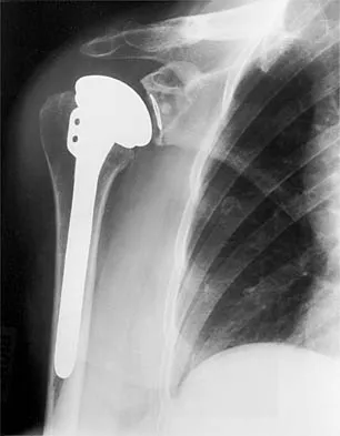
Figure 1 shows the radiograph of a 71-year-old man who has had increasing pain and weakness in his shoulder for the past 3 years. Nonsurgical management has failed to provide relief. Examination shows 130 degrees of active forward flexion and intact external rotation strength. During surgery, a 1- x 1-cm rotator cuff tear involving the supraspinatus is encountered. Treatment should include


Explanation

Given the size of the rotator cuff tear, it is likely to be repaired; therefore, the treatment of choice is a total shoulder replacement with rotator cuff repair. Severe rotator cuff insufficiency can lead to early glenoid failure because of superior instability, and glenoid resurfacing should be avoided in those instances. Boyd AD Jr, Thomas WH, Scott RD, Sledge CB, Thornhill TS: Total shoulder arthroplasty versus hemiarthroplasty: Indications for glenoid resurfacing. J Arthroplasty 1990;5:329-336.


Question 3

Which of the following is considered the cause of Milwaukee shoulder, a joint disease similar to rotator cuff arthropathy?


Explanation

Neer and associates focused on mechanical and nutritional factors as the etiology of rotator cuff arthropathy. McCarty and associates, in describing a similar syndrome known as Milwaukee shoulder, focused on an inflammatory cause in proposing the pathogenic role of hydroxyapatite, a basic calcium phosphate. Neer CS II, Craig EV, Fukuda H: Cuff-tear arthropathy. J Bone Joint Surg Am 1983;65:1232-1244.


Question 4

The MRI scan of the shoulder shown in Figure 2 was performed with the arm in abduction and external rotation. The image reveals what condition?


Explanation

Internal impingement of the shoulder is now a well-recognized cause of shoulder pain in the throwing athlete. First described by Walch and associates, it involves contact of the rotator cuff and labrum in the maximally externally rotated and abducted shoulder, such as in the late cocking phase of the throwing motion. Schickendantz and associates have shown this contact to be physiologic in most patients and becoming pathologic with repetitive overhead activity. Schickendantz MS, Ho CP, Keppler L, Shaw BD: MR imaging of the thrower's shoulder: Internal impingement, latissimus dorsi/subscapularis strains, and related injuries. Magn Reson Imaging Clin N Am 1999;7:39-49. Walch G, Boileau P, Noel E, et al: Impingement of the deep surface of the supraspinatus tendon on the posterosuperior glenoid rim: An arthroscopic study. J Shoulder Elbow Surg 1992;1:238-245.


Question 5

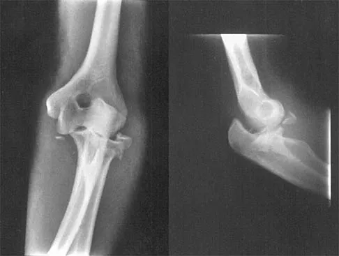
Figure 3 shows the radiographs of a 32-year-old man who fell 12 feet onto his outstretched arm and sustained a fracture-dislocation of the elbow. Initial management consisted of closed reduction of the dislocation. Surgical treatment should now include repair or reduction and fixation of the


Explanation

The radiographs show fractures of the coronoid and radial head. The medial collateral ligament has been avulsed from the ulnar insertion, and there is a valgus opening on the medial side. The lateral collateral ligament is always disrupted in elbow dislocations and fracture-dislocations that occur secondary to falls. This is known as the terrible triad injury (dislocation and fractures of the coronoid and radial head); it has a very poor prognosis because of its propensity for recurrent or persistent instability and late arthritis. The principle in treating this injury is to repair all of the injured parts or protect them with a hinged external fixator until they heal. Norris TR (ed): Orthopaedic Knowledge Update: Shoulder and Elbow. Rosemont, IL, American Academy of Orthopaedic Surgeons, 1997, pp 345-354. Kasser JR (ed): Orthopaedic Knowledge Update 5. Rosemont, IL, American Academy of Orthopaedic Surgeons, 1996, pp 283-294.


Question 6

It is important to avoid which of the following exercises in the immediate postoperative period after humeral head replacement for an acute four-part fracture?


Explanation

It is critical to withhold active range of motion of the shoulder within the first 6 weeks after arthroplasty for acute fracture to prevent tuberosity avulsion. When radiographic and clinical findings show that the tuberosities are healed, active motion may be instituted, usually at 6 to 8 weeks. Immediate passive range-of-motion exercises, including external rotation with a stick, pendulum, and passive elevation, should begin within the limits of the repair on the day of surgery to prevent stiffness. Hartstock LA, Estes WJ, Murray CA, et al: Shoulder hemiarthroplasty for proximal humerus fractures. Orthop Clin North Am 1998;29:467-475.


Question 7

A 38-year-old man has winging of the ipsilateral scapula after undergoing a transaxillary resection of the first rib 3 weeks ago. What is the most likely cause of this finding?


Explanation

During transaxillary resection of the first rib, the long thoracic nerve is at risk as it passes either through or posterior to the middle scalene muscle. Injury to this nerve may occur as the result of overly aggressive retraction of the middle scalene during the procedure. Leffert RD: Thoracic outlet syndrome. J Am Acad Orthop Surg 1994;2:317-325.


Question 8

A 73-year-old man who underwent repair of the left rotator cuff 6 years ago reports good pain relief but notes residual weakness of the left shoulder, especially with overhead tasks. He denies having pain at night and has minimal discomfort with activities of daily living but is dissatisfied with his shoulder strength. Radiographs show an acromiohumeral interval of 2 mm. Appropriate management should consist of


Explanation

An exercise program to strengthen the deltoid and remaining rotator cuff will most likely offer the best results. Revision rotator cuff surgery yields better results in decreasing pain than improving strength and function, and this patient has only minimal pain. Tendon transfers, involving the use of the latissimus dorsi or teres major, have been used when the rotator cuff is deemed irreparable but are not indicated in elderly patients with minimal symptoms. Bigliani LU, Cordasco FA, McIlveen SJ, Musso ES: Operative treatment of failed repairs of the rotator cuff. J Bone Joint Surg Am 1992;74:1505-1515. DeOrio JK, Cofield RH: Results of a second attempt at surgical repair of a failed initial rotator-cuff repair. J Bone Joint Surg Am 1984;66:563-567.


Question 9

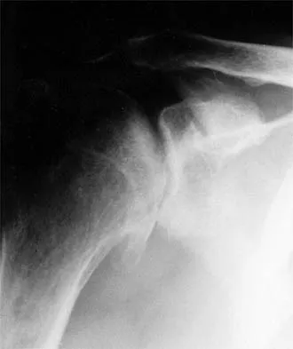
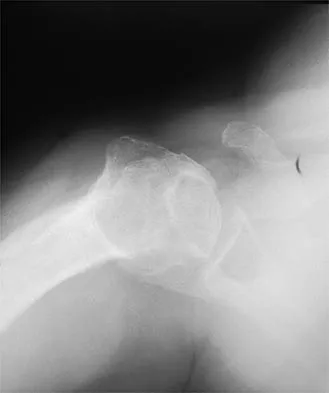
A 45-year-old woman has had progressive right shoulder pain for the past 6 months. She notes that the pain disrupts her sleep, she has pain at rest that requires the use of narcotic analgesics, and she has limited use of her left shoulder for most activities of daily living. History reveals the use of corticosteroids for systemic lupus erythematosus. Examination shows diminished range of motion. Radiographs of the right shoulder are shown in Figures 4a and 4b. Treatment should consist of


Explanation

Humeral arthroplasty provides excellent pain relief and function for stage IV osteonecrosis with humeral collapse. In late disease with glenoid involvement (stage V), total shoulder arthroplasty is preferred. Some authors have reported satisfactory results with core decompression of the humeral head for early stages of osteonecrosis, but results for stage IV osteonecrosis are less satisfactory when compared with those for humeral arthroplasty. Cruess RL: Steroid-induced avascular necrosis of the head of the humerus: Natural history and management. J Bone Joint Surg Br 1976;58:313-317. LePorte DM, Mont MA, Mohan V, Pierre-Jacques H, Jones LC, Hungerford DS: Osteonecrosis of the humeral head treated by core decompression. Clin Orthop 1998;355:254-260.


Question 10

The relocation test is most reliable for diagnosing anterior subluxation of the glenohumeral joint when


Explanation

The relocation test is most accurate when true apprehension is produced with the arm in combined abduction and external rotation and then relieved when posterior pressure is placed on the humeral head. Pain with this test is a less specific response and may occur with other shoulder disorders such as impingement.


Question 11

A 16-year-old high school pitcher notes acute pain on the medial side of his elbow during a pitch. Examination that day reveals medial elbow tenderness, pain with valgus stress, mild swelling, and loss of extension. Plain radiographs show closed physes and no fracture. Which of the following diagnostic studies will best reveal his injury?


Explanation

The history and findings are consistent with a diagnosis of a sprain of the medial collateral ligament (MCL) of the elbow; therefore, contrast-enhanced MRI is considered the most sensitive and specific study for accurately showing this injury. Arthroscopic visualization of the MCL is limited to the most anterior portion of the anterior bundle only; complete inspection of the MCL using the arthroscope is not possible. CT without the addition of contrast is of no value in this situation. Use of a technetium Tc 99m bone scan is limited to aiding in the diagnosis of occult fracture, a highly unlikely injury in this patient. There are no clinical indications for electromyography. Timmerman LA, Andrews JR: Undersurface tear of the ulnar collateral ligament in baseball players: A newly recognized lesion. Am J Sports Med 1994;22:33-36. Timmerman LA, Schwartz ML, Andrews JR: Preoperative evaluation of the ulnar collateral ligament by magnetic resonance imaging and computed tomography arthrography: Evaluation of 25 baseball players with surgical confirmation. Am J Sports Med 1994;22:26-32.


Question 12

Figures 5a and 5b show the radiographs of a 45-year-old patient. What is the most likely diagnosis?


Explanation

Glenoid dysplasia is an uncommon anomaly that usually has a benign course but may result in shoulder pain, arthritis, or multidirectional instability. Shoulder pain and instability often improve with shoulder strengthening exercises. Wirth MA, Lyons FR, Rockwood CA Jr: Hypoplasia of the glenoid: A review of sixteen patients. J Bone Joint Surg Am 1993;75:1175-1184.


Question 13

A 14-year-old boy sustains a twisting injury to his right shoulder and recalls feeling a snap during a wrestling match. Examination shows hesitancy to raise the arm away from the side, diffuse tenderness and swelling of the upper arm, and no evidence of neurovascular compromise. Figures 6a and 6b show an AP radiograph and MRI scan. What is the most likely diagnosis?


Explanation

While difficult to appreciate on the AP radiograph of the shoulder, the increased physeal signal demonstrated on the axial MRI scan is consistent with a nondisplaced growth plate fracture. A comparison radiograph of the left shoulder also could be considered and the injured shoulder evaluated for physeal widening. Proximal humeral fractures in children are somewhat unusual, representing less than 1% of all fractures seen in children and only 3% to 6% of all epiphyseal fractures. Physeal injuries are classified according to the Salter-Harris classification scheme. Salter-Harris type I fractures represent approximately 25% of physeal injuries to the proximal humerus in adolescents. The proximal humeral physis is responsible for 80% of the longitudinal growth of the humerus; therefore, there is tremendous potential for remodeling of fractures in this region. Management for nondisplaced Salter-Harris type I fractures is limited to a short period of immobilization followed by a gradual return to activities as clinical symptoms resolve. Curtis RJ, Rockwood CA Jr: Fractures and dislocations of the shoulder in children, in Rockwood CA Jr, Matsen FA III (eds): The Shoulder. Philadelphia, PA, WB Saunders, 1990, pp 991-1007.


Question 14

Figure 7 shows the radiograph of an otherwise healthy 65-year-old man who injured his right dominant shoulder while skiing 18 months ago. He did not seek treatment at the time of the injury. He now reports intermittent soreness when playing golf but has no other limitations. Examination reveals full range of motion and no tenderness, but he has slight pain with a crossed arm adduction stress test. He is neurologically intact. Initial management should consist of


Explanation

The radiograph shows a displaced type II distal clavicle fracture with nonunion. Because the patient's symptoms are minimal, the injury can be treated like a grade III acromioclavicular separation. Present management should consist of ice, anti-inflammatory drugs, activity modification, and perhaps physical therapy. If nonsurgical management fails to provide relief, the surgical options are varied with no uniformity in the literature regarding surgical treatment of this injury. Beaty JH (ed): Orthopaedic Knowledge Update 6. Rosemont, IL, American Academy of Orthopaedic Surgeons, 1999, pp 271-286.


Question 15

Figure 8 shows the AP radiograph of a 33-year-old woman who sustained a midshaft clavicle fracture from a motorcycle accident 15 months ago. She continues to have significant pain with activities of daily living. Management should consist of


Explanation

The patient has a symptomatic painful atrophic midclavicular nonunion, and the treatment of choice is rigid internal fixation with a dynamic compression plate and autogenous bone grafting. A tension band effect is desired and achieved by placing the plate superiorly. Excellent success rates of 90% to 100% have been reported using this technique. Intramedullary screw fixation without bone grafting has a decreased success rate. Partial claviculectomy is not a preferred option. Jupiter JB, Leffert RD: Non-union of the clavicle: Associated complications and surgical management. J Bone Joint Surg Am 1987;69:753-760.


Question 16

A 62-year-old patient with rheumatoid arthritis has had pain and instability of the elbow following total elbow replacement 2 years ago. A complete work-up, including aspiration and cultures, is negative. Figures 9a and 9b show the AP and lateral radiographs. Treatment should consist of


Explanation

The patient has aseptic loosening of the original semiconstrained prosthesis and significant proximal ulnar bone destruction; therefore, the treatment of choice is revision arthroplasty using a semiconstrained design. Although orthotic stabilization could be used, it will not provide long-term pain relief. Resection arthroplasty after removal of the components may lead to painful instability. Elbow arthrodesis would be difficult with the bone stock loss and is not considered the best option. Two main contraindications to the use of an unconstrained prosthesis are significant bone loss and previous use of a hinged or semiconstrained prosthesis. An ulnar allograft could be combined with the use of a semiconstrained long-stemmed ulnar prosthesis as a treatment modification. Ewald FC, Simmons ED Jr, Sullivan JA, et al: Capitellocondylar total elbow replacement in rheumatoid arthritis: Long-term results. J Bone Joint Surg Am 1993;75:498-507.


Question 17

A 21-year-old football player reports increasing pain and a deformity involving his chest after colliding with another player during a scrimmage. Imaging studies confirm an anterior sternoclavicular dislocation. Management should consist of


Explanation

For the patient with an anterior sternoclavicular dislocation, the most appropriate initial treatment should be symptomatic. Surgical options are usually contraindicated because the incidence of intraoperative and postoperative complications is high. A deformity from an anterior sternoclavicular dislocation is usually well tolerated. Return to play is allowed when symptoms resolve. Rockwood CA Jr: Disorders of the sternoclavicular joint, in Rockwood CA Jr, Matsen FA III (eds): The Shoulder. Philadelphia, PA, WB Saunders, 1998, vol 1, pp 477-525.


Question 18

During total shoulder replacement for rheumatoid arthritis, fracture of the humeral shaft occurs. An intraoperative radiograph shows a displaced short oblique fracture at the tip of the prosthesis. At this point, the surgeon should


Explanation

The risk of intraoperative fracture in osteopenic rheumatoid bone is significant. Fractures may occur with dislocation of the head and canal reaming, especially while extending and externally rotating the shoulder. If the fracture occurs at the distal tip of the prosthesis, the use of a long-stemmed prosthesis to bypass the fracture site and supplementation with wire cables has been reported with good results. Wright TW, Cofield RH: Humeral fractures after shoulder arthroplasty. J Bone Joint Surg Am 1995;77:1340-1346. Boyd AD Jr, Thornhill TS, Barnes CL: Fractures adjacent to humeral protheses. J Bone Joint Surg Am 1992;74:1498-1504.


Question 19

What is the most common contracture deformity of the spastic shoulder secondary to a cerebrovascular accident?


Explanation

The resultant spasticity and weakness (paresis) following a cerebrovascular accident leads to muscle imbalance that commonly results in contracture of the shoulder in adduction, internal rotation, and varying degrees of forward flexion. In addition, the elbow is usually flexed and the forearm pronated. Braun RM, Botte MJ: Treatment of shoulder deformity in acquired spasticity. Clin Orthop 1999;368:54-65.


Question 20

A 21-year-old collegiate pitcher has had pain in his dominant shoulder for the past 3 months despite management consisting of rest, rehabilitation, and an analysis of throwing mechanics. An arthroscopic photograph from the posterior portal is shown in Figure 10. The biceps anchor to the bone was not detached to probing. Treatment of the lesion to the left of the cannula should consist of arthroscopic


Explanation

The lesion is a variation of a type I superior labrum anterior and posterior lesion; therefore, appropriate treatment is simple debridement. Biceps tenodesis or release is not indicated because the biceps tendon and anchor are intact. There is no indication for labral repair or capsulorraphy. Beaty JH (ed): Orthopaedic Knowledge Update 6. Rosemont, IL, American Academy of Orthopaedic Surgeons, 1999, pp 261-270.


Question 21

After humeral head replacement for four-part fractures, what is the most commonly reported difficulty?


Explanation

Results show that patients who underwent humeral head replacement for fracture almost routinely report pain relief, but functional reports vary. The most commonly reported difficulty is the use of weight in the overhead position with wide variation in active elevation. Factors found to affect active elevation include age, humeral offset, greater tuberosity positioning, and four-part (as compared with three-part) fractures. Goldman RT, Koval KJ, Cuomo F, Gallagher MA, Zuckerman JD: Functional outcome after humeral head replacement for acute three- and fourth-part proximal humeral fractures. J Shoulder Elbow Surg 1995;4:81-86.


Question 22

Figures 11a and 11b show the AP and lateral radiographs of a 32-year-old patient on hemodialysis who has increasing elbow pain and a visibly growing mass over the extensor surface. Figure 11c shows the photomicrograph of the biopsy specimen. What is the most likely diagnosis?


Explanation

The radiographic findings are classic for tumoral calcinosis; they are not consistent with myositis ossificans, fungal granuloma, or hemochromatosis. The condition typically appears as large aggregations of dense calcified lobules confined to the surrounding soft tissues. Hyperphosphatemia is a fundamental factor in many patients with this condition. Tumoral calcinosis also occurs in the setting of chronic renal failure when mineral homeostasis is not controlled. The histologic appearance is essentially a foreign body granuloma reaction. Multilocular cysts with purplish amorphous material are surrounded by thick connective tissue capsules. The fibrous walls contain numerous foreign body giant cells. Surgical excision is indicated if the tumor causes discomfort or interferes with function. Sisson HA, Murray RO, Kemp HBS (eds): Orthopaedic Diagnosis: Clinical, Radiological and Pathological Coordinates. New York, NY, Springer-Verlag, 1984.


Question 23

A 52-year-old man who was a former high school pitcher now reports loss of elbow flexion and extension with pain at the extremes of motion. Nonsurgical management has failed to provide relief. Examination reveals movement from 50 degrees to 110 degrees and is painful only at the limits of motion. A radiograph is shown in Figure 12. Treatment should consist of


Explanation

Based on the history, examination, and radiograph, the patient has typical degenerative arthritis of the elbow. This condition is found almost exclusively in men, and there is almost universally a history of repetitive heavy use or overuse of the elbow. Patients report pain at terminal extension and usually have a flexion contracture. Radiographs reveal osteophytes on the coronoid and olecranon and in the coronoid and olecranon fossae. The osteophytes are often associated with loose bodies that sometimes are attached to the soft tissues. Treatment should consist of removal of all loose bodies and impinging osteophytes using open technique or by arthroscopy. The capsular contractures should be released at the same time. Kasser JR (ed): Orthopaedic Knowledge Update 5. Rosemont, IL, American Academy of Orthopaedic Surgeons, 1996, pp 283-294. Morrey BF: Primary degenerative arthritis of the elbow: Treatment by ulnohumeral arthroplasty. J Bone Joint Surg Br 1992;74:409-413. Redden JF, Stanley D: Arthroscopic fenestration of the olecranon fossa in the treatment of osteoarthritis of the elbow. Arthroscopy 1993;9:14-16.


Question 24

A 79-year-old woman with polyarticular rheumatoid arthritis has had progressively increasing right shoulder pain for the past year, and nonsurgical management has failed to provide relief. Her neurologic examination is entirely normal, but she is unable to elevate her arm against gravity. An AP radiograph is shown in Figure 13. Treatment should consist of


Explanation

Because the patient has end-stage rheumatoid arthritis with glenoid and rotator cuff deficiency, humeral arthroplasty is the treatment of choice. When a patient has an intact rotator cuff and there is sufficient glenoid bone stock to implant a glenoid component, total shoulder arthroplasty is the preferred method because it appears to provide more predictable pain relief. Glenohumeral arthrodesis is generally avoided when there is a functional deltoid or rotator cuff. Open synovectomy is appropriate in early rheumatoid disease before articular changes are present. Anterior acromioplasty with coracoacromial ligament resection is avoided in patients with rheumatoid arthritis because this procedure compromises the coracoacromial arch and may result in anterosuperior instability. Neer CS II, Watson KC, Stanton FJ: Recent experience in total shoulder replacement. J Bone Joint Surg Am 1982;64:319-337. Neer CS II: Glenohumeral arthroplasty, in Neer CS II (ed): Shoulder Reconstruction. Philadelphia, PA, WB Saunders, 1990, pp 143-271. Pollock RG, Deliz ED, McIlveen ST, et al: Prosthetic replacement in rotator cuff deficient shoulders. J Shoulder Elbow Surg 1992;1:173-186.


Question 25

A 22-year-old woman has had progressive upper extremity weakness for the past several years. History reveals no pain in her neck or shoulders. Examination reveals scapular winging of both shoulders and weakness in external rotation. She can abduct to only 120 degrees bilaterally, and there is mild supraspinatus weakness. She is otherwise neurologically intact with normal sensation and reflexes; however, she has difficulty whistling. A clinical photograph is shown in Figure 14. What is the most likely diagnosis?


Explanation

Progressive weakness is a common sign with a large differential diagnosis. Nerve, muscle, and joint problems should be excluded when a patient has diffuse weakness and atrophy. Fascioscapulohumeral dystrophy is a rare disease characterized by facial muscle weakness and proximal shoulder muscle weakness. The weakness is usually bilateral, and scapular winging is common. If the scapular winging becomes pronounced, elevation of the shoulder can be affected. In severe cases, scapulothoracic fusion or pectoralis muscle transfer to the scapula may be indicated. Duchenne muscular dystrophy is typically severe and progressive. The other diagnoses are not compatible with the history or the physical findings. Shapiro F, Specht L: The diagnosis and orthopaedic treatment of inherited muscular diseases of childhood. J Bone Joint Surg Am 1993;75:439-454.


Question 26

An 72-year-old woman presents with severe shoulder pain and inability to actively elevate her arm above 50 degrees. Radiographs demonstrate severe superior migration of the humeral head with articulation directly against the acromion, with subchondral sclerosis but no significant glenohumeral osteoarthritis. What is the most appropriate definitive surgical management?


Explanation

Reverse total shoulder arthroplasty is the treatment of choice for elderly patients with massive, irreparable rotator cuff tears and pseudoparalysis. It utilizes the deltoid to elevate the arm by medializing the center of rotation and increasing the deltoid lever arm.

Question 27

A 22-year-old collegiate rugby player presents with recurrent anterior shoulder instability. A 3D CT scan demonstrates a 28% anterior glenoid bone defect. Which of the following is the most appropriate surgical treatment?


Explanation

For anterior glenoid bone loss exceeding 20-25% in a high-demand athlete, an arthroscopic soft-tissue repair has an unacceptably high failure rate. A bony augmentation, such as a Latarjet procedure, is indicated to restore glenoid track stability.

Question 28

A 65-year-old man undergoes open reduction and internal fixation with a locking plate for a 3-part proximal humerus fracture. Two months postoperatively, radiographs show varus collapse of the humeral head and intra-articular screw cutout. Which of the following technical errors most likely contributed to this failure?


Explanation

Failure to restore medial cortical support (the medial calcar) in proximal humerus fractures significantly increases the risk of postoperative varus collapse. Medial support can be achieved by anatomic reduction, impaction of the shaft into the head, or using an endosteal fibular strut graft.

Question 29

A 35-year-old man sustains a closed midshaft humerus fracture. Initial emergency department examination reveals normal distal neurovascular function. Following the application of a coaptation splint and closed reduction, the patient is unable to actively extend his wrist or fingers. What is the most appropriate next step in management?


Explanation

A secondary radial nerve palsy that develops immediately after a closed reduction attempt of a humeral shaft fracture is an absolute indication for surgical exploration. This presentation suggests the nerve has been incarcerated in the fracture site during reduction.

Question 30

A 40-year-old man presents to the emergency department after suffering a first-time generalized tonic-clonic seizure. He complains of right shoulder pain and his arm is locked in internal rotation. An axillary radiograph reveals a posterior shoulder dislocation with an impaction fracture of the anterior humeral head (reverse Hill-Sachs lesion) involving 30% of the articular surface. What is the most appropriate surgical management?


Explanation

For reverse Hill-Sachs lesions involving 20% to 40% of the articular surface, transferring the lesser tuberosity and subscapularis into the defect (modified McLaughlin procedure) prevents engagement on the posterior glenoid rim. Defects >40% typically require arthroplasty.

Question 31

A 45-year-old heavy laborer presents with deep shoulder pain and a sensation of mechanical catching. MRI arthrogram reveals an isolated Type II SLAP tear. He has failed 6 months of supervised physical therapy and NSAIDs. What is the most reliable surgical option to relieve his symptoms and allow return to work?


Explanation

In older patients (typically >40 years) or heavy laborers, biceps tenodesis provides more reliable pain relief and better functional outcomes than SLAP repair. SLAP repairs in this demographic have higher rates of postoperative stiffness and persistent pain.

Question 32

The stability of the glenohumeral joint relies heavily on balanced force couples. Which of the following muscular combinations forms the critical force couple that provides a compressive force across the joint in the transverse (axial) plane?


Explanation

The transverse (axial) force couple consists of the subscapularis anteriorly and the infraspinatus/teres minor posteriorly. This couple centers the humeral head dynamically in the glenoid during arm elevation.

Question 33

During a classic posterior approach to the shoulder for internal fixation of a scapular neck fracture, the internervous plane is developed. Which of the following nerves is at greatest risk of injury during overzealous medial retraction of the superior muscle in this interval?


Explanation

The posterior approach utilizes the interval between the infraspinatus (suprascapular nerve) and teres minor (axillary nerve). Medial retraction of the infraspinatus places the suprascapular nerve at risk as it courses around the spinoglenoid notch.

Question 34

A 75-year-old woman sustains a highly comminuted 4-part proximal humerus fracture. She lives independently and is highly active. Radiographs show significant displacement of the tuberosities and a high likelihood of humeral head ischemia. Which of the following surgical options offers the most predictable return of forward elevation?


Explanation

Reverse total shoulder arthroplasty is the preferred treatment for displaced 4-part proximal humerus fractures in the elderly. It provides a more reliable return of active forward elevation compared to hemiarthroplasty, which heavily relies on perfect tuberosity healing.

Question 35

A 24-year-old weightlifter feels a "pop" in his anterior axilla while performing a heavy bench press. Examination reveals an asymmetric axillary fold and weakness in internal rotation. If surgical repair of the ruptured structure is undertaken, where should the tendon be anatomically reattached?


Explanation

The patient has a pectoralis major rupture, which typically occurs during eccentric loading (e.g., bench press). The anatomic insertion of the pectoralis major is on the lateral lip of the bicipital groove of the humerus.

Question 36

A 30-year-old overhead athlete presents with insidious onset, vague posterior shoulder pain. Examination reveals normal strength in forward elevation and internal rotation, but objective weakness in external rotation. An MRI reveals a paralabral cyst in the spinoglenoid notch. What is the underlying pathology causing this weakness?


Explanation

A cyst in the spinoglenoid notch typically results from a posterior superior labral tear and compresses the suprascapular nerve distal to the innervation of the supraspinatus. This results in isolated denervation and weakness of the infraspinatus (external rotation).

Question 37

A 28-year-old woman presents with bilateral shoulder pain and feelings of instability. She has a positive sulcus sign, generalized ligamentous hyperlaxity, and apprehension with extreme ranges of motion but no history of acute dislocation. What is the mainstay of initial treatment?


Explanation

This patient has multidirectional instability (MDI). The cornerstone of treatment for MDI is an extended course of physical therapy focusing on strengthening the periscapular stabilizers and the rotator cuff to provide dynamic joint stability.

Question 38

A 38-year-old male presents with a persistent, mobile, and painful nonunion of a midshaft transverse humerus fracture 8 months after nonoperative management in a functional brace. He has no neurovascular deficits. What is the gold standard surgical treatment?


Explanation

The gold standard for atrophic or symptomatic long-standing humeral shaft nonunions is open reduction, rigid internal fixation with compression plating, and the addition of autologous bone graft (such as from the iliac crest) to provide both mechanical stability and biological stimulus.

Question 39

Historically, the anterior humeral circumflex artery was believed to be the primary blood supply to the humeral head. Recent anatomic studies have demonstrated that the primary arterial supply to the proximal humerus actually arises from which of the following vessels?


Explanation

Recent quantitative perfusion studies have overturned historical teaching, proving that the posterior humeral circumflex artery provides the vast majority (up to 64%) of the blood supply to the humeral head.

Question 40

According to the Hamada classification of rotator cuff tear arthropathy, what radiographic finding defines Stage 3 disease?


Explanation

In the Hamada classification, Stage 1 is an AHI > 6mm. Stage 2 is an AHI < 6mm. Stage 3 involves "acetabularization" or concave erosion of the acromion undersurface. Stage 4 includes glenohumeral arthritis, and Stage 5 includes humeral head collapse.

Question 41

A 21-year-old collegiate athlete is evaluated for recurrent anterior shoulder instability. MRI reveals a Hill-Sachs lesion that is determined to be "off-track". What is the primary clinical significance of an "off-track" lesion in surgical planning?


Explanation

An "off-track" Hill-Sachs lesion means the defect will engage the anterior glenoid rim when the arm is abducted and externally rotated. This requires addressing the defect directly, either via remplissage or a bony augmentation procedure, to prevent recurrent instability.

Question 42

A 45-year-old man undergoes treatment for a humerus shaft fracture using an antegrade intramedullary nail. Which of the following is the most commonly reported long-term complication unique to this specific surgical approach compared to plating?


Explanation

Antegrade intramedullary nailing of the humerus is heavily associated with postoperative shoulder pain and rotator cuff dysfunction, primarily due to violation of the rotator cuff insertion and superior hardware prominence.

Question 43

A 48-year-old man sustains an acute, traumatic anterior shoulder dislocation. After successful closed reduction in the emergency department, he is noted to have persistent, profound weakness in active forward elevation and external rotation, though axillary nerve sensation is intact. What is the most likely diagnosis?


Explanation

In patients over 40 years old, an acute traumatic anterior shoulder dislocation is highly associated with an acute rotator cuff tear. Profound weakness in elevation and external rotation post-reduction mandates advanced imaging (MRI) to rule out a massive cuff tear.

Question 44

Following a traumatic anterior shoulder dislocation, a 65-year-old patient develops numbness over the lateral aspect of the shoulder and an inability to actively abduct the arm past 15 degrees. Which peripheral nerve is most likely injured?


Explanation

The axillary nerve is the most commonly injured nerve during anterior shoulder dislocations. Injury leads to deltoid paralysis (loss of abduction past 15 degrees) and loss of sensation over the lateral shoulder (regimental badge area).

Question 45

A 22-year-old competitive rugby player presents with his fourth anterior shoulder dislocation. Advanced imaging demonstrates a 26% anterior glenoid bone loss. Which of the following is the most appropriate surgical management to minimize the risk of recurrence?


Explanation

In the setting of recurrent anterior instability with critical glenoid bone loss (>20-25%), a bone-block procedure such as the Latarjet is indicated. Soft-tissue repairs alone (e.g., Bankart) have an unacceptably high failure rate in this scenario.

Question 46

A 74-year-old woman presents with severe right shoulder pain and an inability to actively elevate her arm past 40 degrees. Passive elevation is preserved to 150 degrees.

Radiographs show superior migration of the humeral head articulating with the acromion. Which of the following is the most appropriate treatment?


Explanation

This patient has rotator cuff arthropathy with pseudoparalysis (inability to actively elevate the arm despite full passive motion). Reverse total shoulder arthroplasty is the treatment of choice as it relies on the deltoid to restore active elevation.

Question 47

A 35-year-old man sustains a closed midshaft humerus fracture. Upon presentation in the emergency department, his radial nerve function is intact. He undergoes a closed reduction and splinting. Post-reduction examination reveals a new-onset complete loss of wrist extension and thumb extension. What is the most appropriate next step in management?


Explanation

A primary radial nerve palsy (present at injury) is typically observed. However, a secondary radial nerve palsy that develops immediately after a closed reduction is an absolute indication for surgical exploration due to the high risk of nerve entrapment in the fracture site.

Question 48

A 55-year-old man presents with anterior shoulder pain after a fall. Physical examination reveals a positive belly-press test and negative lift-off test. Passive range of motion testing is most likely to reveal which of the following compared to the contralateral normal shoulder?


Explanation

A positive belly-press test with a negative lift-off test suggests an isolated tear of the upper subscapularis. A physical examination hallmark of a subscapularis tear is increased passive external rotation due to the loss of the anterior restraints.

Question 49

Recent anatomic injection studies have redefined the primary arterial blood supply to the humeral head. Which of the following vessels provides the most significant perfusion to the humeral head?


Explanation

Historically, the arcuate branch of the anterior humeral circumflex artery was thought to be the primary supply. Recent literature demonstrates that the posterior humeral circumflex artery provides the most robust vascular supply to the humeral head via rich intraosseous anastomoses.

Question 50

During arthroscopy for a 45-year-old overhead athlete, you identify a partial articular-sided supraspinatus tendon avulsion (PASTA) lesion. Measurement with a probe indicates the footprint exposed is 8 mm medial-to-lateral. What is the most widely accepted surgical approach for this specific lesion size?


Explanation

The normal medial-to-lateral dimension of the supraspinatus footprint is approximately 14-16 mm. A tear exposing 8 mm represents >50% footprint involvement, which is the generally accepted threshold for surgical repair (either in-situ or via takedown).

Question 51

A 28-year-old weightlifter presents with chronic, vague posterior shoulder pain. Examination demonstrates normal forward elevation, normal strength in internal rotation, and symmetric active external rotation in adduction. However, he has marked weakness in external rotation when the arm is abducted to 90 degrees. MRI demonstrates a paralabral cyst. At what anatomic location is this cyst most likely compressing the nerve?


Explanation

Isolated weakness of the infraspinatus (tested via external rotation in 90 degrees of abduction) indicates compression of the suprascapular nerve at the spinoglenoid notch. Compression at the suprascapular notch would affect both the supraspinatus and infraspinatus.

Question 52

A 40-year-old male sustains a midshaft humerus fracture. He is managed nonoperatively in a functional brace. Which of the following radiographic parameters exceeds the acceptable limits for conservative management of a humeral shaft fracture?


Explanation

Acceptable alignment criteria for nonoperative management of a humeral shaft fracture include <20 degrees of anterior/posterior bowing, <30 degrees of varus/valgus angulation, and <3 cm of shortening. A 35-degree varus deformity is unacceptable and warrants surgical fixation.

Question 53

A 24-year-old female presents with recurrent anterior shoulder instability. Arthroscopy reveals an engaging Hill-Sachs lesion. Which of the following best describes the biomechanics of an engaging Hill-Sachs lesion?


Explanation

An engaging Hill-Sachs lesion is a posterolateral humeral head defect that aligns parallel to the anterior glenoid rim and "drops in" or engages during abduction and external rotation, leading to instability. This typically requires a remplissage or bone grafting procedure.

Question 54

During an arthroscopic stabilization for anterior shoulder instability, the surgeon notes an anterior labroligamentous periosteal sleeve avulsion (ALPSA) lesion. How does this differ anatomically from a classic Bankart lesion?


Explanation

In an ALPSA lesion, the anterior labrum is detached from the glenoid rim, but the anterior scapular periosteum remains intact, allowing the labroligamentous complex to displace and heal medially on the glenoid neck. A classic Bankart lesion involves a complete tear of the periosteum.

Question 55

A 68-year-old man presents with a massive, retracted rotator cuff tear. On physical examination, he is unable to maintain his arm in external rotation against gravity when the arm is supported in 90 degrees of abduction, causing his hand to drop to his mouth when attempting to blow a horn. This "Hornblower's sign" is most indicative of severe fatty infiltration in which of the following muscles?


Explanation

A positive Hornblower's sign (inability to actively maintain external rotation in 90 degrees of abduction) is highly specific for a tear and advanced fatty infiltration of the teres minor. The infraspinatus is primarily responsible for external rotation with the arm at the side.

Question 56

When utilizing a proximal humerus locking plate for a comminuted surgical neck fracture, placement of the medial calcar screws is critical to prevent which of the following modes of construct failure?


Explanation

Placement of inferomedial calcar screws into the inferomedial quadrant of the humeral head provides critical mechanical support to the medial column. This resists the deforming forces of the pectoralis major and rotator cuff, significantly reducing the risk of varus collapse.

Question 57

During the open Latarjet procedure, the coracoid process is transferred to the anterior glenoid neck. Which of the following correctly describes the handling of the subscapularis muscle to allow exposure and transfer of the bone block?


Explanation

The classic Latarjet procedure approaches the anterior glenoid by performing a horizontal split through the mid-substance of the subscapularis muscle, separating the upper two-thirds from the lower one-third. This preserves the musculotendinous unit and dynamic stability.

Question 58

A 19-year-old female gymnast presents with bilateral shoulder pain and a sensation of slipping with overhead activities. Examination reveals positive sulcus signs bilaterally, positive apprehension and relocation tests, and generalized ligamentous laxity.

What is the initial treatment of choice for this condition?


Explanation

This patient has multidirectional instability (MDI). The cornerstone and initial treatment of choice for MDI is a prolonged, structured physical therapy program focusing on strengthening the periscapular stabilizers and rotator cuff musculature.

Question 59

Biomechanically, active shoulder elevation can be maintained in the setting of a massive supraspinatus tear if the "transverse force couple" remains intact. Which two muscles primarily comprise this critical transverse force couple?


Explanation

The transverse force couple balances anterior and posterior forces to keep the humeral head centered on the glenoid during active elevation. It is primarily composed of the subscapularis anteriorly and the infraspinatus (and teres minor) posteriorly.

Question 60

A 32-year-old male is evaluated for a locked posterior shoulder dislocation following a generalized seizure. CT imaging reveals an anteromedial humeral head impression defect (reverse Hill-Sachs lesion) involving 25% of the articular surface. Which of the following is the most appropriate surgical intervention?


Explanation

For a reverse Hill-Sachs lesion between 20% and 40% of the articular surface, filling the defect with the subscapularis tendon alone (McLaughlin) or the lesser tuberosity (modified McLaughlin) is the standard treatment to restore stability and prevent the defect from engaging the posterior glenoid.

Question 61

Following rotator cuff repair, tendon-to-bone healing progresses through distinct biological phases. During the early proliferative phase (1 to 3 weeks post-repair), which type of collagen is predominantly synthesized to form the initial scar tissue matrix?


Explanation

During the early proliferative phase of tendon healing, fibroblasts predominantly synthesize Type III collagen, which provides early, disorganized scaffolding. This is gradually remodeled into the stronger, more organized Type I collagen over subsequent months.

Question 62

When evaluating a proximal humerus fracture, which of the following radiographic criteria is the strongest predictor of impending avascular necrosis (AVN) of the humeral head?


Explanation

Hertel's criteria for predicting ischemia of the humeral head include a short calcar length (< 8 mm) attached to the articular segment, disruption of the medial hinge, and basicervical fracture lines. These strongly correlate with high rates of AVN.

Question 63

A 60-year-old active man with symptomatic long head of the biceps tendinosis undergoes an arthroscopic biceps tenotomy. Compared to a biceps tenodesis, which of the following outcomes is significantly more likely to occur following a tenotomy?


Explanation

Biceps tenotomy is simpler and has a quicker recovery, but it carries a higher risk of aesthetic "Popeye" deformity and subjective cramping pain in the biceps muscle belly compared to tenodesis. Strength differences (grip or elbow flexion) are generally minimal or not clinically significant.

Question 64

A 58-year-old female sustains a displaced two-part surgical neck fracture of the proximal humerus. She complains of numbness over the lateral aspect of her shoulder. Which nerve is most likely injured, and what is its correct anatomic relationship in the shoulder?


Explanation

The axillary nerve is the most commonly injured nerve in proximal humerus fractures, presenting with lateral deltoid numbness. It exits the axilla posteriorly through the quadrilateral space, bordered superiorly by the teres minor, inferiorly by the teres major, medially by the long head of the triceps, and laterally by the humeral shaft.

Question 65

Which of the following best describes the biomechanical alterations achieved by a standard Grammont-style reverse total shoulder arthroplasty?


Explanation

A Grammont-style reverse total shoulder arthroplasty medializes and inferiorizes the center of rotation. This effectively lengthens the deltoid moment arm and increases its tension, allowing it to compensate for a deficient rotator cuff.

Question 66

A 22-year-old rugby player presents with recurrent anterior shoulder instability. A 3D CT scan reveals a 25% anterior glenoid bone loss. What is the most appropriate surgical treatment?


Explanation

Glenoid bone loss greater than 20-25% in the setting of recurrent anterior shoulder instability is an indication for a bony augmentation procedure like the Latarjet. Soft tissue repairs have unacceptably high failure rates in this setting.

Question 67

Based on recent quantitative anatomic studies, which of the following arterial branches provides the majority of the blood supply to the humeral head?


Explanation

Recent quantitative anatomic studies (e.g., Hettrich et al.) have demonstrated that the posterior circumflex humeral artery provides 64% of the blood supply to the humeral head. This challenges the historical belief that the anterior circumflex humeral artery was the dominant blood supply.

Question 68

A 55-year-old manual laborer presents with a chronic, massive, irreparable posterosuperior rotator cuff tear. He has severe weakness in external rotation and active elevation, but subscapularis function remains intact. He has minimal glenohumeral osteoarthritis. Which of the following is the most appropriate tendon transfer?


Explanation

Latissimus dorsi or lower trapezius tendon transfers are indicated for massive, irreparable posterosuperior rotator cuff tears in younger, active patients. An intact or repairable subscapularis is highly recommended for a successful latissimus dorsi transfer.

Question 69

A 35-year-old man sustains a closed spiral fracture of the distal third of the humeral shaft (Holstein-Lewis fracture). In the emergency department, he is noted to have an inability to extend his wrist and fingers. What is the most appropriate initial management?


Explanation

A primary radial nerve palsy in the setting of a closed humeral shaft fracture is generally managed nonoperatively initially, as most palsies resolve spontaneously. Operative exploration is strictly indicated if a new palsy develops after an initially normal exam following closed reduction.

Question 70

A 45-year-old man presents with anterior shoulder pain and weakness after falling onto an outstretched arm. Physical examination reveals a positive bear-hug test and increased passive external rotation of the shoulder. Which structure is most likely injured?


Explanation

The bear-hug test, belly-press test, and lift-off test evaluate subscapularis function. A tear of the subscapularis often presents with increased passive external rotation due to the loss of its anterior restraint.

Question 71

A 30-year-old man is evaluated in the emergency department after experiencing a first-time generalized tonic-clonic seizure. He has severe shoulder pain and his arm is locked in internal rotation. An AP radiograph shows a 'lightbulb' sign. What is the most likely diagnosis?


Explanation

Posterior shoulder dislocations classically occur following seizures, electrocution, or severe trauma, presenting with the arm locked in internal rotation. The AP radiograph may show a 'lightbulb' sign due to internal rotation of the humeral head making it appear symmetrically spherical.

Question 72

A 72-year-old woman presents with pseudoparalysis of the right shoulder and severe glenohumeral osteoarthritis secondary to a chronic, massive rotator cuff tear. Which of the following is considered an absolute contraindication to performing a reverse total shoulder arthroplasty?


Explanation

Reverse total shoulder arthroplasty biomechanically relies on a functioning deltoid muscle to elevate the arm. A non-functioning deltoid (e.g., due to severe axillary nerve palsy) is an absolute contraindication to the procedure.

Question 73

Which of the following radiographic criteria in a proximal humerus fracture is generally considered a strong indication for surgical intervention rather than nonoperative management?


Explanation

Displacement of the greater tuberosity greater than 5 mm is generally an indication for surgery in a proximal humerus fracture. Superior displacement of the tuberosity can lead to severe subacromial impingement and blocked external rotation.

Question 74

A 28-year-old man undergoes magnetic resonance arthrography (MRA) for recurrent anterior shoulder instability. The radiologist notes a 'J-sign' on the coronal fluid-sensitive sequences, representing a capsuloligamentous injury. What specific lesion does this finding describe?


Explanation

Humeral Avulsion of the Glenohumeral Ligament (HAGL) produces a 'J-sign' on MRI arthrography. The normal U-shaped axillary pouch drops down into a J-shape due to the detachment of the inferior glenohumeral ligament from the humeral neck.

Question 75

A 42-year-old woman presents with a 7-month history of a painful midshaft humerus fracture initially treated with a functional brace. Radiographs show an atrophic nonunion with minimal callus. What is the most appropriate surgical treatment?


Explanation

Atrophic humeral shaft nonunions require rigid stability and biological stimulation. Open reduction with dynamic compression plating and autologous bone grafting is the gold standard, achieving very high union rates.

Question 76

A 19-year-old female gymnast complains of bilateral shoulder pain and a feeling of looseness. Examination reveals a positive sulcus sign that does not reduce with external rotation. What is the most appropriate initial management?


Explanation

The initial management for multidirectional instability (MDI) is a prolonged trial (at least 6 months) of physical therapy focusing on strengthening the dynamic stabilizers. Surgical stabilization is reserved for cases strictly refractory to conservative care.

Question 77

A 38-year-old elite volleyball player has isolated atrophy of the infraspinatus muscle and weakness in external rotation. An MRI reveals a paralabral cyst. At which anatomic location is the nerve most likely compressed?


Explanation

Compression of the suprascapular nerve at the spinoglenoid notch (often by a paralabral cyst associated with a posterior SLAP tear) results in isolated infraspinatus weakness. Compression more proximally at the suprascapular notch affects both the supraspinatus and infraspinatus.

Question 78

When performing an anterolateral approach (deltoid split) for plating a proximal humerus fracture, how far distal to the lateral edge of the acromion is it generally safe to split the deltoid before placing the axillary nerve at significant risk?


Explanation

The axillary nerve courses transversely across the deep surface of the deltoid approximately 5 to 7 cm distal to the lateral edge of the acromion. Splitting the deltoid longitudinally beyond 5 cm places the nerve at significant risk of iatrogenic injury.

Question 79

Which of the following represents the primary biomechanical goal of a Superior Capsular Reconstruction (SCR) using a dermal allograft for a massive, irreparable rotator cuff tear?


Explanation

Superior Capsular Reconstruction (SCR) utilizes a graft to replace the deficient superior capsule. Its primary biomechanical function is to serve as a static restraint, preventing superior migration of the humeral head and restoring the glenohumeral fulcrum.

Question 80

A 25-year-old baseball pitcher presents with deep shoulder pain during the late cocking phase of throwing. He undergoes shoulder arthroscopy and is diagnosed with a Type II SLAP lesion. Which of the following best describes this pathology?


Explanation

A Type II SLAP lesion is defined by the detachment of both the superior labrum and the origin of the long head of the biceps tendon from the superior glenoid. It is a common cause of pain in overhead athletes and often requires surgical repair.

Question 81

A 65-year-old female sustains a closed, highly displaced proximal humerus fracture after a fall. Following closed reduction and splinting, she is noted to have decreased sensation over the lateral aspect of her shoulder and is unable to actively contract her deltoid. Injury to which of the following nerves is most likely responsible for these findings?


Explanation

The axillary nerve is the most commonly injured nerve in proximal humerus fractures and anterior shoulder dislocations. It supplies motor innervation to the deltoid and teres minor, and sensation to the lateral shoulder (superior lateral cutaneous nerve of the arm).

Question 82

A 75-year-old man presents with chronic shoulder pain and inability to raise his arm above 60 degrees. Examination demonstrates intact deltoid function but a positive drop arm sign. Radiographs reveal superior migration of the humeral head with articulation against the acromion and severe glenohumeral osteoarthritis.

Which of the following is the most appropriate surgical treatment?


Explanation

This patient has rotator cuff tear arthropathy with pseudoparalysis. A reverse total shoulder arthroplasty (RTSA) is the treatment of choice as it relies on the intact deltoid to elevate the arm by medializing the center of rotation and increasing the deltoid moment arm.

Question 83

A 22-year-old rugby player presents with recurrent anterior shoulder instability. A 3D CT scan of the shoulder reveals 28% bone loss of the anterior-inferior glenoid width. Which of the following is the most appropriate surgical management?


Explanation

Anterior glenoid bone loss exceeding 20-25% in the setting of recurrent instability is an indication for a bony augmentation procedure. The Latarjet procedure (coracoid transfer) addresses the bone defect and provides a sling effect via the conjoint tendon.

Question 84

A 35-year-old male presents to the emergency department after a witnessed generalized tonic-clonic seizure. His shoulder is locked in internal rotation, and he has severe pain with attempted external rotation. Standard AP radiographs appear surprisingly normal. Which of the following represents the most likely pathognomonic lesion on advanced imaging or axillary radiograph?


Explanation

Seizures or electrical shocks classically cause posterior shoulder dislocations, which lock the arm in internal rotation. The characteristic impaction fracture on the anteromedial humeral head is a reverse Hill-Sachs lesion.

Question 85

During a deltopectoral approach for open reduction and internal fixation of a proximal humerus fracture, the cephalic vein is identified. Which of the following correctly describes the internervous plane utilized in this surgical approach?


Explanation

The deltopectoral approach utilizes the internervous plane between the deltoid (innervated by the axillary nerve) and the pectoralis major (innervated by the medial and lateral pectoral nerves).

Question 86

A 26-year-old elite volleyball attacker presents with vague posterior shoulder pain and isolated weakness in external rotation. Active forward flexion and abduction are symmetric to the contralateral side. MRI demonstrates a paralabral cyst. At which of the following anatomic locations is the nerve compression most likely occurring?


Explanation

Compression of the suprascapular nerve at the spinoglenoid notch affects only the infraspinatus, causing isolated external rotation weakness. Compression at the suprascapular notch would affect both the supraspinatus (abduction) and infraspinatus (external rotation).

Question 87

A 30-year-old male sustains a closed, spiral fracture of the distal third of the humeral shaft (Holstein-Lewis fracture) following an arm-wrestling match. On examination, he is unable to actively extend his wrist or fingers. Radiographs show acceptable alignment. What is the most appropriate initial management?


Explanation

Primary radial nerve palsy in the setting of a closed humeral shaft fracture is typically a neurapraxia and does not initially require surgical exploration. Expectant management with functional bracing and observation is standard, as >85% spontaneously recover.

Question 88

Which of the following muscle pairings constitutes the primary transverse plane force couple responsible for maintaining the humeral head centered on the glenoid during active shoulder motion?


Explanation

The transverse force couple is composed of the subscapularis anteriorly and the infraspinatus/teres minor posteriorly. This balance is critical for glenohumeral stability and allows active elevation even in the presence of a supraspinatus tear.

Question 89

An 18-year-old male pitcher complains of right shoulder pain during the late cocking and early acceleration phases of throwing. Examination shows a positive O'Brien's test. Nonoperative management has failed. MRI reveals a detachment of the superior labrum and biceps anchor from the glenoid. Which of the following is the most appropriate surgical treatment?


Explanation

In young, active overhead athletes, a symptomatic Type II SLAP tear that fails conservative management is best treated with arthroscopic repair of the superior labrum. Biceps tenodesis is generally reserved for older patients or revision settings.

Question 90

A 28-year-old weightlifter feels a sudden 'pop' and sharp pain in his anterior shoulder during a heavy bench press. Examination reveals loss of the anterior axillary fold contour and significant weakness with resisted internal rotation and adduction. What is the recommended treatment for optimal return of strength?


Explanation

Pectoralis major ruptures commonly occur at the humeral insertion during heavy eccentric loading (e.g., bench press). Surgical repair to the humeral shaft provides the best outcomes for strength and cosmetic restoration in young, active patients.

Question 91

A 68-year-old female presents with a 4-part proximal humerus fracture.

According to Hertel's criteria, which of the following radiographic findings is most predictive of humeral head ischemia and subsequent avascular necrosis?


Explanation

Hertel identified specific predictors for humeral head ischemia (AVN) following proximal humerus fractures. The most significant predictors include a metaphyseal calcar length < 8 mm, disruption of the medial hinge, and an anatomic neck fracture pattern.

Question 92

A 25-year-old male with recurrent anterior shoulder instability undergoes an MR arthrogram. The imaging shows extravasation of contrast inferiorly and a characteristic 'J-sign' of the axillary pouch, without a definitive Bankart lesion. What is the most likely diagnosis?


Explanation

A HAGL lesion represents an avulsion of the inferior glenohumeral ligament from its humeral insertion. The classic MR arthrogram finding is the 'J-sign', whereby contrast extends inferiorly due to the lack of the normal axillary pouch containment.

Question 93

A 55-year-old diabetic woman complains of insidious onset of severe right shoulder pain and progressive loss of motion over the past 4 months. Examination shows significant limitations in both active and passive range of motion in forward flexion, abduction, and external rotation. Radiographs are unremarkable. What is the primary pathophysiologic mechanism of this condition?


Explanation

This patient has adhesive capsulitis (frozen shoulder), highly associated with diabetes. The hallmark is global loss of both active and passive motion due to fibroblastic proliferation and contracture, specifically involving the coracohumeral ligament and rotator interval.

Question 94

A 52-year-old male trips and falls on an outstretched hand. He now presents with right shoulder pain and weakness. On examination, he has 20 degrees of increased passive external rotation compared to the left shoulder and a positive 'lift-off' test. Which structure is most likely injured?


Explanation

The subscapularis is the primary internal rotator of the shoulder. A tear results in weakness of internal rotation (positive lift-off or belly press test) and an increase in passive external rotation due to the loss of anterior soft-tissue restraint.

Question 95

An 8-month follow-up radiograph of a 45-year-old female treated nonoperatively for a midshaft humerus fracture shows a persistent fracture line with sclerotic, rounded bone ends and no bridging callus. Which of the following is the most appropriate surgical intervention for this atrophic nonunion?


Explanation

Atrophic nonunions lack adequate biological healing potential and require both rigid mechanical stability and biological augmentation. ORIF with compression plating and autogenous bone grafting (e.g., iliac crest) is the gold standard.

Question 96

A 17-year-old female gymnast complains of bilateral vague shoulder pain. Examination reveals a positive sulcus sign, extreme generalized ligamentous laxity (Beighton score 8/9), and apprehension with both anterior and posterior translation, but no history of acute dislocation. What is the initial treatment of choice?


Explanation

Multidirectional instability (MDI) typically presents in hyperlax individuals without a distinct traumatic event. The mainstay of initial treatment is prolonged physical therapy focusing on dynamic stabilizers (rotator cuff and periscapular muscles).

Question 97

A 60-year-old male with a massive, retracted, irreparable tear of the supraspinatus and infraspinatus tendons presents with chronic pain and weakness. The subscapularis and deltoid are entirely intact, and there is no evidence of glenohumeral arthritis. He desires to improve his active external rotation. Which of the following tendon transfers is most appropriate?


Explanation

Latissimus dorsi tendon transfer is indicated for younger, active patients with massive, irreparable posterosuperior rotator cuff tears, intact subscapularis, and no arthritis. It restores active elevation and external rotation.

Question 98

A 24-year-old powerlifter presents with an insidious onset of pain on top of his left shoulder. The pain is exacerbated by bench pressing and dips. Examination shows localized tenderness over the acromioclavicular (AC) joint and pain with cross-body adduction. Radiographs reveal widening of the AC joint and subchondral cysts in the lateral clavicle. What is the most likely diagnosis?


Explanation

Distal clavicle osteolysis ('weightlifter's shoulder') is caused by repetitive microtrauma to the AC joint, commonly seen in young male athletes who lift heavy weights. Radiographs classicially show resorption and microcystic changes of the distal clavicle.

Question 99

A 78-year-old female sustains a minimally displaced, 1-part proximal humerus fracture after a ground-level fall. Which of the following management strategies will best optimize her functional outcome and minimize complications?


Explanation

Minimally displaced proximal humerus fractures are treated nonoperatively. Early progressive range of motion (starting at 1-2 weeks) is crucial to prevent profound shoulder stiffness (adhesive capsulitis), which is the most common complication in this demographic.

Question 100

A collegiate baseball pitcher presents with posterior shoulder pain. Examination reveals a 25-degree loss of internal rotation (GIRD) compared to the contralateral side. Pathologic GIRD is widely believed to be primarily driven by which of the following anatomic changes?


Explanation

Glenohumeral internal rotation deficit (GIRD) in throwing athletes is primarily caused by contracture of the posteroinferior capsule, responding to repetitive eccentric loads during the deceleration phase of throwing. Treatment involves sleeper stretches.

None

Clinic OS
Medically Verified Content by
Prof. Clinic OS
Consultant Orthopedic & Spine Surgeon
Chapter Index