العربية
Part of the Master Guide

100 High-Yield Orthopedic MCQs: Shoulder & Elbow | Mock Exam 251

Orthopedic Anatomy MCQs (Set 1): Shoulder, Knee, Spine | AAOS & ABOS Exam Prep

23 Apr 2026 54 min read 115 Views
Anatomy 2000 MCQs - Part 1

Key Takeaway

This high-yield question set for AAOS, ABOS, and OITE exams meticulously covers essential orthopedic anatomy. It features detailed MCQs on upper extremity musculoskeletal structures, lower extremity ligaments and joints, and critical spinal column neurovascular anatomy for comprehensive board preparation.

Orthopedic Anatomy MCQs (Set 1): Shoulder, Knee, Spine | AAOS & ABOS Exam Prep

Comprehensive 100-Question Exam


00:00

Start Quiz

Question 1

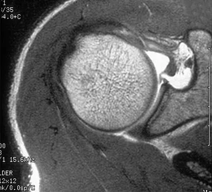
A patient has right shoulder pain. Figure 1a shows a gadolinium-enhanced transverse MRI scan at the level of the coracoid. Figure 1b shows an arthroscopic view of the anterior structures from a posterior portal. These images reveal which of the following findings?


Explanation

The area shown in the arthroscopic view and MRI scan is referred to as a Buford complex and represents a normal labral variant. It consists of a thickened, cord-like middle glenohumeral ligament, a superior labral attachment of the middle glenohumeral ligament just anterior to the biceps tendon, and absence of the anterosuperior labrum. This combination of findings can be confusing and may simulate labral pathology. Mistaken repair of the lesion back to the glenoid rim can result in significant loss of external rotation. A Bankart lesion would be located at the inferior anterior glenoid rim. The subscapularis is seen anterior to the labrum. Normal variations that occur in the anterosuperior labrum can simulate pathology. Gusmer PB, Potter HG, Schatz JA, et al: Labral injuries: Accuracy of detection with unenhanced MR imaging of the shoulder. Radiology 1996;200:519-524. Griffin LY (ed): Orthopaedic Knowledge Update: Sports Medicine. Rosemont, IL, American Academy of Orthopaedic Surgeons, 1994, pp 47-63.

Question 2

What muscle attaches to the site shown by the arrow in Figure 2?


Explanation

The latissimus dorsi inserts on the humerus metaphysis between the pectoralis major (posterior) and teres major (anterior). Teres minor inserts on the base of the greater tuberosity. Pectoralis minor does not insert on the humerus. Williams PL, Warwick R, Dyson M, Bannister LH: Neurology, in Gray's Anatomy, ed 37. Edinburgh, Scotland, Churchill Livingstone, 1989, pp 1131-1132.


Question 3

Figures 3a and 3b show the inversion stress radiographs of a patient's ankle. What is the most likely ligament injury pattern?


Explanation

The radiographic findings show 30 degrees of talar tilt (severe) and 10 mm of anterior translation that typically involves laxity of both of the major lateral ligaments of the ankle (anterior talofibular and calcaneofibular). There is no evidence of deltoid laxity. Harper MC: Stress radiographs in the diagnosis of lateral instability of the ankle and hindfoot. Foot Ankle 1992;13:435-438.


Question 4

Posterior sternoclavicular dislocations are most commonly associated with which of the following complications?


Explanation

Posterior sternoclavicular dislocations are commonly associated with tracheal compression, which can be a life-threatening condition requiring immediate reduction. The other listed complications are less common. Brooks AL, Henning GD: Injury to the proximal clavicular epiphysis, abstracted. J Bone Joint Surg Am 1972;54:1347-1348.


Question 5

An AP radiograph of the pelvis is shown in Figure 4. What muscle attaches to the avulsed fragment of bone identified by the arrow?


Explanation

The radiograph reveals an avulsion of the ischial apophysis, most likely the result of violent contraction of the attached hamstring tendons (semimembranosus, semitendinosus, and long head of the biceps femoris). The short head of the biceps femoris arises from the linea aspera on the posterior femur. The pectineus and adductor longus attach to the pubic portion of the pelvis. The piriformis runs from the sacrum to the femur. Woodburne RT (ed): Essentials of Human Anatomy. New York, NY, Oxford University Press, 1978, pp 542-545.


Question 6

A patient with an acromioclavicular dislocation has a very prominent distal clavicle. Examination reveals that the deformity increases rather than reduces with an isometric shoulder shrug. Which of the following structures is most likely intact?


Explanation

Severely displaced acromioclavicular injuries disrupt the deltotrapezial fascia and muscular origin in addition to the ligaments (acromioclavicular and coracoclavicular or trapezoid and conoid). When the deltoid is still attached to the clavicle, an isometric shoulder shrug will tend to reduce the displacement. When the deltoid is detached but the trapezius is attached, this manuever will increase the deformity and surgery may be indicated.


Question 7

Figures 5a and 5b show axial and coronal MRI images of the left ankle of a patient with lateral ankle pain. What is the most likely diagnosis?


Explanation

The figures show a longitudinal split within the peroneus brevis tendon as it courses posterior to the fibula. The peroneus longus tendon has been driven between the medial and lateral components of the peroneus brevis tendon. Peroneal split syndrome is a cause of lateral ankle pain but may be less asymptomatic in the elderly. It may be associated with tendon subluxation following a tear of the superior peroneal retinaculum.


Question 8

Which of the following anatomic structures is often difficult to visualize during elbow arthroscopy?


Explanation

The ulnar collateral ligament is often difficult to visualize during elbow arthroscopy. It can be seen clearly in only 10% to 30% of elbow arthroscopies. All of the other structures should be easily and thoroughly seen and palpated during elbow arthroscopy. Johnson LL: Arthroscopic Surgery: Principles and Practice. St Louis, MO, CV Mosby, 1988.


Question 9

The quadrilateral space in the shoulder contains which of the following structures?


Explanation

The quadrilateral or quadrangular space of the shoulder is formed laterally by the humerus, proximally by the subscapularis (and teres minor viewed from posterior), distally by the teres major, and medially by the long head of triceps. The posterior humeral circumflex artery and axillary nerve pass through it. The axillary artery is more proximal. The radial nerve and profunda brachii pass through a triangular space more inferior. The circumflex scapular artery passes through a triangular space more medial. Hollinshead WH: Textbook of Anatomy, ed 3. Hagerstown, MD, Harper and Row, 1974, pp 205-206.


Question 10

Based on the MRI scan shown in Figure 6, the abnormal signal is seen in what carpal bone?


Explanation

The MRI scan reveals an abnormal signal in the trapezoid, which lies adjacent to the capitate in the distal carpal row. The tumor is a giant cell tumor of bone. Cooney WP, Linscheid RL, Dobyns JH: The Wrist: Diagnosis and Operative Treatment. St Louis, MO, Mosby-Year Book, 1998, vol 1, pp 278-282. Green DP, Hotchkiss RN, Pederson WC (eds): Operative Hand Surgery, ed 4. New York, NY, Churchill Livingstone, 1999, pp 2238-2240. bar based on these measurements is shown in Figure 54d. Initial treatment should consist of 1- bony bar resection and distal fibula epiphysiodesis. 2- bony bar resection and corrective osteotomy. 3- bony bar resection and physiodesis of the opposite distal tibial physis. 4- corrective osteotomy and a limb-lengthening procedure. 5- corrective osteotomy and physiodesis of the opposite distal tibial physis. 2 54a 54b 54c 54d Mapping of a physeal bar from biplane polytomography or CT helps to identify lesions that should be treated surgically and aids in planning the surgical approach and resection. Criteria for surgical excision are at least 2 years of longitudinal growth remaining and involvement of no more than 50% of the physis. Osteotomy is required if angular deformity is greater than 20 degrees. Although this physeal bar is large, it is slightly less than 50% of the total area of the physis. Limb lengthening in this case should be reserved for failure of bar resection. Physiodesis of the opposite distal tibia at this age would result in disproportionate shortening of both tibiae. Carlson WO, Wenger DR: A mapping method to prepare for surgical excision of a partial physeal arrest. J Pediatr Orthop 1984;4:232-238.


Question 11

The recurrent motor branch of the median nerve innervates which of the following muscles?


Explanation

The recurrent motor branch of the median nerve supplies the thenar muscles (abductor pollicis brevis, flexor pollicis brevis, and opponens pollicis) that are primarily responsible for thumb opposition. The nerve can be injured in carpal tunnel release. A branch of the nerve also supplies the first lumbrical. The adductor pollicis and the interossei are supplied by the ulnar nerve. Last RJ: Anatomy: Regional and Applied, ed 6. London, England, Churchill Livingstone, 1978, p 109.


Question 12

Which of the following nerves innervates the muscle that originates from the middle third of the dorsal surface of the lateral border of the scapula, as shown in Figure 7?


Explanation

Teres minor originates from the middle third of the dorsal surface of the lateral border of the scapula. It is supplied by the axillary nerve (C5). Williams PL, Warwick R, Dyson M, Bannister LH: Myology, in Gray's Anatomy, ed 37. Edinburgh, Scotland, Churchill Livingstone, 1989, pp 611-615.


Question 13

Based on the MR arthrogram of the elbow shown in Figure 8, which of the following structures is torn?


Explanation

Based on the MR arthrogram in which gadolinium (bright on T1-weighted images) was injected into the joint space prior to imaging, the study shows a tear of the anterior band of the ulnar collateral ligament (UCL). The disruption in the distal end of the UCL is outlined by contrast. A small collection of contrast extravasation into the flexor musculature further confirms the presence of a tear. The UCL has a broad-based attachment on the medial epicondyle and has a pointed or tapered attachment distally on the ulna. Most UCL tears occur distally at the ulnar (coronoid) attachment. MR arthrography provides improved sensitivity compared to conventional MRI, without contrast, for the detection of UCL pathology, particularly in the subacute or chronic setting. After the soft-tissue edema and joint fluid associated with the injury have resolved, the torn end of the ligament may lie in contact with its adjacent attachment and create a false-negative appearance. In this patient, a noncontrasted MR arthrogram showed no tear, yet the tear is apparent with intra-articular contrast and distention. MR arthrography of the elbow also may be useful in detecting intra-articular bodies or in evaluation for loose osteochondral fragments or flaps. Morrey BF: Acute and chronic instability of the elbow. J Am Acad Orthop Surg 1996;4:117-128.


Question 14

A 26-year-old man has recurrent right knee pain. Figures 9a and 9b show consecutive sagittal T2-weighted MRI scans, and Figure 9c shows a coronal T1-weighted MRI scan. What is the most likely diagnosis?


Explanation

A discoid meniscus is a large disk-like meniscus. It is seen in the lateral meniscus in 3% of the population; a discoid medial meniscus is much less common. It can be identified on the coronal view by noting meniscal tissue extending into the tibial spine at the intercondylar notch. The average width of a normal meniscus is less than 11 mm. A bow-tie appearance should not be seen on more than two consecutive sagittal images because the conventional thickness of the sagittal slices is 3 mm and the interval between two consecutive slices is 1.5 mm. Two sagittal slices will cover a 9-mm thickness. A discoid meniscus can be diagnosed on the sagittal views by noting a bow-tie appearance on more than two consecutive images. Helms CA: MR image of the knee, in Fundamentals of Skeletal Radiology, ed 2. Philadelphia, PA, WB Saunders, 1995, pp 172-191.


Question 15

The gluteus maximus is innervated by which of the following nerves?


Explanation

The inferior gluteal nerve supplies the gluteus maximus muscle. The superior gluteal nerve supplies the gluteus medius, gluteus minimus, and tensor fascia lata muscles. The femoral nerve supplies the quadriceps, sartorius, and pectineus muscles. The pudendal nerve is primarily a sensory nerve.


Question 16

The dorsal (Thompson) approach to the proximal forearm uses which of the following intermuscular intervals?


Explanation

The Thompson posterior approach is used in treatment of fractures of the proximal radius. Dissection is carried out through the interval between the extensor carpi radialis brevis (radial nerve) and the extensor digitorum communis (posterior interosseous nerve). To identify this interval, the forearm is pronated and the mobile lateral wad of muscles (the ulnar-most belly is the extensor carpi radialis brevis) is grasped with the thumb and finger and pulled from the much less mobile mass of the extensor digitorum communis. The furrow created is marked with a skin marker for subsequent skin incision. The skin incision follows a line from the lateral epicondyle of the humerus to a point corresponding to the middle of the posterior aspect of the wrist. Distally, the intermuscular plane is between the extensor carpi radialis brevis and the extensor pollicis longus. Crenshaw AH Jr: Surgical techniques and approaches, in Canale ST (ed): Campbell's Operative Orthopaedics, ed 9. St Louis, MO, Mosby-Year Book, 1998, vol 1, pp 128-129. Hoppenfeld S, deBoer P: Posterior approach to the radius, in Surgical Exposures in Orthopaedics: The Anatomic Approach, ed 2. Philadelphia, PA, Lippincott-Raven, 1992, pp 136-146.


Question 17

A 45-year-old man who smokes reports the rapid onset of color changes and coolness in the fingers. Examination shows an abnormal Allen test. Plain radiographs of the hand and wrist are normal. Which of the following studies will best aid in diagnosis?


Explanation

The patient has symptoms typical of Raynaud's phenomenon secondary to underlying vascular disease. The next most appropriate step in the management of this patient should be to perform contrast angiography on the involved upper extremity to look for proximal or distal arterial lesions or insufficiencies. MRI and contrast CT are not as specific as angiography for the identification of vascular lesions of the upper extremity. Although patients with primary Raynaud's vasospastic disease can have normal angiographic findings, they typically are younger than age 40 years, are female, and have normal results on an Allen test. Green DP, Hotchkiss RN, Pederson WC (eds): Operative Hand Surgery, ed 4. New York, NY, Churchill Livingstone, 1999, pp 2288-2290.


Question 18

A purulent flexor tenosynovitis of the thumb may communicate with the small finger flexor through which of the following structures?


Explanation

Only the flexor sheaths of the thumb and small finger are continuous from the digit through the carpal canal and into the distal forearm. If one of the sheaths ruptures from synovitis, it may contaminate the other sheath through Parona's space in the distal forearm. This potential space lies superficial to the pronator quadratus and deep to the flexor tendons. Green DP, Hotchkiss RN, Pederson WC (eds): Operative Hand Surgery, ed 4. New York, NY, Churchill Livingstone, 1999, pp 1044-1045.


Question 19

Which of the following nerves travels with the deep palmar arch?


Explanation

The ulnar nerve divides alongside the pisiform, and the deep branch supplies the three hypothenar muscles and crosses the palm with the deep palmar arch to supply the two ulnar lumbricals, all interossei, and finally the adductor pollicis. The superficial branch supplies the ulnar digital branches to the small and ring fingers. The median nerve branches are more superficial in the palm near the superficial palmar arch. Last RJ: Anatomy: Regional and Applied, ed 6. London, England, Churchill Livingstone, 1978, p 109.


Question 20

Figures 10a through 10c show the plain radiograph and MRI scans of a 41-year-old man who has right hip pain. What is the most likely diagnosis?


Explanation

Transient osteoporosis is a self-limited painful but reversible disorder. Although first described in pregnant women, it is more common in young to middle-aged men. The radiograph shows loss of mineralization in the right hip relative to the left side. There is no osseous destruction or cortical expansion typical of metastasis or giant cell tumor. The process is confined to the femoral side of the joint unlike rheumatoid arthritis, which would be centered in the joint. Osteonecrosis is better defined with sharp but irregularly shaped margins, and there is no double-line sign. The MRI scans reveal diffuse edema in the femoral head and neck that is atypical for osteonecrosis. Transient osteoporosis may recur in the same or opposite hip.


Question 21

Figure 11 shows the anatomic dissection of the medial side of the knee joint after removal of the superficial fascia. The arrow is pointing to what structure?


Explanation

The semitendinosus and gracilis tendons lie beneath the superficial fascia and superficial to the medial collateral ligament. The semitendinosus is located more inferior to the gracilis tendon. The sartorius is more posterior and distal as is the medial collateral ligament. The semimembranosus is posterior. Pagnani MJ, Warner JJ, O'Brien SJ, Warren RF: Anatomic considerations in harvesting the semitendinosus and gracilis tendons and a technique of harvest. Am J Sports Med 1993;21:565-571.


Question 22

Figure 12 shows a lateral radiograph of the elbow. What is the most likely diagnosis?


Explanation

The figure shows a supracondylar process, which is a normal anatomic variant. An osteochondroma tends to occur more toward the end of bones, and the medullary space of the underlying bone extends into the base of the osteochondroma. The presence of a supracondylar process is usually asymptomatic. However, the ligament of Struthers that always extends from the supracondylar process to the medial epicondyle can result in median nerve entrapment secondary to trauma. Last RJ: Anatomy: Regional and Applied, ed 6. London, England, Churchill Livingstone, 1978, pp 132-133.


Question 23

Which of the following nerves is most likely responsible for symptoms associated with plantar fasciitis?


Explanation

The first branch of the lateral calcaneal nerve innervates the abductor digiti minimi. It is reported to be trapped at the interval between the abductor hallucis and the quadratus plantae muscles.


Question 24

A 16-year-old cheerleader reports an ache in the right shoulder and arm that is worse after activity. She denies any history of acute trauma. Examination reveals a positive sulcus sign and an AP glide test with a posterior and anterior apprehension sign. To confirm a diagnosis of multidirectional instability, which of the following imaging studies is most appropriate?


Explanation

Multidirectional instability is a common finding in young female athletes. The anatomic structures are all intact but are hypermobile; therefore, CT and bone scans and scapular Y-views are often normal. Obtaining a weighted or AP stress view while applying downward traction on the arm will document instability and hypermobility of the joint. MRI generally is not indicated in this condition. Ultrasound is used primarily for rotator cuff pathology. Neer CS II, Foster CR: Inferior capsular shift for involuntary inferior and multidirectional instability of the shoulder: A preliminary report. J Bone Joint Surg Am 1980;62:897-908.


Question 25

Which of the following findings is seen in the chest radiograph shown in Figure 13?


Explanation

Orthopaedic surgeons are often responsible for interpreting radiographs of general examinations such as the chest radiograph shown. For accurate interpretation, it is important to systematically review all of the information available on the radiograph. Using this approach, the fracture of the left proximal humerus is readily recognized. Linear air soft-tissue density at the lung periphery would suggest a pneumothorax, but this finding is not shown on the radiograph. The upper thoracic spine is well aligned. The sternoclavicular and distal clavicles are normal.


Question 26

Which of the following structures forms the superior border of the lumbar intervertebral foramen?


Explanation

The lumbar intervertebral foramen is bordered superiorly by the inferior notch of the superior pedicle. Anteriorly it is bordered by the vertebral body and disc, inferiorly by the superior notch of the inferior pedicle, and posteriorly by the facet joint.

Question 27

Which of the following best describes the blood supply to the adult medial meniscus?


Explanation

In the adult knee, only the peripheral 10% to 30% of the medial meniscus is vascularized by the superior and inferior medial genicular arteries. The inner portions are avascular and rely on diffusion from synovial fluid.

Question 28

A patient presents with weakness in shoulder abduction and external rotation following a posterior shoulder dislocation. MRI shows compression of a nerve passing through the quadrangular space. What are the borders of this space?


Explanation

The quadrangular space is bordered by the teres minor (superiorly), teres major (inferiorly), long head of the triceps (medially), and the surgical neck of the humerus (laterally). It contains the axillary nerve and posterior circumflex humeral artery.

Question 29

During an anterior approach to the thoracolumbar spine, care must be taken to avoid injury to the artery of Adamkiewicz. On which side and at what spinal levels does this artery most commonly originate?


Explanation

The artery of Adamkiewicz provides the major blood supply to the anterior spinal artery of the lower spinal cord. It most commonly arises on the left side between the T9 and L1 vertebral levels.

Question 30

Which of the following structures is considered a primary static stabilizer of the posterolateral corner (PLC) of the knee?


Explanation

The primary static stabilizers of the posterolateral corner (PLC) are the lateral collateral ligament (LCL), the popliteus tendon, and the popliteofibular ligament. The biceps femoris is a dynamic stabilizer.

Question 31

The coracoacromial ligament is a key structure in subacromial impingement syndrome. What are its attachments?


Explanation

The coracoacromial ligament attaches the coracoid process to the anterior undersurface of the acromion, forming the coracoacromial arch. This arch is the primary rigid roof under which the rotator cuff must pass.

Question 32

A patient sustains a whiplash injury and is suspected of having craniocervical instability. The alar ligaments primarily limit which of the following movements?


Explanation

The alar ligaments connect the sides of the dens to the medial aspect of the occipital condyles. They act as primary restraints to limit axial rotation and lateral bending of the occipitocervical junction.

Question 33

When performing an anterior drawer test at 90 degrees of knee flexion, which bundle of the anterior cruciate ligament (ACL) is the primary restraint to anterior tibial translation?


Explanation

The ACL has two main bundles: the anteromedial (AM) and posterolateral (PL). The AM bundle is tightest in flexion and is the primary restraint to anterior translation at 90 degrees, while the PL bundle is tightest in extension.

Question 34

A 28-year-old volleyball player presents with isolated atrophy of the infraspinatus muscle and normal supraspinatus strength. Entrapment of the suprascapular nerve is most likely occurring at which anatomical location?


Explanation

Entrapment of the suprascapular nerve at the spinoglenoid notch affects only the infraspinatus. Entrapment at the suprascapular notch (proximal) would cause weakness and atrophy in both the supraspinatus and infraspinatus.

Question 35

The orientation of the facet joints in the subaxial cervical spine most closely approximates which of the following planes?


Explanation

The subaxial cervical facet joints are oriented at approximately 45 degrees to the axial plane and parallel to the coronal plane. This alignment facilitates the large degree of flexion, extension, and rotation in the cervical spine.

Question 36

The medial patellofemoral ligament (MPFL) is a primary restraint to lateral patellar displacement. Where is its femoral attachment located?


Explanation

The femoral footprint of the MPFL (Schöttle's point) is located between the medial epicondyle and the adductor tubercle. It provides the primary restraint to lateral patellar subluxation at 0 to 30 degrees of flexion.

Question 37

During arthroscopy for a SLAP tear, the surgeon notes that the long head of the biceps tendon originates entirely from the posterior labrum. What is this normal anatomic variant called?


Explanation

The long head of the biceps usually originates from the supraglenoid tubercle and superior labrum. A posterior dominant origin, where the bulk of the tendon attaches to the posterior labrum, is a recognized normal variant.

Question 38

Discogenic back pain is mediated by sensory fibers in the outer annulus fibrosus. Which nerve provides the primary innervation to the posterior aspect of the lumbar intervertebral disc?


Explanation

The sinuvertebral nerve (nerve of Luschka) is a recurrent nerve that branches from the ventral ramus and sympathetic plexus. It re-enters the spinal canal to innervate the posterior annulus fibrosus, posterior longitudinal ligament, and ventral dura.

Question 39

The popliteus muscle acts to unlock the knee from a fully extended position. What is its specific action on the tibia during early knee flexion in an open kinetic chain?


Explanation

In an open kinetic chain, the popliteus internally rotates the tibia on the femur to unlock the extended knee, initiating flexion. In a closed kinetic chain, it externally rotates the femur on the fixed tibia.

Question 40

During a deltopectoral approach to the shoulder, the axillary nerve is at risk when passing near the inferior capsule. At its closest point, approximately what is the distance from the axillary nerve to the inferior glenoid rim?


Explanation

The axillary nerve runs inferior to the glenohumeral joint capsule before passing through the quadrangular space. It courses approximately 10 to 15 mm inferior to the inferior border of the glenoid.

Question 41

A patient presents with profound weakness in knee extension and loss of sensation over the anterior thigh following a retroperitoneal hematoma. Which nerve roots form the nerve most likely affected?


Explanation

The femoral nerve innervates the quadriceps and supplies sensation to the anterior thigh. It is formed by the dorsal divisions of the ventral rami of L2, L3, and L4 within the psoas major muscle.

Question 42

The posterior cruciate ligament (PCL) consists of two functional bundles. Which bundle is most taut in full knee extension?


Explanation

The PCL is composed of the larger anterolateral (AL) bundle and the smaller posteromedial (PM) bundle. The PM bundle is taut in full extension, while the AL bundle is taut in flexion.

Question 43

Which of the following nerves provides innervation to the upper and lower portions of the subscapularis muscle?


Explanation

The subscapularis muscle is innervated by the upper and lower subscapular nerves, which are branches of the posterior cord of the brachial plexus. The lower subscapular nerve also innervates the teres major.

Question 44

During a posterior cervical spine fusion, screw placement into the lateral mass of C7 must be done carefully to avoid vascular injury. Why is the vertebral artery generally not at risk within the C7 transverse foramen?


Explanation

The vertebral artery typically enters the transverse foramen at the C6 level, bypassing the C7 transverse foramen in about 90% of individuals. Thus, it usually travels anterior to the C7 transverse process.

Question 45

The iliotibial (IT) band inserts onto Gerdy's tubercle. During knee motion, the IT band shifts relative to the lateral femoral epicondyle. At what angle of knee flexion does the IT band typically snap posteriorly over the epicondyle?


Explanation

The iliotibial band lies anterior to the lateral femoral epicondyle in extension and shifts posteriorly during knee flexion. This snapping or shifting typically occurs at approximately 20 to 30 degrees of knee flexion.

Question 46

A 28-year-old professional volleyball player presents with insidious onset of painless weakness in shoulder external rotation. On examination, abduction strength is 5/5, but external rotation is 3/5. At which of the following anatomical sites is the affected nerve most likely compressed?


Explanation

The suprascapular nerve innervates the supraspinatus and infraspinatus muscles. Entrapment at the spinoglenoid notch affects only the branch to the infraspinatus, causing isolated external rotation weakness, whereas entrapment at the suprascapular notch affects both muscles.

Question 47

During a posterolateral corner (PLC) reconstruction of the knee, anatomic femoral tunnel placement is critical. Which of the following describes the correct anatomic relationship of the popliteus tendon attachment on the lateral femur relative to the lateral collateral ligament (LCL) attachment?


Explanation

On the lateral femoral epicondyle, the popliteus tendon inserts in a sulcus that is positioned anterior and inferior to the origin of the lateral collateral ligament (LCL).

Question 48

A 45-year-old male presents with severe right leg radiculopathy. MRI of the lumbar spine reveals a far lateral (extraforaminal) disc herniation at the L4-L5 level. Which nerve root is most likely compressed by this specific pathology?


Explanation

In the lumbar spine, a far lateral or foraminal disc herniation compresses the exiting nerve root at that level. Therefore, an L4-L5 far lateral herniation compresses the exiting L4 nerve root.

Question 49

Historically, the anterior circumflex humeral artery was considered the primary blood supply to the humeral head. Based on modern quantitative cadaveric perfusion studies, which vessel is now recognized as providing the predominant blood supply to the articular segment of the proximal humerus?


Explanation

Recent anatomical and perfusion studies have demonstrated that the posterior circumflex humeral artery provides the dominant blood supply (approximately 64%) to the humeral head, challenging older literature.

Question 50

The anterior cruciate ligament (ACL) is composed of two primary bundles that function synergistically during knee range of motion. Which of the following statements most accurately describes the biomechanics of these bundles?


Explanation

The anteromedial (AM) bundle of the ACL tightens in flexion to control anterior translation, while the posterolateral (PL) bundle tightens in extension to provide primary rotational stability.

Question 51

A trauma patient sustains a highly comminuted cervical spine fracture. A CT angiogram is ordered to evaluate the vertebral artery. In a normal anatomic variant, at which cervical level does the vertebral artery typically first enter the transverse foramen?


Explanation

The vertebral artery typically branches from the first part of the subclavian artery and enters the transverse foramen at the C6 level, traveling superiorly toward the foramen magnum.

Question 52

A 32-year-old weightlifter presents with vague posterior shoulder pain and numbness over the lateral deltoid. MRI confirms a mass in the quadrilateral space compressing the axillary nerve. Which muscle forms the superior border of this anatomic space?


Explanation

The quadrilateral space is bounded superiorly by the teres minor (and subscapularis anteriorly), inferiorly by the teres major, medially by the long head of the triceps, and laterally by the surgical neck of the humerus.

Question 53

Failure to recognize and repair a posterior medial meniscus root tear leads to biomechanical consequences equivalent to a total meniscectomy. Where is the exact anatomical insertion of the posterior horn of the medial meniscus root?


Explanation

The posterior root of the medial meniscus attaches to the posterior intercondylar fossa of the tibia, located directly anterior to the tibial attachment of the posterior cruciate ligament (PCL).

Question 54

During posterior spinal fusion for scoliosis, a surgeon places pedicle screws in the thoracic spine. Which level of the thoracic spine typically has the greatest transverse pedicle angle (most medial angulation)?


Explanation

The transverse pedicle angle dictates the medial trajectory for pedicle screw placement. This angle is greatest at T1 (up to 30 degrees medially) and progressively decreases to become nearly sagittal (0-5 degrees) at T12.

Question 55

A surgeon is performing an open reduction internal fixation of a proximal humerus fracture using a lateral deltoid-splitting approach. To avoid iatrogenic injury to the axillary nerve, the deltoid split should safely not extend distally beyond what average distance from the lateral edge of the acromion?


Explanation

The axillary nerve courses transversely across the deep surface of the deltoid approximately 5 to 7 cm distal to the lateral margin of the acromion. A deltoid split is generally kept within 4-5 cm to remain in the safe zone.

Question 56

A 16-year-old female undergoes medial patellofemoral ligament (MPFL) reconstruction for recurrent patellar instability. Anatomic femoral graft placement is critical to avoid altering joint contact pressures. What is the native anatomic origin of the MPFL on the femur?


Explanation

The MPFL originates on the medial femur in a distinct saddle-like depression located between the adductor tubercle superiorly and the medial femoral epicondyle inferiorly.

Question 57

A trauma surgeon is placing an S1 iliosacral screw for a vertically unstable pelvic fracture. If the guidewire is placed too anteriorly and breaches the anterior cortex of the sacral ala, which nerve root is most directly at risk of injury?


Explanation

The L5 nerve root descends anteriorly across the sacral ala to join the lumbosacral trunk. An anteriorly misplaced S1 iliosacral screw directly threatens this nerve root.

Question 58

A patient presents with pronounced medial winging of the scapula after a direct blow to the lateral chest wall. The injured nerve originates from which of the following brachial plexus structures?


Explanation

Medial scapular winging is caused by serratus anterior paralysis due to injury of the long thoracic nerve. This nerve arises directly from the anterior rami of the C5, C6, and C7 nerve roots.

Question 59

A 35-year-old male sustains a severe knee trauma resulting in a proximal fibula fracture and complete common peroneal nerve palsy. On physical examination, which of the following specific sensory deficits is expected alongside a foot drop?


Explanation

The common peroneal nerve wraps around the fibular neck and bifurcates into the deep and superficial peroneal nerves. Injury here causes foot drop (motor) and sensory loss over the dorsolateral foot (superficial) and first dorsal web space (deep).

Question 60

A patient suffers a stab wound to the thoracic spine resulting in a classic Brown-Séquard syndrome. Which of the following physical examination findings is characteristic of this anatomic spinal cord injury?


Explanation

Brown-Séquard syndrome involves hemisection of the spinal cord. It presents with ipsilateral motor (corticospinal tract) and proprioceptive (dorsal column) loss, and contralateral loss of pain and temperature (spinothalamic tract).

Question 61

During a pectoralis major tendon repair for a complete rupture at the humerus, the surgeon must identify the distinct sternal and clavicular heads. Which statement correctly describes the complex anatomic insertion of the pectoralis major tendon on the humerus?


Explanation

The pectoralis major tendon twists 180 degrees before inserting onto the lateral lip of the bicipital groove. The sternal head twists to insert proximally and deep, while the clavicular head remains superficial and inserts more distally.

Question 62

During placement of a pedicle screw in the lumbar spine, an inferior cortical breach of the pedicle places which of the following structures at highest risk of immediate injury?


Explanation

The exiting nerve root travels immediately inferior to the pedicle in the lumbar spine. An inferior breach of the pedicle directly risks injury to the exiting root of that specific level.

Question 63

When performing a posterior cruciate ligament (PCL) reconstruction, the surgeon must accurately identify the native anatomic footprint. Which of the following correctly describes the femoral attachment of the PCL?


Explanation

The PCL attaches to the anterolateral aspect of the medial femoral condyle. In contrast, the ACL attaches to the posteromedial aspect of the lateral femoral condyle.

Question 64

A patient presents with weakness in external rotation and abduction following a posterior shoulder dislocation. MRI reveals a paralabral cyst compressing the quadrilateral space. Which of the following defines the superior border of this anatomic space?


Explanation

The quadrilateral space is bordered superiorly by the teres minor, inferiorly by the teres major, medially by the long head of the triceps, and laterally by the humeral shaft. It contains the axillary nerve and posterior circumflex humeral artery.

Question 65

During an anterior cervical discectomy and fusion (ACDF), excessive lateral dissection over the longus colli muscle places the vertebral artery at risk. At which cervical level does the vertebral artery most commonly enter the transverse foramen?


Explanation

The vertebral artery typically branches from the subclavian artery and enters the transverse foramen at the C6 level in approximately 90% of individuals. It is highly variable but rarely enters at C7.

Question 66

When reconstructing the posterolateral corner (PLC) of the knee, understanding the anatomic relationship of the femoral attachments is critical. Which of the following correctly describes the origin of the lateral collateral ligament (LCL) relative to the popliteus tendon on the lateral femoral epicondyle?


Explanation

On the lateral femoral epicondyle, the LCL origin is located proximal and posterior to the popliteus tendon insertion. This relationship is critical for anatomic PLC reconstruction.

Question 67

A 28-year-old volleyball player presents with isolated weakness in shoulder external rotation. Abduction strength is normal. An MRI confirms a paralabral cyst. At which of the following locations is the nerve compression most likely occurring?


Explanation

Compression at the spinoglenoid notch affects only the infraspinatus branch of the suprascapular nerve, causing isolated external rotation weakness. Compression at the suprascapular notch would also affect the supraspinatus, causing additional abduction weakness.

Question 68

During a posterior cervical foraminotomy at C5-C6, the surgeon aggressively retracts the lateral aspect of the facet joint. Which of the following anatomical structures is most at risk of iatrogenic injury in the extraforaminal space?


Explanation

The vertebral artery runs in the transverse foramen, which lies immediately anterior to the exiting cervical nerve roots. Overly aggressive lateral dissection during a posterior foraminotomy puts the vertebral artery at significant risk.

Question 69

A 28-year-old overhead athlete presents with isolated weakness in external rotation of the shoulder but normal abduction. An MRI confirms nerve entrapment by a paralabral cyst. At which anatomical location is the entrapment most likely occurring?


Explanation

Entrapment of the suprascapular nerve at the spinoglenoid notch selectively affects the infraspinatus muscle, leading to isolated external rotation weakness. Entrapment at the suprascapular notch would affect both the supraspinatus and infraspinatus.

Question 70

During an anatomic reconstruction of the posterolateral corner (PLC) of the knee, identifying the fibular collateral ligament (FCL) footprint is critical. What is the anatomical relationship of the FCL femoral footprint to the popliteus tendon insertion?


Explanation

The femoral attachment of the FCL is located slightly proximal and posterior to the popliteus tendon attachment on the lateral femoral epicondyle. Recognizing this spatial relationship is essential for anatomic tunnel placement.

Question 71

A surgeon is performing a lateral transpsoas approach to the L4-L5 disc space. Postoperatively, the patient reports severe weakness in hip flexion and knee extension, along with anterior thigh numbness. Which of the following nerves was most likely injured?


Explanation

The femoral nerve (L2-L4) courses through the posterior aspect of the psoas major at the L4-L5 level. It innervates the iliopsoas and quadriceps, making it vulnerable to retraction injury during the transpsoas approach.

Question 72

A 35-year-old male sustains a Type III acromioclavicular (AC) joint separation requiring surgical reconstruction. To accurately recreate the coracoclavicular ligaments, the surgeon must identify their native footprints. What is the average distance from the distal end of the clavicle to the conoid and trapezoid tuberosities, respectively?


Explanation

The conoid tuberosity is located approximately 45 mm medial to the distal clavicle, while the trapezoid tuberosity is more lateral, at roughly 30 mm. Anatomic reconstruction relies on accurate placement of these drill holes.

Question 73

Which of the following accurately describes the regional vascularity and healing potential of the meniscus in a young adult knee?


Explanation

The outer 10-30% of the meniscus (the red-red zone) receives its blood supply from the perimeniscal capillary plexus originating from the medial and lateral genicular arteries. The inner avascular zone relies on diffusion from synovial fluid.

Question 74

When placing an iliosacral screw for a zone 1 sacral fracture, the surgeon must stay within the 'alar safe zone.' An errant guidewire placed anteriorly through the sacral ala primarily endangers which neural structure?


Explanation

The L5 nerve root courses directly anterior to the sacral ala as it descends to join the sacral plexus. An anterior cortical breach during iliosacral screw placement places the L5 root at high risk for injury.

Question 75

A patient presents with a paralabral cyst compressing a nerve in the quadrangular space of the shoulder. Which blood vessel accompanies the compressed nerve through this specific anatomical space?


Explanation

The quadrangular space transmits the axillary nerve and the posterior humeral circumflex artery. The circumflex scapular artery travels through the adjacent triangular space.

Question 76

The anterior cruciate ligament (ACL) is composed of two functional bundles. During a physical examination, when the knee is in full extension, how are these bundles oriented relative to each other in terms of tension?


Explanation

In full knee extension, the posterolateral (PL) bundle is tight and provides the primary restraint to anterior tibial translation. The anteromedial (AM) bundle becomes tightest in deeper knee flexion.

Question 77

During pedicle screw insertion in the midthoracic spine (T6-T8) for adolescent idiopathic scoliosis, a medial cortical breach of the pedicle occurs. Which of the following structures is at the most immediate risk of injury?


Explanation

A medial breach of the thoracic pedicle directs the instrumentation into the spinal canal, placing the spinal cord at direct risk. Lateral breaches threaten the lung, pleura, and segmental vessels.

Question 78

The superficial medial collateral ligament (sMCL) is a key static stabilizer of the knee. Proximal to its primary attachment on the medial epicondyle, where does its distal tibial attachment firmly insert?


Explanation

The primary distal attachment of the superficial MCL is located deep to the pes anserinus tendons, roughly 4-5 cm distal to the medial joint line. This broad footprint provides significant valgus stability.

Question 79

When exposing the posterior arch of C1 and the lateral masses of C2 for atlantoaxial fusion, a large neurovascular structure is routinely encountered crossing the posterior aspect of the C1-C2 joint. Which structure must be mobilized caudally or transected to achieve lateral mass exposure?


Explanation

The C2 nerve root and its dorsal root ganglion exit and course directly posterior to the C1-C2 facet joint. It often obstructs the starting point for C1 lateral mass screws and must be retracted caudally or transected.

Question 80

A 24-year-old weightlifter ruptures his pectoralis major tendon. During an open anatomic repair, the surgeon isolates the sternal and clavicular heads. Which of the following describes the insertion of the sternal head relative to the clavicular head on the lateral lip of the bicipital groove?


Explanation

The pectoralis major tendon undergoes a 180-degree twist before insertion. The inferior (sternal) fibers twist to insert proximal and deep (posterior) to the superior (clavicular) fibers.

Question 81

A patient sustains a high-energy knee dislocation (KD-III). Vascular surgery is consulted due to an absent dorsalis pedis pulse. The popliteal artery is exceptionally prone to traction injury in this scenario because it is tethered at which two anatomical landmarks?


Explanation

The popliteal artery is rigidly fixed proximally as it exits the adductor hiatus (Hunter's canal) and distally as it passes under the tendinous arch of the soleus. This lack of mobility makes it highly susceptible to stretch and intimal tearing during gross knee dislocations.

Question 82

The primary blood supply to the supraspinatus tendon is derived from branches of which of the following arteries?


Explanation

The suprascapular artery courses superior to the transverse scapular ligament and provides the primary vascular supply to the supraspinatus and infraspinatus muscles and their tendinous insertions.

Question 83

A 28-year-old overhead athlete presents with posterior shoulder pain and deltoid weakness. MRI demonstrates atrophy of the teres minor. Entrapment of the involved nerve occurs in a space bounded laterally by which of the following structures?


Explanation

The axillary nerve is entrapped in the quadrilateral space. The borders of this space are the teres minor (superior), teres major (inferior), long head of the triceps (medial), and the surgical neck of the humerus (lateral).

Question 84

A 45-year-old male presents with right leg pain radiating to the dorsum of his foot and weakness in ankle dorsiflexion. MRI reveals a far lateral (extra-foraminal) disc herniation at the L4-L5 level. Which nerve root is most likely compressed?


Explanation

In the lumbar spine, a far lateral (extra-foraminal) disc herniation compresses the exiting nerve root at the same level. Therefore, an L4-L5 far lateral herniation compresses the L4 nerve root.

Question 85

A 30-year-old male sustains a twisting injury to his knee. Examination reveals increased external tibial rotation at 30 degrees of knee flexion, but symmetrical rotation at 90 degrees of flexion compared to the contralateral side. Which of the following structures is most likely injured?


Explanation

Increased external rotation at 30 degrees of flexion with normal rotation at 90 degrees indicates an isolated posterolateral corner (PLC) injury, which involves the popliteofibular ligament, LCL, and popliteus tendon. Combined PLC and PCL injuries typically show increased rotation at both 30 and 90 degrees.

Question 86

During an arthroscopic stabilization procedure for anterior shoulder instability, the surgeon performs a rotator interval closure. Which of the following structures form the superior and inferior boundaries of this interval, respectively?


Explanation

The rotator interval is a triangular anatomic space in the anterosuperior shoulder bordered superiorly by the supraspinatus and inferiorly by the subscapularis. It contains the coracohumeral ligament, superior glenohumeral ligament, and the long head of the biceps tendon.

Question 87

During a thoracolumbar corpectomy, the surgeon must be mindful of the artery of Adamkiewicz to prevent anterior spinal cord syndrome. From which of the following regions does this vessel most commonly arise?


Explanation

The artery of Adamkiewicz is the major blood supply to the anterior lower two-thirds of the spinal cord. It most commonly arises from the left side of the aorta between the T8 and L1 vertebral levels.

Question 88

A 22-year-old female undergoes ACL reconstruction. The surgeon drills the femoral tunnel independently to accurately recreate the anatomic footprint of the ACL. Which of the following accurately describes the biomechanical function of the anteromedial (AM) bundle of the native ACL?


Explanation

The ACL consists of two main bundles. The anteromedial (AM) bundle is primarily tight in flexion and provides anterior-posterior stability, while the posterolateral (PL) bundle is tight in extension and provides rotational stability.

Question 89

A 26-year-old male volleyball player presents with painless weakness of his hitting arm. Physical examination reveals isolated atrophy of the infraspinatus fossa with normal supraspinatus bulk and strength. An MRI is likely to show a paralabral cyst compressing a nerve at which of the following anatomic locations?


Explanation

The suprascapular nerve innervates the supraspinatus and then passes through the spinoglenoid notch to innervate the infraspinatus. Entrapment at the spinoglenoid notch results in isolated infraspinatus weakness, whereas entrapment at the suprascapular notch affects both muscles.

Question 90

A 40-year-old patient with rheumatoid arthritis presents with neck pain and myelopathy. Flexion-extension radiographs demonstrate atlantoaxial instability. Which of the following ligaments is the primary restraint to anterior translation of the atlas on the axis?


Explanation

The transverse ligament of the atlas, a key component of the cruciform ligament, is the primary stabilizer preventing anterior translation of C1 on C2. The alar ligaments primarily function to limit axial rotation.

Question 91

A 35-year-old male is undergoing an arthroscopic medial meniscectomy. The surgeon carefully assesses the periphery of the posterior horn to avoid injury to vital structures. Which of the following structures lies immediately posteromedial to the posterior horn of the medial meniscus?


Explanation

The semimembranosus tendon has several important insertions on the posteromedial corner of the knee, placing it immediately posteromedial to the posterior horn of the medial meniscus. The popliteal artery is located directly posterior to the posterior capsule, more centrally.

Question 92

During an open Latarjet procedure, the surgeon identifies the musculocutaneous nerve to protect it during coracoid transfer. Approximately how far distal to the tip of the coracoid does the musculocutaneous nerve typically enter the coracobrachialis?


Explanation

The musculocutaneous nerve typically enters the coracobrachialis muscle approximately 5 to 8 cm distal to the tip of the coracoid process. Retraction in this area must be meticulously controlled to avoid neuropraxia.

Question 93

A spine surgeon is performing a posterior cervical foraminotomy at C5-C6. To avoid injury to the vertebral artery, the surgeon must be aware of its typical anatomic course. The vertebral artery typically enters the transverse foramen at which cervical level?


Explanation

The vertebral artery most commonly enters the transverse foramen at the C6 vertebral level. It then ascends through the transverse foramina of C6 through C1 before entering the foramen magnum.

Question 94

A 16-year-old female with recurrent patellar dislocations is scheduled for medial patellofemoral ligament (MPFL) reconstruction. The femoral origin of the MPFL (Schöttle's point) is best described anatomically as being located:


Explanation

The femoral footprint of the MPFL is located in a saddle-shaped depression between the adductor tubercle (superiorly) and the medial epicondyle (inferiorly). Proper anatomic placement is critical to restore patellar tracking without over-constraining the joint.

Question 95

A 32-year-old bodybuilder sustains a rupture of the pectoralis major tendon while bench-pressing. During surgical repair, the surgeon isolates the two heads of the muscle. Which of the following accurately describes the insertion of the sternal head relative to the clavicular head?


Explanation

The pectoralis major tendon rotates 90 degrees before inserting on the lateral lip of the bicipital groove. This rotation causes the inferiorly arising sternal head to form the posterior lamina, inserting deep and superior to the clavicular head.

Question 96

When placing S1 pedicle screws for spinopelvic fixation, bicortical purchase may be desired for increased pull-out strength. If the screw penetrates the anterior sacral cortex at the level of the S1 promontory too medially, which of the following structures is at greatest risk of injury?


Explanation

The middle sacral artery and vein run centrally along the anterior aspect of the sacrum. A bicortical S1 screw placed too medially risks injuring these vessels, whereas overly lateral placement jeopardizes the internal iliac vessels and the L5 nerve root.

Question 97

A 45-year-old male sustains a posterior knee dislocation. The surgeon is highly concerned about an intimal tear of the popliteal artery. Which of the following anatomic characteristics places the popliteal artery at particularly high risk during knee dislocation?


Explanation

The popliteal artery is firmly tethered proximally at the adductor hiatus (Hunter's canal) and distally at the fibrous arch of the soleus. This rigid fixation makes it highly susceptible to traction and intimal injury during extreme excursions of the knee.

None

Clinic OS
Medically Verified Content by
Prof. Clinic OS
Consultant Orthopedic & Spine Surgeon
Chapter Index