العربية
Part of the Master Guide

100 High-Yield Orthopedic MCQs: Shoulder & Elbow | Mock Exam 251

Upper Extremity Orthopedic MCQs (Set 4): Shoulder, Elbow & Wrist Trauma | ABOS & OITE Board Review

23 Apr 2026 60 min read 100 Views
Upper Extremity 2008 MCQs - Part 4

Key Takeaway

This high-yield question set for the AAOS, ABOS, and OITE exams covers critical aspects of upper extremity orthopedics. Topics include shoulder girdle fractures and dislocations, common elbow and forearm trauma, and relevant wrist and hand pathologies, providing comprehensive preparation for board certification.

Upper Extremity Orthopedic MCQs (Set 4): Shoulder, Elbow & Wrist Trauma | ABOS & OITE Board Review

Comprehensive 100-Question Exam


00:00

Start Quiz

Question 1

Outcome measures should have established psychometric properties of reliability, validity, and responsiveness. Reliability refers to which of the following?


Explanation

The recent JBJS article by Kocher and associates defines the different psychometric properties that are used in outcome measures. Reliability is a measure of how reproducible a test is. This can be interobserver reliability (ie, reliability between people), or intraobserver reliability (ie, reliability for the same person doing the outcome measure at different occasions).

Question 2

With the arm abducted 90 degrees and fully externally rotated, which of the following glenohumeral ligaments resists anterior translation of the humerus?


Explanation

With the arm in the abducted, externally rotated position, the anterior band of the inferior glenohumeral ligament complex moves anteriorly, preventing anterior humeral head translation. Both the coracohumeral ligament and the superior glenohumeral ligament restrain the humeral head to inferior translation of the adducted arm, and to external rotation in the adducted position. The middle glenohumeral ligament is a primary stabilizer to anterior translation with the arm abducted to 45 degrees. The posterior band of the inferior glenohumeral ligament complex resists posterior translation of the humeral head when the arm is internally rotated. Harryman DT II, Sidles JA, Harris SL, et al: The role of the rotator interval capsule in passive motion and stability of the shoulder. J Bone Joint Surg Am 1992;74:53-66.

Question 3

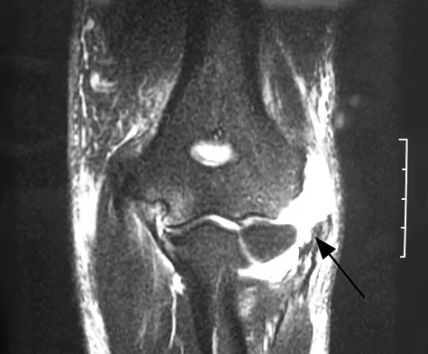
Figure 37 shows a coronal T2-weighted MRI scan. What is the name of the labeled torn structure?


Explanation

The labeled structure is the LCL, and it is avulsed from the lateral humeral epicondyle. This is the most common site of injury for the LCL. The biceps and brachialis tendon insertions are not well visualized in this section. The MCL and flexor/pronator origin are intact. Potter HG, Weiland AJ, Schatz JA, et al: Posterolateral rotatory instability of the elbow: Usefulness of MR imaging in diagnosis. Radiology 1997;204:185-189.


Question 4

The best candidate for a reverse total shoulder arthroplasty is a patient with rotator cuff tear arthropathy with


Explanation

Reverse total shoulder arthroplasty is relatively contraindicated in patients with acromial stress fractures and rheumatoid arthritis. A patient with active forward elevation to 130 degrees is better treated with a hemiarthroplasty because the motion already exceeds the average forward elevation attained in most studies using the reverse prosthesis. A centered case of rotator cuff tear arthropathy is also better treated with a hemiarthroplasty, especially in patients with a large external rotation lag sign because the reverse prosthesis has been shown to decrease active external rotation. However, hemiarthroplasties have not performed well in patients with anterior superior escape and in this group of patients, the reverse prosthesis is best. Rittmeister M, Kerschbaumer M: Grammont reverse total shoulder arthroplasty in patients with rheumatoid arthritis and nonreconstructible rotator cuff lesions. J Shoulder Elbow Surg 2001;10:17-22. Visotosky JL, Basamania C, Seebauer L, et al: Cuff tear arthropathy: Pathogenesis, classification, and algorithm for treatment. J Bone Joint Surg Am 2004;86:35-40.

Question 5

Which of the following findings is a contraindication to isolated percutaneous pinning of a distal radius fracture?


Explanation

Intrafocal pinning allows the Kirschner wires to be placed through a site of comminution and then drilled through intact cortex. Generally Kapandji intrafocal pinning is done for dorsal comminuted extra-articular dorsal bending fractures, but it also may be used to elevate and buttress radial comminution. Simple intra-articular fractures can also be treated with pinning alone. Intrafocal pinning works best as a dorsal or radial buttress to prevent shortening. When there is volar comminution, the fracture is prone to shortening and supplemental external fixation or plating is recommended. Trumble TE, Wagner W, Hanel DP, et al: Intrafocal (Kapandji) pinning of distal radius fractures with and without external fixation. J Hand Surg Am 1998;23:381-394. Choi KY, Chan WS, Lam TP, et al: Percutaneous Kirschner-wire pinning for severely displaced distal radial fractures in children: A report of 157 cases. J Bone Joint Surg Br 1995;77:797-801.

Question 6

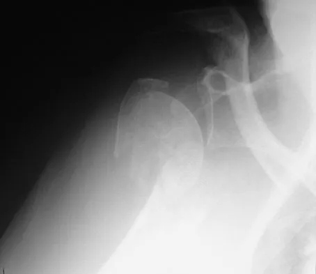
Figure 38 shows the radiograph of a 75-year-old woman who has had right shoulder pain, difficulty sleeping on the affected arm, and difficulties performing activities of daily living for the past 6 weeks. Initial nonsurgical management includes analgesics, a subacromial cortisone injection, and gentle range-of-motion exercises. However, these modalities have failed to provide relief, and the patient reports that she is unable to elevate her arm. Her pain is worse and she would like the most reliable treatment method for pain relief and functional improvement. What is the best surgical treatment?


Explanation

The authors of several studies conducted in Europe have reported promising results in the short- and medium-term with use of a reversed or inverted shoulder implant. The most recent investigation, a multicenter study in Europe in which 77 patients (80 shoulders) with glenohumeral osteoarthritis and a massive rupture of the rotator cuff were treated with the Delta III prosthesis, described an improvement in the mean constant score of 42 points, an increase of 65 degrees in forward elevation, and minimal or no pain in 96% of the patients. Hemiarthroplasty, the "nonconstrained" option, has long been the standard of care for rotator cuff tear arthropathy. However, careful examination of the literature reveals that the results have not been uniform. Favard L, Lautmann S, Sirveaux F, et al: Hemiarthroplasty versus reverse arthroplasty in the treatment of osteoarthritis with massive rotator cuff tear, in Walch G, Boileau P, Mole D (eds): 2000 Shoulder Prosthesis Two to Ten Year Follow-Up. Montpellier, France, Sauramps Medical, 2001, pp 261-268. Frankle M, Siegal S, Pupello D, et al: The reverse shoulder prosthesis for glenohumeral arthritis associated with severe rotator cuff deficiency: A minimum two-year follow-up study of sixty patients. J Bone Joint Surg Am 2005;87:1697-1705.


Question 7

An extended head hemiarthroplasty (rotator cuff tear arthropathy head) has what theoretic advantage when compared to a standard hemiarthroplasty?


Explanation

The theoretic advantage of a metal-to-bone articulation with the acromion is that there is a greater arc in which a smooth metal surface contacts the glenoid and acromion. This may improve pain and function, but no studies have evaluated this to date. One study showed results comparable to that of a standard hemiarthroplasty. There are no other biomechanic advantages. Visotsky JL, Basamania C, Seebauer L, et al: Cuff tear arthropathy: Pathogenesis, classification, and algorithm for treatment. J Bone Joint Surg Am 2004;86:35-40.

Question 8

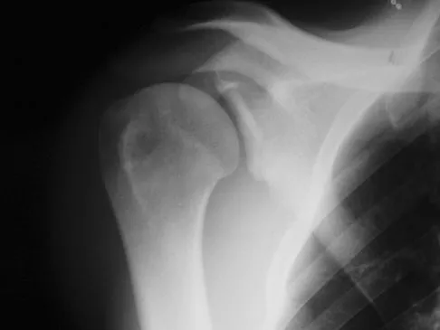
A 21-year-old right hand-dominant male collegiate swimmer reports painful clicking in the right shoulder. He states that he can occasionally feel his shoulder "slip out" when he is working out. AP, true AP, and axillary radiographs are shown in Figures 39a through 39c. What is the next most appropriate step in management?


Explanation

The radiographs show glenoid hypoplasia. The common radiographic findings of glenoid hypoplasia include an inferior and posterior glenoid deficiency, enlargement of the distal end of the clavicle, and sometimes an indentation in the glenoid. It is usually bilateral and rarely associated with other syndromes; therefore, an echocardiogram, abdominal ultrasound, or a skeletal survey is unnecessary unless the patient has stigmata of a syndrome such as Holt-Oram or Apert's. Although posterior instability has been reported, the results of glenoid osteotomy have been variable and should not be considered initially. Physical therapy is the mainstay of initial management, but the patient should be counseled that this may be a recurrent problem with early osteoarthritis developing in many patients. Radiographs of the contralateral side should be obtained because this is usually bilateral. Wirth MA, Lyons FR, Rockwood CA Jr: Hypoplasia of the glenoid: A review of sixteen patients. J Bone Joint Surg Am 1993;75:1175-1184.


Question 9

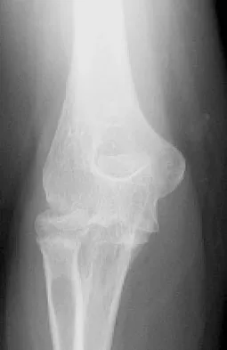
A 55-year-old man sustained an elbow dislocation in a fall. Postreduction radiographs are shown in Figures 40a and 40b. What is the best course of management?


Explanation

The radiographs show an elbow dislocation associated with a comminuted radial head fracture. In the setting of comminution and instability, factures of the radial head are best managed with an arthroplasty rather than open reduction and internal fixation. Resection of the radial head will worsen the instability and is not recommended. Silastic radial head replacements are contraindicated. Hildebrand KA, Patterson SD, King GJ: Acute elbow dislocations: Simple and complex. Orthop Clin North Am 1999;30:63-79.


Question 10

Osteochondritis dissecans of the capitellum is a source of elbow pain and most commonly occurs in what patient population?


Explanation

The etiology of osteochondritis dissecans of the capitellum is somewhat unclear. However, trauma has been implicated in this disease process. Gymnasts who load their upper extremities during tumbling and throwing athletes with repetitive trauma during the throwing motion are common patient subgroups in which osteochondritis dissecans of the elbow is seen. This often occurs in the adolescent age population. Baumgarten TE, Andrews JR, Satterwhite YE: The arthroscopic classification and treatment of osteochondritis dissecans of the capitellum. Am J Sports Med 1998;26:520-523.

Question 11

An 82-year-old woman fell on her right shoulder 2 days ago. She is alert, oriented, and in mild discomfort. Prior to falling, she lived alone and functioned independently. Examination reveals extensive ecchymosis extending to the midhumeral region. Her neurovascular examination is normal. Radiographs are shown in Figures 41a and 41b. What is the most appropriate management?


Explanation

The patient has a displaced four-part proximal humerus fracture. Given her age and the presence of osteopenia, a cemented hemiarthroplasty is the treatment of choice. The glenoid is uninjured so a total shoulder arthroplasty is not indicated. Percutaneous pinning in younger individuals with good bone quality may be indicated but not in an 82-year-old woman with osteopenia. Sling immobilization and immediate pendulum exercises will lead to a nonunion. Sling immobilization for 6 weeks followed by active range of motion will result in a nonunion or malunion with unacceptable functional results. Neer CS II: Displaced proximal humeral fractures: I. Classification and evaluation. J Bone Joint Surg Am 1970;52:1077-1089.


Question 12

Figures 42a and 42b show the radiographs of a 52-year-old man who sustained a fall from a motorcycle 6 months ago and now reports pain and stiffness in his left shoulder. What is the most reliable treatment to improve function and comfort of the shoulder?


Explanation

Appropriate treatment is based on multiple considerations, which include the chronicity of the dislocation, the amount of humeral head involvement, the medical condition, and functional limitations of the patient. It has been shown that shoulder arthroplasty for locked posterior dislocation provides pain relief and improved motion. Transfer of the lesser tuberosity with its attached subscapularis tendon into the defect is recommended for anteromedial humeral defects that are smaller than approximately 40% of the joint surface. Subscapularis transfer as described by McLaughlin and the modification thereof later described by Hawkins and associates in which the lesser tuberosity is transferred into the defect, have yielded good results if the defect is less than 40% of the humeral head. Prosthetic replacement is preferred for larger defects. If the dislocation is less than 3 weeks old and has less than 25% of humeral head involvement, closed reduction with the patient under general anesthesia should be attempted and the stability assessed by internally rotating the arm. If the arm can be safely internally rotated to the abdomen, then 6 weeks of immobilization in an orthosis that maintains the shoulder in slight extension and external rotation can yield a good result. If the dislocation has been present for more than 3 weeks, closed reduction becomes exceedingly difficult. Gerber C, Lambert SM: Allograft reconstruction of segmental defects of the humeral head for the treatment of chronic locked posterior dislocation of the shoulder. J Bone Joint Surg Am 1996;78:376-382. Spencer EE Jr, Brems JJ: A simple technique for management of locked posterior shoulder dislocations: Report of two cases. J Shoulder Elbow Surg 2005;14:650-652. Sperling JW, Pring M, Antuna SA, et al: Shoulder arthroplasty for locked posterior dislocation of the shoulder. J Shoulder Elbow Surg 2004;13:522-527. Hawkins RJ, Neer CS II, Pianta RM, et al: Locked posterior dislocation of the shoulder. J Bone Joint Surg Am 1987;69:9-18.


Question 13

In a patient with rheumatoid arthritis of the wrist, which of the following extensor tendons is most at risk of rupture?


Explanation

The tendon most prone to rupture in a patient with rheumatoid arthritis of the wrist is the extensor digiti quinti. It can be a silent injury since the extensor digitorum communis can provide extension to the fifth finger. The extensor digiti quinti is at high risk since it is overlying the ulnar head where it is prone to attritional rupture (Vaughan-Jackson syndrome). Vaughan-Jackson OJ: Rupture of extensor tendons by attrition at the inferior radioulnar joint: A report of two cases. J Bone Joint Surg Br 1948;30:528-530.

Question 14

A 40-year-old right hand-dominant construction worker has had a 6-month history of aching left shoulder pain that is worse after working a long day. Examination reveals limited range of motion and good strength when compared to his asymptomatic right arm. He has not had any orthopaedic intervention to date. Radiographs are shown in Figures 43a and 43b. What is the most appropriate treatment?


Explanation

The patient is a young laborer with osteoarthritis. Initial treatment should begin with nonsurgical management that may include anti-inflammatory drugs, cortisone injections, and physical therapy to diminish pain and improve motion. The other choices may eventually be necessary but should only follow a course of nonsurgical management. Norris TR (ed): Orthopaedic Knowledge Update: Shoulder and Elbow 2. Rosemont, IL, American Academy of Orthopaedic Surgeons, 2002, pp 257-266.


Question 15

What is the most appropriate surgical treatment for a stage III symptomatic scapholunate advanced collapsed (SLAC) wrist?


Explanation

SLAC is the end result of chronic scapholunate instability. The arthritis follows a predictable pattern. Stage I disease involves cartilage loss between the waist of the scaphoid and the radial styloid. In stage II, the arthritis progresses to include the proximal pole of the scaphoid and the scaphoid fossa of the radius. Finally, stage III goes on to include arthritis of the capitolunate joint. The only treatment option that addresses all of the sites of arthritis is the scaphoid excision and four corner fusion. Ashmead DT IV, Watson HK, Damon C, et al: Scapholunate advanced collapse wrist salvage. J Hand Surg Am 1994;19:741-750.

Question 16

A 25-year-old man shot himself at the base of the right index finger while cleaning his handgun. Examination reveals that the finger is cool and cyanotic. A clinical photograph and radiograph are shown in Figures 44a and 44b. What is the recommended treatment?


Explanation

The gunshot wound has caused injury to multiple systems: bone, vascular, skin, and tendon; therefore, the treatment of choice is amputation. An immediate ray amputation allows for a more rapid return to activities and less time off work. Peimer CA, Wheeler DR, Barrett A, et al: Hand function following single ray amputation. J Hand Surg Am 1999;24:1245-1248.


Question 17

What are the two terminal branches of the lateral cord of the brachial plexus?


Explanation

The lateral cord divides into the musculocutaneous and median nerves. The posterior cord terminates into the axillary and radial nerves. The medial cord divides into the ulnar and median nerves. Hollinshead WH: Anatomy for Surgeons, ed 3. Philadelphia, PA, Harper and Row, 1982, pp 228-236.

Question 18

A 32-year-old patient reports progressively increasing pain and stiffness after undergoing arthroscopic shoulder stabilization 1 year ago. The stabilization procedure was a Bankart repair with anchor fixation and supplemented with the heat probe. Radiographs are shown in Figures 45a and 45b. What is the most likely diagnosis?


Explanation

Postshoulder stabilization chondrolysis is a rare but devastating complication. It has been implicated with the use of the radiofrequency heat probe in some patients. Levine WN, Clark AM Jr, D'Alessandro DF, et al: Chondrolysis following arthroscopic thermal capsulorrhaphy to treat shoulder instability: A report of two cases. J Bone Joint Surg Am 2005;87:616-621.


Question 19

A 35-year-old man who is an avid weight lifter competing in local tournaments reports new onset pain and loss of motion in his dominant right shoulder. Examination reveals joint line tenderness, active elevation to 100 degrees, and external rotation to 10 degrees. His contralateral shoulder reveals 170 degrees forward elevation and 50 degrees external rotation. Radiographs are shown in Figures 46a and 46b. What is the next most appropriate step in management?


Explanation

New onset pain and stiffness in the young arthritic shoulder is a difficult problem to treat. Initial management should be aimed at reducing pain and improving motion in all planes. This patient's activities and age preclude a shoulder arthroplasty at this time. If nonsurgical management fails to provide relief, then arthroscopic debridement and capsular release may be beneficial. Norris TR (ed): Orthopaedic Knowledge Update: Shoulder and Elbow 2. Rosemont, IL, American Academy of Orthopaedic Surgeons, 2002, pp 257-266.


Question 20

A 23-year-old man who is a competitive overhead athlete has shoulder pain. Based on the pathology shown in Figure 47, what treatment option would yield the highest satisfaction and return to overhead sports?


Explanation

The patient has a classic type II SLAP tear that will respond best to arthroscopic repair. Labral debridement has been shown to lead to predictably poor results, and biceps tenodesis and tenotomy may be appropriate for an older patient who is not a competitive overhead athlete. Snyder SJ, Karzel RP, Del Pizzo W, et al: SLAP lesions of the shoulder. Arthroscopy 1990;6:274-279.


Question 21

Acute redislocation of the glenohumeral joint is a complication that occurs following a first-time dislocation. This is most often seen with


Explanation

Redislocation following acute dislocation occurs in approximately 3% of patients. This redislocation tends to occur in middle-aged and elderly patients. A higher incidence of redislocation occurs when there are accompanying fractures of the glenoid rim and the greater tuberosity. Robinson CM, Kelly M, Wakefield AE: Redislocation of the shoulder during the first six weeks after a primary anterior dislocation: Risk factors and results of treatment. J Bone Joint Surg Am 2002;84:1552-1559.

Question 22

A 20-year-old college pitcher reports medial elbow pain after 3 innings of hard throwing. He recalls no injury and reports no pain with light throwing. The examination shown in the clinical photograph in Figure 48 reproduces the elbow pain. What is the most likely diagnosis?


Explanation

The milking test, as seen in the photograph, elicits pain when a tear is present in the medial collateral ligament. Complete rupture is possible but unlikely when there is no history of trauma and the patient is able to throw pain-free for several innings. Subluxation of the ulnar nerve and triceps tendon subluxation present as a painful snapping over the medial aspect of the elbow. Williams RJ III, Urquhart ER, Altchek DW: Medial collateral ligament tears in the throwing athlete. Instr Course Lect 2004;53:579-586.


Question 23

A 51-year-old woman is seen for evaluation of chronic supraspinatus and infraspinatus tendon tears. Three years ago, in an attempted repair the surgeon was unable to repair the supraspinatus and infraspinatus tendon tears. Currently she has a marked amount of pain, reduced range of motion, and weakness. Examination reveals anterosuperior escape. Radiographs show no signs of arthritic changes. You are considering a latissimus dorsi tendon transfer. During the discussion, you mention that


Explanation

Latissimus dorsi tendon transfer is considered a surgical option for treatment in patients with chronic supraspinatus and infraspinatus tendon tears. Preoperative subscapularis function is necessary for good clinical results. Additionally, men with active elevation to shoulder level and active external rotation to 20 degrees have predictably good results. Women with active shoulder elevation limited to below chest level have poor results from this procedure and should not be considered candidates. Postoperatively they lack pain control, active elevation, and active external rotation. Muscular atrophy in the latissimus dorsi does not occur, and glenohumeral arthritic changes frequently develop postoperatively. Gerber C, Maquieira G, Espinosa N: Latissimus dorsi transfer for the treatment of irreparable rotator cuff tears: Factors affecting outcome. J Bone Joint Surg Am 2006;88:113-120.

Question 24

A patient undergoes an arthroscopic debridement for lateral epicondylitis. Postoperatively she reports pain and a sense of clicking of the elbow. Examination reveals apprehension to supination, load, and extension. What structure has been injured resulting in the clinical presentation?


Explanation

The patient has an iatrogenic injury to the lateral ulnar collateral ligament following the arthroscopic procedure. Failure to adhere to known anatomic landmarks can lead to this devastating complication. The examination findings are classic for posterolateral elbow instability. Koval KJ (ed): Orthopaedic Knowledge Update 7. Rosemont, IL, American Academy of Orthopaedic Surgeons, 2002, p 318.

Question 25

A patient with refractory long head biceps pain in the shoulder undergoes biceps tenotomy. The patient is concerned about possible postoperative deformity and loss of supination strength. Which of the following techniques provides the strongest initial fixation to prevent distal migration?


Explanation

Recent articles have looked at the cyclic load failure and ultimate load failure of biceps tenodesis techniques. The interference screw has proved superior to bone tunnel, suture anchor, and soft-tissue tenodesis techniques in laboratory cadaveric testing. Whether this is clinically relevant or not is still unknown. Ozalay M, Akpinar S, Karaeminogullari O, et al: Mechanical strength of four different biceps tenodesis techniques. Arthroscopy 2005;21:992-998.

Question 26

A 45-year-old male falls on an outstretched hand and sustains a 'terrible triad' injury of the elbow. During surgical intervention, what is the most appropriate standard sequence of repair?


Explanation

The standard surgical algorithm for a terrible triad injury works from deep to superficial and medial to lateral: fixation of the coronoid first, followed by radial head repair or replacement, and finally LCL repair. The MCL is typically only addressed if the elbow remains unstable after the lateral-sided repair.

Question 27

A 22-year-old male sustains a minimally displaced fracture of the scaphoid waist. Which of the following best describes the primary blood supply to the proximal pole of the scaphoid?


Explanation

The primary blood supply to the scaphoid is from the dorsal carpal branch of the radial artery, which enters the distal 80% of the bone and provides retrograde blood flow to the proximal pole. This tenuous retrograde supply increases the risk of avascular necrosis in proximal pole fractures.

Question 28

Recent quantitative anatomic studies have redefined the primary arterial supply to the articular segment of the proximal humerus. Which vessel provides the majority of the blood supply to the humeral head?


Explanation

While older literature emphasized the anterior humeral circumflex artery (arcuate branch), recent studies (e.g., Hettrich et al.) demonstrate that the posterior humeral circumflex artery provides approximately 64% of the blood supply to the humeral head.

Question 29

In a Galeazzi fracture-dislocation, which of the following radiographic or anatomic findings is the strongest predictor of distal radioulnar joint (DRUJ) instability following anatomic fixation of the radius?


Explanation

Radius fractures located within 7.5 cm of the articular surface are associated with a high rate of DRUJ instability. Fractures proximal to this 7.5 cm threshold uncommonly result in DRUJ disruption.

Question 30

A 25-year-old athlete sustains a direct blow to the shoulder and subsequently presents with medial winging of the scapula. Injury to which of the following nerves is the most likely cause?


Explanation

Medial scapular winging is characteristic of serratus anterior palsy, which is innervated by the long thoracic nerve. Lateral winging is typically associated with trapezius dysfunction due to spinal accessory nerve injury.

Question 31

A 6-year-old child presents with a Bado Type I Monteggia fracture-dislocation. What is the direction of the radial head dislocation in this injury pattern?


Explanation

A Bado Type I Monteggia lesion involves a fracture of the proximal or middle third of the ulna with an anterior dislocation of the radial head. It is the most common type of Monteggia fracture in children.

Question 32

Which of the following represents an acceptable radiographic parameter for the nonoperative management of a distal radius fracture in an active adult?


Explanation

Acceptable radiographic parameters for nonoperative management of a distal radius fracture typically include radial shortening < 5 mm, dorsal tilt < 10 degrees past neutral (or < 15 degrees), and articular step-off < 2 mm.

Question 33

Which of the following is the most significant clinical or radiographic risk factor for nonunion of a midshaft clavicle fracture treated nonoperatively?


Explanation

Fracture shortening greater than 2 cm and 100% displacement are the most significant risk factors for nonunion and symptomatic malunion in completely displaced midshaft clavicle fractures.

Question 34

In a purely ligamentous lesser arc perilunate dislocation of the wrist, which carpal bone classically remains anatomically aligned with the distal radius on a true lateral radiograph?


Explanation

In a perilunate dislocation, the lunate remains contained within the lunate fossa of the distal radius, while the capitate and the rest of the carpus dislocate dorsally. In a lunate dislocation, the lunate is displaced anteriorly ('spilled teacup').

Question 35

A 35-year-old female presents with an elbow injury. Radiographs reveal a coronal shear fracture of the capitellum that includes the lateral trochlear ridge (McKee modification of Bryan and Morrey Type IV). What is the recommended treatment?


Explanation

Type IV capitellum fractures involve the lateral trochlear ridge, making the elbow highly unstable if the fragment is removed. Open reduction and internal fixation (typically with headless compression screws) is required to restore radiocapitellar and ulnohumeral stability.

Question 36

During a single-incision anterior approach for a distal biceps tendon repair, injury to which nerve is the most frequently reported complication?


Explanation

The lateral antebrachial cutaneous nerve is the most commonly injured nerve during the single-incision anterior approach for distal biceps repair. The posterior interosseous nerve (PIN) is more at risk during a two-incision approach.

Question 37

A 22-year-old athlete sustains a traumatic anterior shoulder dislocation. During preoperative planning, an 'engaging' Hill-Sachs lesion is identified. Which of the following defines an engaging Hill-Sachs lesion?


Explanation

An 'engaging' Hill-Sachs lesion occurs when the orientation of the humeral head defect becomes parallel to the anterior glenoid rim when the shoulder is positioned in abduction and external rotation, allowing the defect to lever out and cause dislocation.

Question 38

A 40-year-old male sustains an Essex-Lopresti injury characterized by a comminuted radial head fracture, interosseous membrane tear, and DRUJ disruption. If the radial head is unreconstructible, what is the most appropriate management?


Explanation

In an Essex-Lopresti injury, the longitudinal stabilizers of the forearm are compromised. Excision of the radial head is strictly contraindicated as it will lead to proximal radius migration. The appropriate treatment is radial head arthroplasty combined with DRUJ stabilization.

Question 39

In the natural history of a scaphoid nonunion advanced collapse (SNAC) wrist, where are the degenerative changes primarily located during Stage II?


Explanation

In a SNAC wrist, Stage I involves arthritis between the radial styloid and the distal scaphoid fragment. Stage II progresses to involve the scaphocapitate joint. Stage III involves the capitolunate joint.

Question 40

An 80-year-old female sustains a highly comminuted, osteoporotic intercondylar distal humerus fracture (AO/OTA 13-C3). What is the primary advantage of total elbow arthroplasty (TEA) over ORIF in this demographic?


Explanation

TEA provides a more predictable functional outcome and permits immediate post-operative mobilization in elderly patients with poor bone stock, bypassing the high failure rates of ORIF in comminuted osteoporotic fractures. However, TEA requires lifelong lifting restrictions.

Question 41

A spiral fracture of the distal third of the humeral shaft (Holstein-Lewis fracture) is most commonly associated with a primary injury to which of the following nerves?


Explanation

A Holstein-Lewis fracture is a spiral fracture of the distal third of the humeral shaft. It is associated with a high incidence of radial nerve palsy as the nerve becomes tethered as it passes through the lateral intermuscular septum.

Question 42

A 35-year-old male presents with a 'terrible triad' injury of the elbow after a fall from a ladder. When performing surgical stabilization, what is the generally accepted optimal sequence of repair for the injured structures?


Explanation

Standard surgical management of terrible triad injuries proceeds from deep to superficial (or anterior to posterior). The accepted sequence is fixation of the coronoid first, followed by the radial head (repair or replace), and finally the LCL complex.

Question 43

A 22-year-old athlete sustains a proximal pole scaphoid fracture. Which of the following vascular structures provides the primary retrograde blood supply to the proximal pole, explaining the high risk of avascular necrosis (AVN) in this fracture pattern?


Explanation

The dorsal carpal branch of the radial artery enters the scaphoid at the distal pole and provides retrograde blood supply to the proximal pole. Fractures at the proximal pole disrupt this supply, resulting in a high rate of AVN.

Question 44

Review the clinical scenario provided. A 68-year-old female sustains a 4-part proximal humerus fracture.

Which of the following is the most reliable radiographic predictor of humeral head ischemia in this setting?


Explanation

Hertel et al. identified specific predictors of humeral head ischemia. A posteromedial metaphyseal head extension (calcar length) of less than 8 mm and disruption of the medial hinge are the most reliable predictors of AVN.

Question 45

When utilizing the volar approach to the distal radius (Modified Henry approach) for internal fixation of a volar Barton's fracture, the surgical interval is developed between which two anatomical structures?


Explanation

The modified Henry approach utilizes the internervous plane between the median nerve (FCR) and radial nerve (brachioradialis). Specifically, the interval is developed between the FCR tendon (retracted ulnarly) and the radial artery (retracted radially).

Question 46

A 28-year-old male sustains a Galeazzi fracture-dislocation. After plate fixation of the radial shaft, the distal radioulnar joint (DRUJ) remains unstable. Which muscle provides the primary deforming force causing volar and ulnar translation of the distal radius fragment?


Explanation

In a Galeazzi fracture, the brachioradialis pulls the proximal fragment proximally, while the pronator quadratus pulls the distal fragment in a volar and ulnar direction, leading to the characteristic deformity.

Question 47

A 19-year-old male falls on his shoulder and sustains a midshaft clavicle fracture. Which of the following findings is an absolute indication for operative management?


Explanation

Absolute indications for clavicle fracture fixation include open fractures, neurovascular compromise, and severe skin tenting that implies impending skin breakdown. Shortening and displacement are considered relative indications.

Question 48

A 25-year-old male sustains an acute anterior shoulder dislocation. Post-reduction, you suspect an axillary nerve injury. What area should be evaluated to assess the sensory distribution of the axillary nerve?


Explanation

The axillary nerve provides sensation to the lateral aspect of the shoulder via the superior lateral brachial cutaneous nerve. It is the most commonly injured nerve in anterior shoulder dislocations.

Question 49

A 45-year-old female presents with an Essex-Lopresti injury characterized by a comminuted Mason Type III radial head fracture and distal radioulnar joint (DRUJ) dislocation. Regarding the management of the radial head, which of the following is most appropriate?


Explanation

In an Essex-Lopresti injury, the interosseous membrane is disrupted. Excision of the radial head is strictly contraindicated as it will lead to proximal migration of the radius. If the radial head is unreconstructable, a radial head arthroplasty is required.

Question 50

A patient falls from a height and sustains a Mayfield Stage IV perilunate dislocation. Review the provided reference image.

What is the characteristic position of the lunate in a Stage IV injury?


Explanation

In Mayfield Stage IV (lunate dislocation), the dorsal radiocarpal ligament ruptures, and the lunate is extruded volarly into the carpal tunnel. This commonly causes acute median nerve compression.

Question 51

A 60-year-old male undergoes tension band wiring for a transverse olecranon fracture. Which of the following is the most frequent complication associated with this specific surgical technique?


Explanation

The most common complication of tension band wiring for olecranon fractures is prominent and symptomatic hardware, which necessitates hardware removal in up to 40-80% of patients once the fracture has healed.

Question 52

A 35-year-old female sustains a complex elbow fracture. Radiographs reveal a coronal shear fracture of the capitellum that extends medially to involve the majority of the trochlea.

According to the Bryan and Morrey classification, what type of fracture is this?


Explanation

The McKee modification (Type 4) of the Bryan and Morrey classification describes a coronal shear fracture of the capitellum that extends to include a significant portion of the trochlea.

Question 53

A trauma surgeon is performing a transolecranon approach for the open reduction and internal fixation of an intercondylar distal humerus fracture (AO type 13-C3). To optimize healing and joint stability, what is the preferred osteotomy shape and orientation?


Explanation

A chevron-shaped osteotomy with the apex directed distally is preferred. This shape maximizes the surface area for healing and provides intrinsic rotational stability to the osteotomy site upon repair.

Question 54

A 7-year-old child presents with a Bado Type I Monteggia fracture-dislocation. Based on this specific fracture pattern, what associated neurological deficit is most likely to be observed on physical examination?


Explanation

Bado Type I Monteggia fractures (anterior dislocation of the radial head with anterior angulation of the ulna) are most commonly associated with posterior interosseous nerve (PIN) neuropraxia due to the anterior displacement of the radial head.

Question 55

In an elderly patient undergoing reverse total shoulder arthroplasty for a 4-part proximal humerus fracture, which of the following factors correlates most strongly with improved postoperative external rotation and overall patient satisfaction?


Explanation

Greater tuberosity healing to the humeral shaft or implant in reverse total shoulder arthroplasty for fractures is associated with significantly improved external rotation and better overall functional outcomes.

Question 56

Which of the following describes the typical mechanism of injury for a 'terrible triad' of the elbow?


Explanation

A terrible triad injury of the elbow (elbow dislocation, radial head fracture, coronoid fracture) typically occurs from a fall on an outstretched hand resulting in an axial load, valgus stress, and forearm supination. This causes sequential failure of structures from lateral to medial.

Question 57

A 65-year-old woman undergoes volar plate fixation for a distal radius fracture. Six months postoperatively, she presents with an inability to actively flex the interphalangeal joint of her thumb. Placement of the plate distal to which of the following anatomical landmarks is the primary risk factor for this complication?


Explanation

The watershed line is the bony ridge on the volar distal radius. Plate placement distal to this line significantly increases the risk of flexor pollicis longus (FPL) tendon attrition and subsequent rupture.

Question 58

Which of the following is considered an absolute indication for open reduction and internal fixation of an acute midshaft clavicle fracture?


Explanation

Skin tenting with impending necrosis is an absolute indication for operative fixation of a clavicle fracture to prevent progression to an open fracture. Shortening and severe displacement are relative indications.

Question 59

A 40-year-old male falls from a height and sustains a highly comminuted, unsalvageable radial head fracture, accompanied by distal radioulnar joint (DRUJ) instability. What is the most appropriate management of the radial head?


Explanation

This patient has an Essex-Lopresti injury (radial head fracture, interosseous membrane disruption, DRUJ instability). Radial head arthroplasty is required to restore longitudinal stability; excision alone leads to proximal radial migration and chronic wrist pain.

Question 60

A 22-year-old male sustains a proximal pole scaphoid fracture. Which of the following arterial branches is responsible for the retrograde blood supply to the proximal pole, making it highly susceptible to avascular necrosis?


Explanation

The scaphoid receives its primary blood supply from the dorsal carpal branch of the radial artery, which enters at the distal pole and waist, providing retrograde flow to the proximal pole.

Question 61

During evaluation of a severe shoulder injury, a radiograph

shows superior displacement of the clavicle by 150% relative to the acromion. Which of the following best describes the pathoanatomy of this Rockwood Type V acromioclavicular injury?


Explanation

A Rockwood Type V injury involves tearing of the acromioclavicular (AC) and coracoclavicular (CC) ligaments, along with extensive stripping of the deltotrapezius fascia, leading to severe superior displacement of the distal clavicle.

Question 62

A 35-year-old female presents with an isolated coronal shear fracture of the capitellum with no posterior comminution. If open reduction and internal fixation with headless compression screws is planned, what is the biomechanically optimal screw trajectory?


Explanation

For isolated capitellar coronal shear fractures, screws placed from posterior to anterior provide superior biomechanical stability compared to anterior-to-posterior screws and avoid articular cartilage penetration.

Question 63

Which of the following factors is most predictive of avascular necrosis following a proximal humerus fracture?


Explanation

According to the Hertel criteria, a calcar length of less than 8 mm attached to the articular segment is a strong predictor of ischemia and subsequent avascular necrosis. Other predictors include disruption of the medial hinge and anatomic neck fractures.

Question 64

A 45-year-old falls on an outstretched hand and sustains a terrible triad injury of the elbow. Which of the following is the recommended surgical sequence of fixation?


Explanation

The standard protocol for treating a terrible triad injury of the elbow progresses from deep to superficial structures. This involves coronoid fixation or repair, followed by radial head replacement or fixation, and finally lateral collateral ligament (LCL) repair.

Question 65

A 60-year-old woman undergoes volar locked plating of a distal radius fracture. Postoperatively, she is unable to actively flex the interphalangeal joint of her thumb. This complication is most commonly associated with which of the following?


Explanation

Flexor pollicis longus (FPL) tendon rupture is a well-known complication of volar locked plating. It most frequently occurs when the plate is placed distal to the watershed line, causing attritional wear of the tendon.

Question 66

A 28-year-old manual laborer presents to the emergency department following a high-energy fall. Radiographs show a perilunate dislocation. Which of the following nerve palsies is most commonly associated with this injury?


Explanation

Acute median nerve compression in the carpal tunnel is the most common neurologic complication associated with lunate and perilunate dislocations. If symptoms of acute carpal tunnel syndrome are present, emergent reduction and possible carpal tunnel release are indicated.

Question 67

A 35-year-old man presents with chronic radial-sided wrist pain 6 months after a fall. Radiographs reveal a scapholunate gap of 4 mm and a widened scapholunate angle. What is the classic radiographic sign seen on the AP view of a complete scapholunate ligament tear?


Explanation

The "Terry Thomas" sign refers to the abnormal widening of the scapholunate interval (greater than 3 mm) seen on the AP or PA view of the wrist. This indicates a disruption of the scapholunate interosseous ligament.

Question 68

A 25-year-old male presents with a painful wrist. He sustained a scaphoid fracture 8 months ago treated nonoperatively. Current imaging demonstrates a scaphoid waist nonunion with a "humpback" deformity. Which of the following is the most appropriate surgical treatment?


Explanation

A humpback deformity involves volar flexion of the distal scaphoid fragment. It is best corrected using a volar approach with structural wedge bone grafting and internal fixation to restore scaphoid length and alignment.

Question 69

Which of the following is considered an absolute indication for operative treatment of an acute midshaft clavicle fracture?


Explanation

Absolute indications for open reduction and internal fixation of a clavicle fracture include open fractures, skin tenting threatening to progress to an open fracture, and associated subclavian vessel or brachial plexus injuries.

Question 70

A 40-year-old woman sustains a coronal shear fracture of the capitellum extending medially to involve the majority of the trochlea. According to the Dubberley classification, what type of fracture is this, and what is the preferred surgical approach?


Explanation

A coronal shear fracture involving the capitellum and the trochlea is a Dubberley Type 3. Due to the extensive medial involvement, a universal posterior approach, often combined with an olecranon osteotomy, is required for adequate visualization and fixation.

Question 71

A 32-year-old male sustains a closed, distal-third spiral humeral shaft fracture (Holstein-Lewis) and presents with an inability to extend his wrist and fingers. What is the most appropriate initial management?


Explanation

Primary radial nerve palsies associated with closed humeral shaft fractures are typically managed expectantly with splinting or functional bracing and observation. Most cases resolve spontaneously within 3 to 4 months.

Question 72

A 25-year-old rugby player sustains an acromioclavicular (AC) joint injury. Radiographs reveal 150% superior displacement of the clavicle relative to the acromion. Which of the following ligaments are disrupted in this Rockwood Type V injury?


Explanation

Rockwood Type V injuries involve severe superior displacement of the clavicle (100-300%). This requires disruption of the AC ligaments, CC ligaments, and significant stripping or detachment of the deltotrapezial fascia.

Question 73

A 6-year-old child presents after a fall with a fracture of the proximal third of the ulna and an anterior dislocation of the radial head. What is the Bado classification of this injury?


Explanation

Bado Type I Monteggia fracture-dislocation is characterized by a fracture of the proximal or middle third of the ulna with an anterior dislocation of the radial head. It is the most common Bado type in pediatric patients.

Question 74

During the surgical treatment of a Galeazzi fracture, after plate fixation of the radius, the distal radioulnar joint (DRUJ) is found to be grossly unstable in supination. What is the most appropriate next step?


Explanation

If the DRUJ remains unstable after anatomic radius fixation in a Galeazzi fracture, it should be stabilized. This is commonly achieved by percutaneous K-wire pinning of the ulna to the radius with the forearm in full supination.

Question 75

A 35-year-old woman sustains a comminuted radial head fracture with more than 3 articular fragments that cannot be anatomically reconstructed. There is an associated disruption of the medial collateral ligament. What is the most appropriate surgical treatment?


Explanation

In the setting of an irreparable radial head fracture with associated elbow instability (such as an MCL tear or interosseous membrane injury), radial head arthroplasty is indicated to restore radiocapitellar stability. Simple excision is contraindicated due to the risk of valgus instability or proximal radial migration.

Question 76

A 20-year-old male presents after a tackle in American football with acute shortness of breath and a palpable void at the medial end of his right clavicle. A CT scan confirms a posterior sternoclavicular dislocation. What is the most appropriate initial management step?


Explanation

Posterior sternoclavicular dislocations are orthopedic emergencies due to potential mediastinal compression. The initial treatment is an attempted closed reduction in the operating room under general anesthesia, with a cardiothoracic surgeon available in case of vascular injury.

Question 77

When utilizing tension band wiring for a transverse olecranon fracture, what is the primary biomechanical principle by which this fixation method promotes bone healing?


Explanation

The tension band principle converts the tensile forces generated on the dorsal cortex by the triceps muscle into compressive forces at the articular surface during elbow flexion. This dynamic compression promotes primary bone healing.

Question 78

A 45-year-old male falls from a ladder and sustains an elbow injury. Radiographs reveal a posterior elbow dislocation, a radial head fracture, and a coronoid fracture. During surgical management of this 'terrible triad' injury, what is the generally accepted sequence of repair?


Explanation

The standard surgical sequence for a terrible triad injury is to repair deep to superficial and inside-out: first the coronoid, then the radial head, followed by the LCL complex. The MCL is only addressed if the elbow remains unstable after the lateral side is secured.

Question 79

A 22-year-old male falls on an outstretched hand and sustains a displaced fracture of the proximal pole of the scaphoid. He is at high risk for avascular necrosis due to the retrograde blood supply. The predominant blood supply to the proximal pole is derived from which of the following?


Explanation

The dorsal carpal branch of the radial artery supplies 80% of the scaphoid via vessels entering the dorsal ridge distally. This creates a retrograde blood flow to the proximal pole, making proximal fractures highly susceptible to avascular necrosis.

Question 80

A 65-year-old female undergoes volar locking plate fixation for a displaced distal radius fracture. Four months postoperatively, she suddenly loses the ability to flex the interphalangeal joint of her thumb. Which of the following technical errors most likely contributed to this complication?


Explanation

Placement of a volar plate distal to the watershed line of the distal radius causes prominence of the hardware against the flexor tendons. This places the flexor pollicis longus (FPL) tendon at high risk for attritional rupture.

Question 81

A 75-year-old woman sustains a severely displaced 4-part proximal humerus fracture. You are considering whether to perform open reduction internal fixation (ORIF) or arthroplasty. Which of the following radiographic factors is the most reliable predictor of humeral head ischemia?


Explanation

Hertel's criteria identify predictors of humeral head ischemia in proximal humerus fractures. The most significant predictors include a posteromedial hinge disruption > 2 mm, a metaphyseal head extension (calcar length) < 8 mm, and an anatomic neck fracture.

Question 82

A 30-year-old mechanic sustains a severe hyperextension injury to his wrist. Radiographs reveal a 'spilled teacup' sign on the lateral view, confirming a lunate dislocation. According to Mayfield's stages of perilunate instability, a complete lunate dislocation represents which stage?


Explanation

Mayfield described four stages of progressive perilunate instability. Stage I is scapholunate dissociation, Stage II adds capitate dislocation, Stage III adds lunotriquetral disruption, and Stage IV is a complete volar dislocation of the lunate.

Question 83

A 24-year-old cyclist falls and sustains a midshaft clavicle fracture. Which of the following is considered an ABSOLUTE indication for operative fixation?


Explanation

Absolute indications for operative fixation of a clavicle fracture include open fractures, fractures with associated neurovascular injury, and severe skin tenting threatening to progress to an open injury. Shortening and displacement are relative indications.

Question 84

In the setting of an acute, simple posterior elbow dislocation, soft tissue disruption classically occurs in a circular progression from lateral to medial (Horii circle). Which structure is typically the FIRST to be injured?


Explanation

According to the Horii circle of instability, elbow ligamentous disruption progresses from lateral to medial. The lateral collateral ligament complex, specifically the LUCL, is the first structure to fail in a typical posterolateral rotatory mechanism.

Question 85

A 35-year-old male undergoes ORIF for a distal-third radial shaft fracture with an associated distal radioulnar joint (DRUJ) dislocation (Galeazzi fracture). Intraoperatively, after rigid fixation of the radius, the DRUJ is found to be reducible but stable ONLY in full supination. What is the most appropriate next step in management?


Explanation

In a Galeazzi fracture, if the DRUJ is reducible and stable in supination after anatomic fixation of the radius, it is appropriate to immobilize the arm in supination for 4 to 6 weeks. K-wire transfixion or open repair is indicated if the joint remains unstable in all positions.

Question 86

A 40-year-old male sustains a high-energy trauma resulting in a scapula fracture. Radiographs demonstrate a transverse fracture line originating at the glenoid fossa and exiting the lateral border of the scapula. According to the Ideberg classification of intra-articular glenoid fractures, which type is this?


Explanation

In the Ideberg classification, Type II is a transverse fracture exiting the lateral scapular border. Type I involves the anterior rim, Type III exits superiorly involving the coracoid, and Type IV exits the medial border.

Question 87

A 55-year-old female presents with an isolated ulnar shaft fracture combined with a dislocation of the radial head. Radiographs show the radial head is dislocated posteriorly. According to the Bado classification of Monteggia fractures, which type does this represent?


Explanation

Bado Type II Monteggia fractures involve posterior dislocation of the radial head with posterior angulation of the ulnar fracture. This pattern is the most common Monteggia lesion seen in adults.

Question 88

During open reduction and internal fixation of a complex intra-articular distal humerus fracture, a transolecranon approach is selected. To minimize damage to the articular cartilage of the proximal ulna, the chevron osteotomy should be directed towards which anatomic landmark?


Explanation

An olecranon osteotomy for distal humerus exposure should be directed to exit through the 'bare area' (non-articular portion) of the greater sigmoid notch. This minimizes damage to the articular cartilage of the ulna.

Question 89

A 72-year-old male presents with profound weakness in external rotation and abduction three weeks after successful closed reduction of an anterior shoulder dislocation. Electromyography reveals normal axillary nerve function. What is the most likely diagnosis?


Explanation

Elderly patients with anterior shoulder dislocations have a very high incidence of concomitant rotator cuff tears (up to 80% in patients > 60 years old). Persistent weakness despite a normal axillary nerve strongly suggests a massive cuff tear.

Question 90

A 48-year-old female was treated non-operatively in a cast for a nondisplaced distal radius fracture. Six weeks later, she reports a sudden inability to actively extend her thumb interphalangeal joint. What is the most appropriate and reliable surgical treatment?


Explanation

Attritional rupture of the EPL tendon is a known complication of nondisplaced distal radius fractures due to ischemia or mechanical wear in Lister's tubercle. The standard and most reliable treatment is an EIP to EPL tendon transfer, as the native tendon ends are typically retracted and degenerated.

Question 91

A 30-year-old male sustains a severely comminuted, unfixable Mason Type III radial head fracture. You plan to perform a radial head excision. This procedure is strictly CONTRAINDICATED without a radial head replacement in the presence of which concurrent injury?


Explanation

Radial head excision without arthroplasty in the setting of a torn interosseous membrane (Essex-Lopresti injury) will lead to proximal migration of the radius, DRUJ dissociation, and severe wrist pain. The radial head must be replaced to maintain longitudinal stability of the forearm.

Question 92

A 28-year-old rugby player sustains a Type III acromioclavicular (AC) joint separation. Which ligaments are structurally disrupted in this specific injury pattern?


Explanation

A Rockwood Type III AC joint separation involves complete tearing of both the acromioclavicular ligaments and the coracoclavicular (conoid and trapezoid) ligaments. The clavicle is typically elevated 25-100% relative to the acromion.

Question 93

In the O'Driscoll classification of coronoid fractures, an anteromedial facet fracture is most commonly associated with which specific mechanism and injury pattern?


Explanation

Anteromedial facet fractures of the coronoid result from a varus force combined with posteromedial rotation. This causes avulsion of the LCL and compression of the anteromedial coronoid facet against the medial trochlea, leading to VPMRI.

Question 94

Following a severe crush injury to the wrist, a patient develops acute carpal tunnel syndrome requiring emergent release. During the procedure, all flexor tendons are inspected. Which of the following tendons is NOT contained within the carpal tunnel?


Explanation

The carpal tunnel contains the median nerve and nine flexor tendons (four FDS, four FDP, and the FPL). The flexor carpi radialis (FCR) runs in its own separate fibro-osseous sheath within the split of the transverse carpal ligament.

Question 95

A 25-year-old male sustains a distal-third spiral fracture of the humeral shaft (Holstein-Lewis fracture). Which nerve is most at risk of entrapment, and what is its anatomic relationship to the intermuscular septum at this level?


Explanation

In a Holstein-Lewis fracture, the radial nerve is tethered as it pierces the lateral intermuscular septum. At this level (distal third of the humerus), it passes from the posterior compartment to the anterior compartment.

Question 96

A 45-year-old female presents with a coronal shear fracture of the capitellum extending into the trochlea (Hahn-Steinthal or Bryan and Morrey Type I).

What is the optimal surgical approach and internal fixation strategy?


Explanation

Capitellum fractures are best managed via an extensile lateral approach. Fixation using headless compression screws placed anterior-to-posterior, buried beneath the articular cartilage, provides optimal biomechanical stability.

Question 97

During an anatomic coracoclavicular (CC) ligament reconstruction for a chronic AC joint separation, the surgeon must replicate the native anatomy. Which statement correctly describes the anatomic insertion of the CC ligaments on the clavicle?


Explanation

The conoid ligament inserts posteromedially on the conoid tubercle of the clavicle, whereas the trapezoid inserts anterolaterally. The conoid is the primary restraint to superior translation, while the trapezoid primarily resists axial compression.

Question 98

A 68-year-old female sustains a displaced proximal humerus fracture. According to the Hertel criteria, which of the following is the most significant radiographic predictor for the development of humeral head avascular necrosis (AVN)?


Explanation

Hertel et al. identified key predictors for humeral head ischemia following proximal humerus fractures. The most reliable predictors include a posteromedial metaphyseal head extension (calcar length) of less than 8 mm, disruption of the medial hinge greater than 2 mm, and an anatomic neck fracture pattern.

Question 99

A 45-year-old male sustains a "terrible triad" injury to his right elbow following a fall. During surgical reconstruction, the coronoid is fixed, the radial head is replaced, and the lateral ulnar collateral ligament (LUCL) is securely repaired to the lateral epicondyle. Intraoperatively, the elbow remains unstable and subluxates in extension. What is the most appropriate next step in management?


Explanation

The standard surgical algorithm for terrible triad injuries involves fixing the coronoid, restoring the radial head, and repairing the lateral collateral ligament complex. If the elbow remains unstable in extension after these structures are addressed, the next step is to repair the medial collateral ligament (MCL) prior to considering an external fixator.

Question 100

A 28-year-old male presents with acute severe wrist pain and median nerve paresthesias following a high-energy motorcycle collision. Radiographs demonstrate a volar dislocation of the lunate into the carpal tunnel. According to Mayfield's progressive stages of perilunate instability, this specific finding represents which stage of the injury pattern?


Explanation

Mayfield's stages of perilunate instability progress sequentially around the lunate: Stage I (scapholunate dissociation), Stage II (capitolunate dislocation), Stage III (lunotriquetral dissociation), and Stage IV (volar lunate dislocation). In Stage IV, the lunate is fully separated from its dorsal attachments and translates volarly into the carpal tunnel, often compressing the median nerve.

None

Clinic OS
Medically Verified Content by
Prof. Clinic OS
Consultant Orthopedic & Spine Surgeon
Chapter Index