العربية
Part of the Master Guide

100 High-Yield Orthopedic MCQs: Shoulder & Elbow | Mock Exam 251

Upper Extremity Orthopedic MCQs (Set 1): Shoulder, Elbow, Hand & Wrist | AAOS/ABOS Exam Prep

27 Apr 2026 52 min read 112 Views
Upper Extremity 2008 MCQs - Part 1

Key Takeaway

This high-yield question set for the AAOS/ABOS exams covers critical upper extremity orthopedic topics. It includes multiple-choice questions on shoulder pathologies, elbow injuries, and various hand & wrist conditions, preparing candidates for board certification and OITE success.

Upper Extremity Orthopedic MCQs (Set 1): Shoulder, Elbow, Hand & Wrist | AAOS/ABOS Exam Prep

Comprehensive 100-Question Exam


00:00

Start Quiz

Question 1

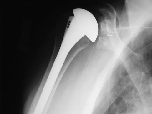
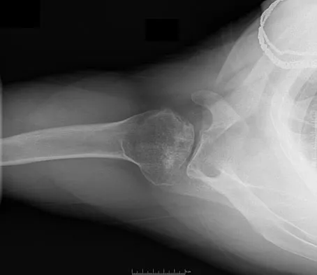
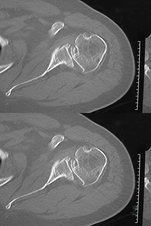
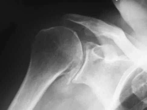
A 68-year-old man had a 3-year history of shoulder pain that failed to respond to nonsurgical management. Examination reveals forward elevation to 120 degrees and external rotation to 30 degrees. True AP and axillary radiographs and an axial CT scan are shown in Figures 1a through 1c. What management option would lead to the best long-term results?


Explanation

The radiographs and CT scan reveal osteoarthritis with posterior subluxation and posterior bone loss. Total shoulder arthroplasty with reaming of the high side to neutralize the glenoid surface has been shown to yield better results than hemiarthroplasty. The amount of bone loss in this patient does not require posterior glenoid augmentation. Reverse total shoulder arthroplasty is indicated for rotator cuff tear arthropathy; therefore, it is not applicable. Arthroscopic debridement has yielded poor results with advanced osteoarthritis and posterior subluxation. Results from glenoid osteotomy have been variable and glenoid osteotomy is not indicated with associated osteoarthritis. Iannotti JP, Norris TR: Influence of preoperative factors on outcome of shoulder arthroplasty for glenohumeral osteoarthritis. J Bone Joint Surg Am 2003;85:251-258.

Question 2

A 66-year-old woman who previously underwent hemiarthroplasty 2 years ago for a fracture continues to have severe pain and loss of motion despite undergoing physical therapy. A radiograph is shown in Figure 2. What is the most likely reason that this patient has failed to improve her motion?


Explanation

The radiograph shows tuberosity malposition. The effect of improper prosthetic placement has also been associated with poor outcomes. However, the malposition of the tuberosity seen on the radiograph clearly explains loss of motion in this patient. It has been demonstrated that the functional results after hemiarthroplasty for three- and four-part proximal humeral fractures appear to be directly associated with tuberosity osteosynthesis. The most significant factor associated with poor and unsatisfactory postoperative functional results was malposition and/or migration of the tuberosities. Factors associated with a failure of tuberosity osteosynthesis in a recent study were poor initial position of the prosthesis, poor position of the greater tuberosity, and women older than age 75 years (most likely with osteopenic bone). Greater tuberosity displacement has been identified by Tanner and Cofield as being the most common complication after prosthetic arthroplasty for proximal humeral fractures. Furthermore, Bigliani and associates examined the causes of failure after prosthetic replacement for proximal humeral fractures and found that although almost all failed cases had multiple causes, the most common single identifiable reason was greater tuberosity displacement. Bigliani LU, Flatow EL, McCluskey G, et al: Failed prosthetic replacement for displaced proximal humeral fractures. Orthop Trans 1991;15:747-748. Boileau P, Krishnan SG, Tinsi L, et al: Tuberosity malposition and migration: Reasons for poor outcomes after hemiarthroplasty for displaced fractures of the proximal humerus. J Shoulder Elbow Surg 2002;11:401-412.


Question 3

Baseball pitchers who have internal impingement will most likely demonstrate what changes in range of motion?


Explanation

Pitchers tend to have a decrease in internal rotation and an increase in external rotation. The increase in external rotation is felt to be multifactorial. An increase in humeral retroversion occurs from repeated throwing. This results in increased soft-tissue stretching and results in a posterior capsular contracture. Meister K, Buckley B, Batts J: The posterior impingement sign: Diagnosis of rotator cuff and posterior labral tears secondary to internal impingement in overhand athletes. Am J Orthop 2004;33:412-415.


Question 4

A 40-year-old woman underwent an arthroscopic acromioplasty and mini-open rotator cuff repair 4 weeks ago. At follow-up examination, the incision is painful, erythematous, and draining fluid. The patient is febrile and has an elevated WBC count. What infectious organism should be under high suspicion of causing this outcome?


Explanation

Proprionobacter acnes has been a leading cause of indolent shoulder infections. During shoulder arthroscopy, the arthroscopic fluid may actually dilute the shoulder preparation and lead to a higher rate of infection during subsequent mini-open rotator cuff repair surgery. The remaining bacteria listed are rarely associated with shoulder infections after arthroscopy. Herrera MF, Bauer G, Reynolds F, et al: Infection after mini-open rotator cuff repair. J Shoulder Elbow Surg 2002;11:605-608.


Question 5

What ligament is the primary stabilizer of the wrist following a proximal row carpectomy?


Explanation

The radioscaphocapitate ligament is the prime stabilizer between the radius and capitate, preventing ulnar translocation of the carpus. Its oblique orientation prevents the carpus from drifting ulnarly. This stout ligament must be protected when excising the scaphoid. Stern PJ, Agabegi SS, Kiefhaber TR, et al: Proximal row carpectomy. J Bone Joint Surg Am 2005;87:166-174.


Question 6

A 30-year-old right hand-dominant woman is seen in the trauma unit after a high-speed motor vehicle accident. She sustained a right shoulder anterior dislocation that is gently reduced under sedation. A CT scan is shown in Figure 3. If left untreated, the patient is at greatest risk for


Explanation

Large, displaced anterior inferior glenoid rim fractures predispose patients to recurrent anterior instability due to loss of the normal concavity compression effect of the glenoid. These defects require open reduction and internal fixation to reestablish shoulder stability. Although intra-articular fractures may lead to arthrosis, recurrent instability is more common. Robinson CM, Kelly M, Wakefield AE: Redislocation of the shoulder during the first six weeks after a primary anterior dislocation: Risk factors and results of treatment. J Bone Joint Surg Am 2002;84:1552-1559.


Question 7

Osteonecrosis of the humeral head is a rare complication seen after dislocation of the glenohumeral joint in skeletally immature patients. When this complication is encountered, treatment should consist of


Explanation

This rare complication occurs after fracture-dislocation and has been seen after surgical stabilization in the adolescent. In most reported cases, prolonged observation has been shown to result in revascularization. Pateder DB, Park HB, Chronopoulos E, et al: Humeral head osteonecrosis after anterior shoulder stabilization in an adolescent: A case report. J Bone Joint Surg Am 2004;86:2290-2293.


Question 8

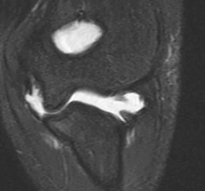
A patient reports persistent anterior shoulder pain following a forceful external rotation injury to the shoulder. An MRI scan is shown in Figure 4. The patient remains symptomatic despite 3 months of nonsurgical management. Treatment should now consist of


Explanation

The MRI scan reveals a subscapularis tear with a biceps that is out of the groove. Treatment in this patient is most predictable if the subscapularis is repaired. The biceps should either be tenodesed or tenotomized since it is unstable. Recentering of the biceps has been found to be unpredictable. Treatment of these lesions has been shown to have better results if the biceps is either released or tenodesed. This prevents recurrent biceps symptoms that can be source of surgical failure. Edwards TB, Walch G, Sirvenaux F, et al: Repair of tears of the subscapularis: Surgical technique. J Bone Joint Surg Am 2006;88:1-10. Deutsch A, Altcheck DW, Veltri DM, et al: Traumatic tears of the subscapularis tendon: Clinical diagnosis, magnetic resonance imaging findings, and operative treatment. Am J Sports Med 1997;25:13-22.


Question 9

A 78-year-old woman falls onto her nondominant left elbow and sustains the injury shown in Figure 5. What treatment option allows her the shortest recovery time and highest likelihood of good function and range of motion?


Explanation

Total elbow arthroplasty has become the treatment of choice for complex, comminuted distal humeral fractures in patients older than age 70 years. It yields a faster recovery with more predictable functional outcomes, although limitations of lifting weight of more than 5 pounds must be followed to avoid loosening. Kamineni S, Morrey BF: Distal humeral fractures treated with noncustom total elbow replacement. J Bone Joint Surg Am 2004;86:940-947.


Question 10

An MRI arthrogram of the elbow is shown in Figure 6. Based on these findings, what is the most likely diagnosis?


Explanation

MRI arthrography is the imaging study of choice for evaluation of medial collateral ligament injuries. Carrino JA, Morrison WB, Zou KH, et al: Noncontrast MR imaging and MR arthrography of the ulnar collateral ligament of the elbow: Prospective evaluation of two-dimensional pulse sequences for detection of complete tears. Skeletal Radiol 2001;30:625-632.


Question 11

A 45-year-old woman awakens with the acute onset of burning left shoulder pain that radiates toward the axilla. She denies any history of trauma. On examination, she is unable to abduct her arm but has full passive shoulder motion. Her sensation is intact. Cervical spine examination reveals full range of motion and a negative Spurling's test. Radiographs and MRI studies are normal for the cervical spine and shoulder. What is the most likely diagnosis?


Explanation

The definition of brachial neuritis or Parsonage-Turner syndrome is a rare disorder of unknown etiology that causes pain or weakness of the shoulder and upper extremity. The loss of active motion excludes cervical C6-7 radiculopathy and impingement. A normal MRI scan and full passive motion exclude a rotator cuff tear and adhesive capsulitis, respectively. Misamore GW, Lehman DE: Parsonage-Turner syndrome (acute brachial neuritis). J Bone Joint Surg Am 1996;78:1405-1408.


Question 12

A 25-year-old woman returns for her first postoperative visit after arthroscopic thermal capsulorrhaphy for recurrent multidirectional instability. Examination reveals that the portals are healed, there is no swelling; and passive range of motion is within the normal range. However, she is unable to actively raise her arm. Shoulder radiographs are normal. What is the most likely cause of these findings?


Explanation

Treatment of shoulder instability with thermal devices has lead to numerous complications including recurrent instability, chondrolysis, stiffness, and capsular necrosis. This patient's findings are consistent with a heat-induced axillary nerve injury. Normal radiographs exclude extensive chondrolysis. Levine WN, Bigliani LU, Ahmad CS: Thermal capsulorrhaphy. Orthopedics 2004;27:823-826.


Question 13

Figure 7 shows a sagittal T1-weighted MRI scan. What muscle/tendon is identified by the arrow?


Explanation

The sagittal T1-weighted MRI scan is useful for interpreting the quality of muscle. The arrow is pointing to the teres minor. Goutallier D, Postel JM, Gleyze P, et al: Influence of cuff muscle fatty degeneration on anatomic and functional outcomes after simple suture of full-thickness tears. J Shoulder Elbow Surg 2003;12:550-554.


Question 14

A 72-year-old man who underwent total shoulder arthroplasty 2 years ago slipped on ice and fell on his shoulder 3 weeks ago. Immediately after falling he was unable to elevate his arm. Motor examination reveals deltoid 5-/5, subscapularis 5-/5, external rotation 4-/5, and supraspinatus 2/5. Radiographs are shown in Figures 8a and 8b. What is the most likely diagnosis?


Explanation

The patient has a traumatic rotator cuff tear. The history of the fall, the weakness on examination, and normal radiographic findings make a traumatic rotator cuff tear the most likely diagnosis. An MRI scan can be obtained to further evaluate the integrity of the rotator cuff. The axillary radiograph shows a reduced, nondislocated total shoulder arthroplasty. His radiographs show a well-seated humeral stem and no signs of loosening. The glenoid is a cemented all-polyethylene component with no evidence of radiolucent lines surrounding the cemented pegs. The polyethylene glenoid component is radiolucent; however, the space between the metallic humeral head and the glenoid bone is the thickness of the polyethylene glenoid component. If the humeral head were directly against the glenoid bone, then catastrophic fracture of the glenoid would be the working diagnosis. Hattrup SJ, Cofield RH, Cha SS: Rotator cuff repair after shoulder replacement. J Shoulder Elbow Surg 2006;15:78-83.


Question 15

A 39-year-old man has had persistent right shoulder pain for the past 6 months. A formal physical therapy program has failed to provide relief, and an injection several months ago provided only short-term relief. Examination reveals a positive Neer and Hawkins test. There is no instability and the neurovascular examination is normal. Arthroscopy reveals a partial rotator cuff tear on the bursal side measuring 60% of the tendon thickness. What is the next most appropriate step in management?


Explanation

Although arthroscopic debridement with or without subacromial decompression is a reasonable response, the patient has positive impingement signs. Several recent studies regarding the surgical treatment of partial rotator cuff tears have demonstrated good to excellent results after repair of tears involving more than 50% of the tendon thickness. This was shown specifically for bursal-sided tears and joint-side tears. Biceps tenotomy is not indicated in a young patient. Matava MJ, Purcell DB, Rudzki JR: Partial-thickness rotator cuff tears. Am J Sports Med 2005;33:1405-1417.


Question 16

The condition shown in Figures 9a and 9b is most likely the result of


Explanation

The clinical photograph and radiograph show gout, which is the result of urate deposition in the joint and soft tissues. Radiographs frequently reveal periarticular erosions. The crystals are intracellular and negatively birefringent under the polarized microscope. Treatment for acute flares include colchicines, indomethacin, and corticosteroids (including injections). Medications such as allopurinol help prevent recurrent flares. Tophi such as that seen in this patient are often confused with and associated with infection. Wortmann RL, Kelley WM: Crystal-induced inflammation: Gout and hyperuricemia, in Harris ED, Budd RC, Firestein GS, et al (eds): Kelley's Textbook of Rheumatology, ed 7. New York, NY, Elsevier Science, 2005, pp 1402-1429. Trumble TE (ed): Hand Surgery Update 3: Hand, Elbow, & Shoulder. Rosemont, IL, American Society for Surgery of the Hand, 2003, pp 433-457.


Question 17

A patient reports hyperesthesia over the base of the thenar eminence following volar locked plating of a distal radius fracture. A standard volar approach of Henry was used. What is the most likely cause of the hyperesthesia?


Explanation

The palmar cutaneous branch of the median nerve separates from the median nerve approximately 4 to 6 cm proximal to the wrist crease and travels between the median nerve and the flexor carpi radialis tendon. It supplies the skin of the thenar region. This nerve is at risk for injury with retraction of the digital flexor tendons in plating the distal radius. Wartenberg's syndrome is compression of the superficial radial nerve which innervates the dorsum of the thumb and the first dorsal web space. Carpal tunnel syndrome causes dysesthesias of the thumb, index, and/or middle fingers. C7 radiculopathy affects the index and middle fingers. Jupiter JB, Fernandez DL, Toh CL, et al: Operative treatment of volar intra-articular fractures of the distal end of the radius. J Bone Joint Surg Am 1996;78:1817-1828.


Question 18

Figures 10a and 10b show the radiographs of a 47-year-old man who reports pain in both shoulders. He has a history of leukemia that was treated with chemotherapy and high-dose cortisone. What is the most reliable treatment option for pain relief in this patient?


Explanation

The radiographs reveal osteonecrosis with collapse. The most reliable and durable treatment for osteonecrosis of the humeral head remains prosthetic shoulder arthroplasty. Osteonecrosis of the humeral head may be seen after the use of steroids, and there is an increasing demand for shoulder arthroplasty in young people because of the use of high-dose steroids in chemotherapy regimes for the treatment of malignant tumors. The indications for most shoulder arthrodeses today include posttraumatic brachial plexus injury, paralytic disorders in infancy, insufficiency of the deltoid muscle and rotator cuff, chronic infection, failed revision arthroplasty, severe refractory instability, and bone deficiency following resection of a tumor in the proximal aspect of the humerus. Clearly, the role of arthroscopy and related minimally invasive techniques in the treatment of humeral head osteonecrosis remains unknown. Hasan SS, Romeo AA: Nontraumatic osteonecrosis of the humeral head. J Shoulder Elbow Surg 2002;11:281-298. Hattrup SJ: Indications, technique, and results of shoulder arthroplasty in osteonecrosis. Orthop Clin North Am 1998;29:445-451.


Question 19

Which of the following surgical devices employed for stabilization of the sternoclavicular joint is associated with the highest incidence of life-threatening complications?


Explanation

Numerous reports have documented serious complications including death from migration of intact or broken Kirschner wires or Steinmann pins into hilar structures such as the heart, pulmonary artery, and the aorta. Gilot GJ, Wirth MA, Rockwood CA: Injuries to the sternoclavicular joint, in Bucholz RW, Heckman JD, Court-Brown C (eds): Fractures in Adults. Philadelphia, PA, Lippincott, Williams and Wilkins, 2006, vol 2, pp 1373-1374.


Question 20

Figure 11a shows the clinical photograph of a 46-year old woman who reports a 3-week history of pain and a "lump" at the base of her neck. She is otherwise in good health and denies any trauma. A 3-D reconstruction CT is shown in Figure 11b. What is the most likely diagnosis?


Explanation

Spontaneous subluxation of the sternoclavicular joint occurs without any significant trauma. It is usually accentuated by placing the extremity in an overhead position. Discomfort usually resolves within 4 to 6 weeks with nonsurgical management. Rockwood CA, Wirth MA: Disorders of the sternoclavicular joint, in Rockwood CA, Matsen FA, Wirth MA, et al (eds): The Shoulder. Philadelphia, PA, WB Saunders, 2004, vol 2, pp 1078-1079.


Question 21

Figure 12a shows the clinical photograph of a 36-year-old man who has left shoulder pain and dysfunction after undergoing a lymph node biopsy 2 years ago. The appearance of the shoulder during abduction and a wall push-up maneuver is shown in Figures 12b and 12c, respectively. Which of the following procedures provides the best pain relief and function?


Explanation

Injury to the spinal accessory nerve can occur after penetrating trauma to the shoulder. Blunt trauma may also cause loss of trapezius function. Most commonly, surgical dissection in the posterior triangle of the neck, such as lymph node biopsy, may expose the nerve to possible damage. Surgical repair of the nerve may be considered up to 1 year after injury; after this time muscle transfer is usually associated with a better functional outcome. Steinman SP, Spinner RJ: Nerve problems in the shoulder, in Rockwood CA, Matsen FA, Wirth MA, et al (eds): The Shoulder. Philadelphia, PA, WB Saunders, 2004, vol 2, pp 1013-1015.


Question 22

What is the most common cause for poor outcomes in patients who undergo total shoulder arthroplasty?


Explanation

In an article in the Journal of Shoulder and Elbow, 431 total shoulder arthroplasties were performed with a cemented all-polyethylene glenoid component between 1990 and 2000. Follow-up averaged 4.2 years. In total, 53 surgical complications occurred in 53 patients (12%). Of these, 32 were major complications (7.4%), with 17 of these requiring reoperation. Index complications in order of frequency included rotator cuff tearing, postoperative glenohumeral instability, and periprosthetic humeral fracture. Notably, glenoid and humeral component loosening requiring reoperation occurred in only one shoulder. Data from the contemporary patient group suggest that there are fewer complications of shoulder arthroplasty and less need for reoperation. Especially striking is the near absence of component revision because of loosening or other mechanical factors. Complications involving the brachial plexus have been reported following total shoulder arthroplasty but are not as common of a cause for failure. Chin PY, Sperling JW, Cofield RH, et al: Complications of total shoulder arthroplasty: Are they fewer or different? J Shoulder Elbow Surg 2006;15:19-22.


Question 23

A 53-year-old man has had a long history of multiple joint symptoms, and he notes that the worst pain is from his left shoulder. A radiograph and MRI scan are shown in Figures 13a and 13b. Prior to surgical treatment of the shoulder, what is the most appropriate work-up?


Explanation

Rheumatoid arthritis is sometimes associated with radiographic evidence of instability of the cervical spine. In a study by Grauer and associates, radiographs of the cervical spine of patients with rheumatoid arthritis who had undergone total joint arthroplasty over a 5-year period were retrospectively reviewed. Nearly one half of the patients had radiographic evidence of cervical instability on the basis of traditional measurements. While radiographic evidence of cervical instability was not infrequent in this population of patients who underwent total joint arthroplasty for rheumatoid arthritis, radiographic predictors of paralysis were much less common. MRI prior to surgery may also be a consideration if the radiographic appearance of the rotator cuff alters the consideration of surgical treatment. In a series of patients undergoing prosthetic arthroplasty for a variety of shoulder disorders, the presence of a rotator cuff tear has been shown to be associated with a less favorable outcome. Most often, the presence of a rotator cuff tear was associated with a diagnosis of rheumatoid or other inflammatory arthritis and the tears were large and generally irreparable. Some case series demonstrated a higher prevalence of loosening of the glenoid component in patients with a large rotator cuff tear associated with superior migration of the humeral head. However, obtaining an MRI scan of the shoulder is not considered the best response since failure to determine cervical instability may result in anesthetic death. Whereas MRI may be helpful in planning reconstruction, it would be a less important priority. Grauer JN, Tingstad EM, Rand N, et al: Predictors of paralysis in the rheumatoid cervical spine in patients undergoing total joint arthroplasty. J Bone Joint Surg Am 2004;86:1420-1424.


Question 24

A 52-year-old man underwent arthroscopic repair of a 1-cm supraspinatus tendon tear 3 weeks ago. He was doing well until he fell down three stairs. One week after the fall he continues to report pain similar to his preoperative pain. An MRI scan reveals a minimally retracted 1-cm supraspinatus tendon tear in the same location as his original tear. Management should now consist of


Explanation

The patient has retorn his rotator cuff repair. This traumatic retear is different from a chronic tear and should be treated similar to an acute rotator cuff tear. Because the patient is younger than age 65 and has a small, single tendon tear, a revision rotation cuff repair is indicated with an expected tendon healing rate of greater than 95%. A physical therapy program is not indicated, and further delay in repair compromises his functional recovery. A cortisone injection is not indicated for this repairable tendon tear. Immobilization will not allow the tendon to heal once it has retorn. A debridement procedure is not indicated on this repairable tendon tear; this procedure is indicated in painful, chronic, irreparable tendon tears. Boileau P, Brassart N, Watkinson DJ, et al: Arthroscopic repair of full-thickness tears of the supraspinatus: Does the tendon really heal? J Bone Joint Surg Am 2005;87:1229-1240. Jost B, Zumstein M, Pfirrmann CWA, et al: Long-term outcome after structural failure of rotator cuff repairs. J Bone Joint Surg Am 2006;88:472-479.


Question 25

A 49-year-old woman with serologically proven rheumatoid arthritis has Larsen grade II radiographic changes in the elbow. Examination reveals a preoperative arc of flexion of less than 90 degrees and there is no instability. Nonsurgical management has failed to provide relief. What is the best treatment option?


Explanation

Larsen grade I and II rheumatoid arthritis is best treated with synovectomy with arthroplasty reserved for later stages, especially in younger patients. Open synovectomy with or without a radial head excision has yielded good results for pain and function, with arthroscopic synovectomies yielding similar results. Arthroscopic synovectomy has been shown to be more effective in restoring function in patients with a flexion arc of less than 90 degrees. Tanaka N, Sakahashi H, Hirose K, et al: Arthroscopic and open synovectomy of the elbow in rheumatoid arthritis. J Bone Joint Surg Am 2006;88:521-525. Horiuchi K, Momohara S, Tomatsu T, et al: Arthroscopic synovectomy of the elbow in rheumatoid arthritis. J Bone Joint Surg Am 2002;84:342-347.


Question 26

A 22-year-old collegiate rugby player presents with recurrent anterior shoulder instability. An MRI reveals a 25% bony Bankart lesion of the anterior glenoid.

Which of the following surgical interventions provides the most reliable long-term stability for this patient?


Explanation

The Latarjet procedure is indicated for patients with recurrent anterior shoulder instability and significant glenoid bone loss (>20-25%). Soft tissue repairs alone in this setting have an unacceptably high failure rate.

Question 27

A 72-year-old woman presents with severe shoulder pain and pseudoparalysis. Radiographs demonstrate severe glenohumeral osteoarthritis with superior migration of the humeral head articulating with the acromion.

Which of the following relies on the deltoid to restore active elevation in this setting?


Explanation

Reverse total shoulder arthroplasty medializes and distalizes the center of rotation, which tensions the deltoid. This allows the deltoid to act as the primary elevator in the setting of rotator cuff tear arthropathy.

Question 28

In the surgical management of a 'terrible triad' injury of the elbow, which of the following structures is typically repaired last to ensure adequate stability?


Explanation

The standard surgical algorithm for a terrible triad injury proceeds from deep to superficial: fixing the coronoid, then the radial head, and finally the LCL complex. The MCL is only repaired if the elbow remains unstable after LCL repair.

Question 29

A 24-year-old man sustains a proximal pole scaphoid fracture. The high risk of avascular necrosis (AVN) in this region is primarily due to which of the following anatomic characteristics?


Explanation

The proximal pole of the scaphoid is entirely covered by articular cartilage and relies on a retrograde blood supply from vessels entering the dorsal ridge distally. Fractures of the proximal pole frequently disrupt this blood supply, causing AVN.

Question 30

A 55-year-old man presents with chronic wrist pain. Radiographs reveal Scapholunate Advanced Collapse (SLAC) Stage III, characterized by arthritis in the capitolunate joint. Which of the following articulations is typically spared in this condition?


Explanation

In SLAC wrist, the radiolunate joint is characteristically spared due to the concentric shape of the lunate fossa and its strong ligamentous supports. This sparing allows for salvage procedures like a four-corner fusion.

Question 31

A 48-year-old manual laborer sustains a Type II SLAP tear. Nonoperative management fails. What is the most appropriate surgical treatment to minimize postoperative stiffness and allow a predictable return to work?


Explanation

In patients older than 40-45 years, biceps tenodesis yields more predictable pain relief and lower complication rates compared to SLAP repair. SLAP repair in older patients has a high risk of postoperative stiffness and persistent pain.

Question 32

When performing a Zone II flexor tendon repair, which of the following surgical techniques allows for the safest implementation of an early active motion rehabilitation protocol?


Explanation

Multi-strand repairs (4- or 6-strand) increase the tensile strength of the repair, allowing it to withstand the forces of early active motion protocols. An epitendinous suture further increases strength and reduces gapping.

Question 33

A 40-year-old weightlifter undergoes an anterior single-incision repair of a distal biceps tendon rupture. Postoperatively, he reports numbness over the lateral aspect of his forearm. Which nerve was most likely injured during the procedure?


Explanation

The lateral antebrachial cutaneous nerve (LACN) is the terminal sensory branch of the musculocutaneous nerve and courses near the cephalic vein in the lateral forearm. It is the most commonly injured nerve during a single-incision anterior distal biceps repair.

Question 34

During a carpal tunnel release, care must be taken to avoid injury to the recurrent motor branch of the median nerve. In the most common anatomical variation (extraligamentous), this branch originates:


Explanation

The extraligamentous type is the most common anatomical variant (approximately 50-80%), where the recurrent motor branch arises distal to the transverse carpal ligament and loops back to innervate the thenar muscles.

Question 35

In an unreduced Type III acromioclavicular (AC) joint separation, which of the following ligaments provides the primary restraint against superior translation of the clavicle?


Explanation

The conoid and trapezoid comprise the coracoclavicular (CC) ligaments. The conoid ligament is located posteromedially and acts as the primary restraint to superior translation of the clavicle.

Question 36

A patient undergoes in situ decompression of the ulnar nerve for cubital tunnel syndrome. During the approach, the roof of the cubital tunnel is incised. What structure primarily forms the roof of this tunnel?


Explanation

The roof of the cubital tunnel is primarily formed by Osborne's ligament, which is the aponeurotic band connecting the humeral and ulnar heads of the flexor carpi ulnaris (FCU).

Question 37

A 25-year-old gymnast complains of ulnar-sided wrist pain after a fall. Examination reveals a positive fovea sign and pain with ulnar deviation. MRI confirms a Palmer Type 1B tear of the Triangular Fibrocartilage Complex (TFCC). What is the appropriate surgical management if conservative treatment fails?


Explanation

A Palmer Type 1B tear is an avulsion of the TFCC from its ulnar insertion (fovea/ulnar styloid). Direct surgical repair (arthroscopic or open) to the fovea is the standard of care to restore distal radioulnar joint (DRUJ) stability.

Question 38

A 22-year-old collegiate rugby player presents with recurrent anterior shoulder instability after a primary traumatic dislocation. A 3D-CT scan reveals 25% anterior glenoid bone loss. What is the most appropriate definitive surgical management?


Explanation

Glenoid bone loss exceeding 20-25% is a classic contraindication for isolated soft tissue repair (Bankart). The Latarjet procedure (coracoid transfer) is the standard of care to restore anterior stability in contact athletes with critical bone loss.

Question 39

A 45-year-old manual laborer presents with chronic wrist pain. Radiographs demonstrate a scaphoid nonunion with radioscaphoid and capitolunate arthritis, but the radiolunate joint is completely preserved. What is the most appropriate surgical intervention?


Explanation

This presentation describes SNAC stage III with capitolunate arthritis, which makes a proximal row carpectomy contraindicated due to capitate wear. Scaphoid excision and four-corner fusion preserves some wrist motion while relying on the spared radiolunate joint.

Question 40

A 35-year-old man falls on an outstretched hand, sustaining a 'terrible triad' injury of the elbow. During surgical reconstruction, what is the generally recommended sequence of repair to restore elbow stability?


Explanation

The standard surgical sequence for a terrible triad injury is repairing deep to superficial. This involves addressing the coronoid first, followed by the radial head (repair or replace), and finally the lateral collateral ligament complex.

Question 41

An active 76-year-old woman sustains a highly comminuted 4-part proximal humerus fracture with a head-split component and severe osteopenia. Which surgical treatment provides the most predictable restoration of forward elevation?


Explanation

Reverse total shoulder arthroplasty provides the most reliable functional outcome and forward elevation for elderly patients with complex, unreconstructible proximal humerus fractures. Hemiarthroplasty relies on predictable tuberosity healing, which is poor in osteopenic bone.

Question 42

A 28-year-old carpenter sustains a Zone II flexor tendon laceration. During repair, the surgeon chooses a 4-strand core suture technique instead of a 2-strand technique. What is the primary biomechanical advantage of this choice?


Explanation

Increasing the number of core suture strands proportionally increases the tensile strength and gap resistance of the repair. This robust repair allows for early active motion protocols, which significantly improve clinical outcomes and decrease adhesions.

Question 43

A 55-year-old woman undergoes volar locked plating for a comminuted distal radius fracture. Six months later, she presents unable to actively flex the interphalangeal joint of her thumb. What technical error during the initial surgery most likely caused this complication?


Explanation

Flexor pollicis longus (FPL) tendon rupture is a known complication of volar plating of the distal radius. It classically occurs when the plate is placed distal to the watershed line, causing mechanical attrition of the tendon against the prominent hardware.

Question 44

A 42-year-old bodybuilder feels a 'pop' in his anterior elbow followed by weakness in supination. He undergoes a classic two-incision distal biceps tendon repair. Which nerve is at greatest risk of injury during the posterolateral dissection of this approach?


Explanation

The posterior interosseous nerve (PIN) is at greatest risk during the two-incision approach. This risk is minimized by keeping the forearm in full pronation during the posterolateral muscle splitting dissection.

Question 45

A 55-year-old man presents with anterior shoulder pain following a fall. Physical examination reveals a positive lift-off test, a positive belly-press test, and passively increased external rotation compared to the contralateral side. What is the most likely diagnosis?


Explanation

A positive lift-off test and belly-press test are highly specific for a subscapularis tendon tear. Increased passive external rotation also occurs due to the loss of the anterior restraint provided by the intact subscapularis.

Question 46

During a limited fasciectomy for Dupuytren's contracture, a surgeon meticulously dissects the spiral cord. The spiral cord alters normal anatomy by displacing the neurovascular bundle in which direction?


Explanation

The spiral cord characteristically displaces the neurovascular bundle proximally, superficially, and centrally (towards the midline of the digit). This distorted anatomy places the digital nerve at extremely high risk of iatrogenic transection.

Question 47

A 32-year-old woman sustains an isolated capitellum fracture that extends medially to include the lateral trochlear ridge (Dubberley Type 1B). What is the preferred surgical approach for optimal open reduction and internal fixation?


Explanation

An extensile lateral approach (such as Kocher or Kaplan) is preferred for capitellar shear fractures. It provides excellent direct visualization of the anterior articular surface of the radiocapitellar joint for precise reduction.

Question 48

A 21-year-old collegiate baseball pitcher complains of vague posterior shoulder pain. Exam reveals 20 degrees of internal rotation and 130 degrees of external rotation. Which anatomic structure is most likely pathologically contracted?


Explanation

Glenohumeral internal rotation deficit (GIRD) in overhead throwing athletes is primarily caused by a contracture of the posteroinferior capsule. Specifically, the posterior band of the inferior glenohumeral ligament is tightened.

Question 49

A 26-year-old motorcyclist falls onto an extended wrist. Lateral radiographs show a 'spilled teacup' sign with the lunate displaced volarly. The capitate is aligned with the radius. What is the correct diagnosis?


Explanation

A volarly displaced and tipped lunate with the capitate maintaining its collinear alignment with the radius describes a lunate dislocation. In a perilunate dislocation, the lunate remains aligned with the radius while the rest of the carpus is dorsally dislocated.

Question 50

A 24-year-old football player grabs an opponent's jersey and feels a pop in his ring finger. He cannot actively flex the DIP joint. Radiographs show a bony avulsion resting at the level of the PIP joint. What is the Leddy and Packer classification and optimal timing for surgery?


Explanation

A 'Jersey finger' where the tendon retracts to the PIP joint level (restrained by an intact vinculum longum) is a Leddy and Packer Type II injury. Surgical repair should ideally be performed within 3 to 4 weeks before definitive tendon retraction and fibrosis occur.

Question 51

A 50-year-old woman with type 1 diabetes mellitus presents with recalcitrant adhesive capsulitis. After 9 months of failed physical therapy and intra-articular corticosteroid injections, she elects for surgery. What is the most appropriate next step in management?


Explanation

Arthroscopic capsular release (often combined with manipulation) is the treatment of choice for refractory adhesive capsulitis. Diabetic patients have a higher rate of recalcitrant disease and often require comprehensive release of the rotator interval and coracohumeral ligament.

Question 52

A 13-year-old male gymnast presents with lateral elbow pain and clicking. MRI demonstrates osteochondritis dissecans (OCD) of the capitellum with an unstable, detached osteochondral fragment resting in situ. What is the most appropriate management?


Explanation

In an adolescent with capitellar OCD demonstrating an unstable or detached fragment on MRI, surgical intervention is indicated. Arthroscopic fragment excision with microfracture of the base, or fragment fixation if amenable, is the standard of care.

Question 53

A 30-year-old manual laborer has progressive dorsal wrist pain. X-rays reveal Kienbock's disease with sclerosis and collapse of the lunate, proximal migration of the capitate, and early secondary arthritic changes. The ulna variance is negative. What is the Lichtman classification and corresponding management?


Explanation

Sclerosis and collapse of the lunate with fixed scaphoid rotation and capitate proximal migration indicates Lichtman Stage IIIB Kienbock's disease. Joint leveling procedures are ineffective at this stage; salvage procedures like proximal row carpectomy or STT fusion are required.

Question 54

A 45-year-old construction worker with chronic anterior shoulder pain exhibits a positive Speed's test. MRI reveals a Type II SLAP lesion with significant macroscopic degeneration of the biceps anchor. What is the most reliable surgical option?


Explanation

In patients over 40 or heavy laborers with Type II SLAP tears and concurrent biceps anchor degeneration, biceps tenodesis provides more reliable pain relief. SLAP repair in this demographic has a high rate of postoperative stiffness and persistent pain.

Question 55

A 72-year-old female presents with chronic right shoulder pain and an inability to actively raise her arm above 60 degrees. Radiographs, similar to those seen in advanced rotator cuff tear arthropathy, demonstrate superior migration of the humeral head with articulation at the acromion.

What is the most appropriate surgical intervention to restore functional elevation in this patient?


Explanation

Reverse total shoulder arthroplasty relies on the deltoid to restore elevation in patients with a deficient rotator cuff. It medializes and distalizes the center of rotation, increasing the deltoid's moment arm and improving function.

Question 56

In the surgical management of a "terrible triad" injury of the elbow, which of the following represents the most accepted sequence of repair after exposing the joint?


Explanation

Standard protocol for the terrible triad includes coronoid fixation, radial head repair or replacement, followed by lateral collateral ligament (LCL) repair. This inside-out sequence restores anterior skeletal stability before addressing the lateral ligamentous restraints.

Question 57

A 55-year-old manual laborer presents with chronic dorsal wrist pain. Radiographs demonstrate advanced narrowing of the capitolunate joint, with relative sparing of the radiolunate joint.

This presentation is most consistent with which stage of Scapholunate Advanced Collapse (SLAC)?


Explanation

SLAC wrist progresses in a predictable pattern: Stage I involves the radial styloid, Stage II involves the entire radioscaphoid facet, and Stage III progresses to the capitolunate joint while typically sparing the radiolunate articulation.

Question 58

A 22-year-old rugby player sustained a closed avulsion of the flexor digitorum profundus (FDP) tendon of the ring finger. Radiographs show no fracture. The tendon is palpable in the palm. What is the Leddy-Packer classification and maximum recommended time frame for primary repair?


Explanation

A Leddy-Packer Type I FDP avulsion involves retraction of the tendon into the palm, disrupting both the vincula longus and brevis. Due to compromised blood supply, surgical repair must be performed within 7-10 days to prevent tendon necrosis.

Question 59

A 45-year-old patient with type 1 diabetes mellitus presents with gradual onset of shoulder stiffness. Examination reveals a significant loss of active and passive external rotation with the arm at the side. What is the primary pathologic mechanism?


Explanation

Adhesive capsulitis is characterized by severe loss of both active and passive range of motion, specifically external rotation. The primary pathology is fibroblastic proliferation and contracture of the rotator interval and the coracohumeral ligament.

Question 60

A 70-year-old man presents with an inability to actively raise his right arm above 40 degrees, despite intact passive range of motion. Radiographs reveal superior migration of the humeral head with articulation against the acromion and severe glenohumeral osteoarthritis.

Which of the following surgical options is the most appropriate management for this patient?


Explanation

Reverse total shoulder arthroplasty is indicated for rotator cuff arthropathy with pseudoparalysis. It medializes and distalizes the center of rotation, increasing the deltoid moment arm to restore elevation.

Question 61

A 25-year-old male presents with a 9-month-old scaphoid waist fracture nonunion. MRI demonstrates no avascular necrosis of the proximal pole, and radiographs show no radiocarpal arthritis (SNAC wrist stage 0). What is the most appropriate surgical management?


Explanation

For scaphoid waist nonunions without avascular necrosis, non-vascularized bone grafting with rigid internal fixation yields high union rates and is the preferred initial treatment. Vascularized grafts are typically reserved for proximal pole nonunions or cases with confirmed AVN.

Question 62

A 35-year-old woman sustains a 'terrible triad' injury of the elbow (elbow dislocation, radial head fracture, and coronoid fracture). Surgical fixation is planned. What is the generally accepted sequence of repair to restore stability?


Explanation

The standard surgical algorithm for a terrible triad injury proceeds from deep to superficial: coronoid fixation first, followed by the radial head, and finally the lateral collateral ligament (LCL) complex. MCL repair is only considered if the elbow remains grossly unstable after these steps.

Question 63

A 45-year-old female with poorly controlled diabetes mellitus presents with a 4-month history of severe shoulder pain and progressively restricted active and passive range of motion. Radiographs are normal. What is the classic pathologic finding associated with this condition?


Explanation

This patient has adhesive capsulitis, which is strongly associated with diabetes. Pathology demonstrates dense fibroblastic proliferation and type III collagen deposition, primarily affecting the rotator interval and coracohumeral ligament.

Question 64

A 32-year-old manual laborer presents with dorsal wrist pain. Radiographs reveal sclerosis of the lunate without collapse, and negative ulnar variance (Lichtman Stage II Kienboeck disease). What is the most appropriate definitive surgical treatment?


Explanation

In Lichtman Stage II Kienboeck disease with negative ulnar variance, a joint-leveling procedure such as a radial shortening osteotomy unloads the lunate and can halt disease progression. Salvage procedures like proximal row carpectomy are reserved for advanced collapse.

Question 65

During a single anterior-incision repair of a distal biceps tendon rupture using an endobutton technique, the patient is at highest risk for iatrogenic injury to which of the following nerves?


Explanation

The lateral antebrachial cutaneous nerve (LABCN) is the most frequently injured nerve during a single-anterior-incision distal biceps repair due to its superficial location and proximity to the cephalic vein. The PIN is more commonly at risk during a two-incision technique.

Question 66

A hand surgeon is repairing a Zone II flexor tendon laceration. To optimize tensile strength for an early active motion protocol while minimizing tendon gliding resistance (work of flexion), what is the optimal suture construct?


Explanation

A 4-strand core suture combined with a running epitendinous suture provides an optimal balance of strength for early active motion while avoiding excessive bulk. Constructs with 6 or 8 strands can increase gliding resistance and risk tendon bunching.

Question 67

A 22-year-old male presents with recurrent anterior shoulder instability. A pre-operative 3D CT scan demonstrates 28% anterior glenoid bone loss. Which of the following procedures provides the most reliable long-term stability?


Explanation

In the setting of significant anterior glenoid bone loss (>20-25%), soft tissue stabilization alone has an unacceptably high failure rate. A bone-block procedure, such as the Latarjet, is required to restore the glenoid articular arc and provide a dynamic sling effect.

Question 68

A patient with severe, longstanding carpal tunnel syndrome undergoes a carpal tunnel release. Due to profound thenar atrophy, an opponensplasty using the flexor digitorum superficialis (FDS) of the ring finger is performed. To optimize the vector for thumb opposition, the transfer should be routed around which structure?


Explanation

The Burkhalter opponensplasty uses the FDS of the ring finger transferred to the abductor pollicis brevis. Routing it around the flexor carpi ulnaris (FCU) acts as a pulley to direct the vector of pull from the pisiform, which is optimal for restoring thumb opposition.

Question 69

A 20-year-old collegiate baseball pitcher is diagnosed with a full-thickness tear of the ulnar collateral ligament (UCL) of the elbow. Reconstruction is planned to restore valgus stability. Which specific ligamentous band must be reconstructed?


Explanation

The anterior band of the anterior bundle of the UCL is the primary restraint to valgus stress at the elbow during the throwing motion. Surgical reconstruction (Tommy John surgery) focuses on restoring this specific anatomical structure.

Question 70

A 28-year-old female presents with persistent ulnar-sided wrist pain after a fall on an outstretched hand. MRI confirms a Palmer Class 1B tear of the triangular fibrocartilage complex (TFCC). After failing conservative management, what is the best surgical intervention?


Explanation

A Palmer Class 1B tear is a peripheral avulsion of the TFCC from its ulnar attachment at the fovea. Because the peripheral TFCC is well-vascularized, it has excellent healing potential and is best treated with arthroscopic or open anatomic repair.

Question 71

During arthroscopic repair of a posterior SLAP tear, the surgeon must be cautious when placing suture anchors or drilling into the posterior glenoid neck. Penetrating the posterior glenoid neck medially by more than what distance puts the suprascapular nerve at significant risk?


Explanation

The suprascapular nerve courses around the spinoglenoid notch approximately 10 to 20 mm medial to the posterior glenoid rim. Drilling or anchor placement that penetrates deeper than 10-15 mm in this area risks direct nerve injury.

Question 72

A 60-year-old man undergoes limited fasciectomy for severe Dupuytren's contracture involving the ring finger. During the dissection, the surgeon carefully traces the neurovascular bundle. Which specific cord is primarily responsible for contracture of the proximal interphalangeal (PIP) joint and typically displaces the digital nerve centrally and superficially?


Explanation

The spiral cord is formed by the pretendinous band, spiral band, lateral digital sheet, and Grayson's ligaments. It is primarily responsible for PIP joint contracture and characteristically displaces the neurovascular bundle centrally, superficially, and proximally, increasing the risk of iatrogenic injury.

Question 73

A 26-year-old man falls from a roof and presents with severe wrist pain. Lateral radiographs show a 'spilled teacup' sign, indicative of a volar lunate dislocation. According to the Mayfield classification of progressive perilunate instability, what is the correct sequence of ligamentous failure?


Explanation

The Mayfield progression of perilunate instability occurs in four sequential stages starting radially and progressing ulnarward: scapholunate (Stage I), capitolunate (Stage II), lunotriquetral (Stage III), and finally volar lunate dislocation (Stage IV).

Question 74

A 50-year-old female presents with severe ulnar neuropathy at the elbow. Intraoperative examination reveals that the ulnar nerve actively subluxates over the medial epicondyle during elbow flexion. What is the most appropriate surgical management?


Explanation

While in situ decompression is effective for most cases of cubital tunnel syndrome, a nerve that subluxates anteriorly with flexion requires anterior transposition. Leaving a subluxating nerve in situ will lead to continued dynamic friction and poor clinical outcomes.

Question 75

A 74-year-old woman presents with severe right shoulder pain and an inability to raise her arm above shoulder level. Radiographs reveal superior migration of the humeral head with an acromiohumeral distance of 4 mm. MRI confirms a massive, retracted rotator cuff tear involving the supraspinatus and infraspinatus with advanced fatty infiltration. What is the most appropriate surgical treatment?


Explanation

Reverse total shoulder arthroplasty (RTSA) is the gold standard for rotator cuff tear arthropathy and massive, irreparable tears with pseudoparalysis in the elderly. It medializes and distalizes the center of rotation, recruiting the deltoid to elevate the arm.

Question 76

A 70-year-old woman presents with severe right shoulder pain and an inability to raise her arm above the horizontal level for the past 6 months. Examination demonstrates active forward elevation to 70 degrees, but passive forward elevation to 150 degrees. Radiographs reveal advanced glenohumeral osteoarthritis with superior migration of the humeral head articulating with the acromion. What is the most appropriate surgical management?


Explanation

Reverse total shoulder arthroplasty relies on a functioning deltoid to restore elevation in patients with rotator cuff tear arthropathy and pseudoparalysis. Anatomic total shoulder arthroplasty is contraindicated due to the deficient rotator cuff, which would lead to eccentric superior loading and early glenoid loosening.

Question 77

A 45-year-old man falls on his outstretched hand and sustains a 'terrible triad' injury of the elbow. He undergoes operative management for a posterolateral elbow dislocation, radial head fracture, and a type II coronoid fracture. Which of the following represents the most appropriate sequence of surgical reconstruction for this injury pattern?


Explanation

The standard surgical sequence for a terrible triad injury of the elbow is fixing deep to superficial: coronoid first, followed by the radial head, and finally repairing the lateral collateral ligament (LCL) complex. Medial collateral ligament repair is generally only performed if the elbow remains unstable after the lateral-sided structures are addressed.

Question 78

A 38-year-old man presents with chronic wrist pain. Radiographs demonstrate a scaphoid nonunion with advanced collapse (SNAC). Imaging shows arthritic changes involving both the radioscaphoid and capitolunate joints, while the radiolunate joint is entirely spared. Which of the following is the most appropriate surgical intervention?


Explanation

In Stage III SNAC wrists (radioscaphoid and capitolunate arthritis), four-corner arthrodesis with scaphoid excision is the preferred treatment. Proximal row carpectomy is contraindicated because the degenerate capitate head would articulate poorly with the lunate fossa, leading to persistent pain.

Question 79

During a flexor tendon repair in Zone II of the hand, maintaining the integrity of the flexor tendon sheath is critical. Which of the following pulley combinations is considered most essential to prevent bowstringing of the flexor tendons?


Explanation

The A2 and A4 pulleys arise directly from the periosteum of the proximal and middle phalanges, respectively, and are the most critical biomechanical pulleys. Loss of these specific pulleys leads to significant flexor tendon bowstringing, causing a loss of active flexion and grip strength.

Question 80

A 22-year-old rugby player presents with recurrent anterior shoulder instability. A CT scan of the shoulder reveals 25% anterior glenoid bone loss. The surgeon plans a Latarjet procedure. This procedure provides dynamic anterior stability through a 'sling effect' primarily involving the interaction between the lower subscapularis and which of the following structures?


Explanation

The Latarjet procedure involves transferring the coracoid process along with the attached conjoined tendon. When the arm is abducted and externally rotated, the conjoined tendon acts as a dynamic sling across the inferior subscapularis and anterior-inferior capsule, providing critical stability.

Question 81

A 20-year-old collegiate baseball pitcher feels a pop in his medial elbow during a pitch. MRI confirms a complete distal avulsion of the ulnar collateral ligament (UCL). The primary restraint to valgus stress at the elbow during the late cocking and early acceleration phases of throwing is the:


Explanation

The anterior band of the anterior bundle of the UCL is the primary restraint to valgus stress at the elbow from 30 to 120 degrees of flexion. This arc of motion corresponds to the high-torque late cocking and early acceleration phases of the overhead throwing motion.

None

Clinic OS
Medically Verified Content by
Prof. Clinic OS
Consultant Orthopedic & Spine Surgeon
Chapter Index