Operative Management and Classification of Ankle Fractures
Key Takeaway
Ankle fractures involve complex disruptions of both bony architecture and ligamentous stabilizers. Successful management mandates precise anatomical restoration of the ankle mortise, ensuring the weight-bearing axis remains perpendicular to the leg. While closed reduction may suffice for stable patterns, open reduction and internal fixation (ORIF) is the gold standard for unstable injuries. This guide details the biomechanics, Lauge-Hansen classification, surgical indications, and step-by-step operative techniques required to optimize functional outcomes and prevent post-traumatic arthrosis.
INTRODUCTION TO ANKLE FRACTURES
Injuries around the ankle joint cause destruction of not only the bony architecture but also, critically, the ligamentous and soft tissue components that confer dynamic and static stability. The ankle is a highly constrained, weight-bearing hinge joint; consequently, only the slightest variation from normal anatomy is compatible with good long-term joint function.
Biomechanical studies have famously demonstrated that a mere 1 millimeter of lateral talar shift reduces the tibiotalar contact area by 42%, exponentially increasing peak contact stresses and predisposing the patient to rapid, debilitating post-traumatic osteoarthritis. Therefore, radiographs obtained after any reduction maneuver must be scrutinized with three absolute requirements in mind:
1. The normal relationships of the ankle mortise must be perfectly restored.
2. The weight-bearing alignment of the ankle must be at a right angle to the longitudinal axis of the leg.
3. The contours of the articular surface must be satisfactorily and anatomically reduced.
The best clinical results are unequivocally obtained by anatomical joint restoration. While closed manipulation and casting may be appropriate for isolated, stable injury patterns, open reduction and internal fixation (ORIF) is the gold standard for unstable fractures. ORIF most reliably ensures anatomical joint restoration, rigid stabilization, and early mobilization, thereby optimizing the environment for osseous union and functional recovery.
SURGICAL ANATOMY AND BIOMECHANICS
Understanding the pathoanatomy of ankle fractures requires a thorough grasp of the ankle mortise. The mortise is formed by the distal tibia (plafond), the medial malleolus, and the lateral malleolus (distal fibula). The talus sits within this mortise, acting as the intercalary segment between the leg and the foot.
The stability of the ankle relies heavily on three distinct ligamentous complexes:
* The Syndesmotic Complex: Comprising the anterior inferior tibiofibular ligament (AITFL), posterior inferior tibiofibular ligament (PITFL), transverse tibiofibular ligament, and the interosseous membrane. This complex maintains the critical relationship between the distal tibia and fibula.
* The Medial Collateral (Deltoid) Ligament: A robust, multi-fascicular structure divided into superficial and deep components. The deep deltoid is the primary medial stabilizer against lateral talar excursion and external rotation.
* The Lateral Collateral Ligaments: Comprising the anterior talofibular ligament (ATFL), calcaneofibular ligament (CFL), and posterior talofibular ligament (PTFL).
Clinical Pearl: The fibula dictates the length and rotation of the lateral column of the ankle. Shortening or malrotation of the lateral malleolus inevitably leads to lateral talar shift and a widened mortise. Anatomical restoration of fibular length is the cornerstone of ankle fracture surgery.
CLASSIFICATION OF ANKLE FRACTURES
Ankle fractures can be classified purely along anatomical lines as monomalleolar, bimalleolar, or trimalleolar. However, mechanistic classifications provide deeper insight into the sequence of tissue failure.
The Lauge-Hansen Classification
The Lauge-Hansen classification remains a cornerstone of orthopedic education. It attempts to associate specific fracture patterns with the mechanism of injury. The nomenclature utilizes two words:
* First Word: The position of the foot at the exact moment of injury (Supination or Pronation).
* Second Word: The direction of the deforming force applied to the talus relative to the tibia (Adduction, Abduction, or Eversion/External Rotation).
Note: In this classification system, the term "eversion" is a historical misnomer; biomechanically, it more correctly refers to external or lateral rotation.
1. Supination-Eversion (External Rotation) (SER)
This is the most common mechanism of ankle fracture, accounting for up to 70% of all cases. The identifying feature is a spiral oblique fracture of the distal fibula.
* Stage I: Disruption of the anterior tibiofibular ligament (AITFL).
* Stage II: Spiral oblique fracture of the distal fibula (typically posteroinferior to anterosuperior).
* Stage III: Disruption of the posterior tibiofibular ligament (PITFL) or an avulsion fracture of the posterior malleolus.
* Stage IV: Fracture of the medial malleolus (usually transverse) or rupture of the deltoid ligament.
2. Supination-Adduction (SA)
Characterized by a transverse fracture of the distal fibula and a relatively vertical fracture of the medial malleolus, driven by talar impaction.
* Stage I: Transverse avulsion-type fracture of the fibula below the level of the joint, or a tear of the lateral collateral ligaments.
* Stage II: Vertical fracture of the medial malleolus due to the adducting talus impacting the medial plafond.
3. Pronation-Abduction (PA)
The pronated foot locks the talus within the mortise, and an abduction force drives the fibula laterally.
* Stage I: Transverse fracture of the medial malleolus or rupture of the deltoid ligament.
* Stage II: Rupture of the syndesmotic ligaments or avulsion fracture of their insertions.
* Stage III: Short, horizontal, oblique fracture of the fibula above the level of the joint (bending fracture).
4. Pronation-Eversion (External Rotation) (PER)
Characterized by a high fibular fracture and a high likelihood of syndesmotic instability.
* Stage I: Transverse fracture of the medial malleolus or disruption of the deltoid ligament.
* Stage II: Disruption of the anterior tibiofibular ligament (AITFL).
* Stage III: Short oblique or spiral fracture of the fibula relatively high above the level of the ankle joint (often a Maisonneuve-type pattern).
* Stage IV: Rupture of the posterior tibiofibular ligament or avulsion fracture of the posterolateral tibia (posterior malleolus).
5. Pronation-Dorsiflexion (PD)
A less common axial loading injury resulting in anterior plafond involvement.
* Stage I: Fracture of the medial malleolus.
* Stage II: Fracture of the anterior margin of the tibia.
* Stage III: Supramalleolar fracture of the fibula.
* Stage IV: Transverse fracture of the posterior tibial surface.
Limitations of the Lauge-Hansen System
While historically significant, modern orthopedic literature has scrutinized the Lauge-Hansen system. Authors have demonstrated considerable interobserver variability when applying this classification. Furthermore, while it is useful for understanding the mechanisms of injury and planning closed reduction maneuvers (which generally reverse the mechanism of injury), it has not been shown to have strong prognostic significance.
Surgical Warning: The Lauge-Hansen classification scheme has demonstrated notable limitations in predicting associated soft tissue injuries when evaluated with advanced imaging like MRI. Surgeons must maintain a high index of suspicion for syndesmotic and deltoid injuries regardless of the strict radiographic stage.
INDICATIONS FOR OPERATIVE INTERVENTION
The decision to proceed with ORIF is dictated by fracture stability and articular congruity. Absolute and relative indications include:
* Displaced Bimalleolar and Trimalleolar Fractures: These are inherently unstable and require fixation.
* Unstable Monomalleolar Fractures: Such as a lateral malleolus fracture with a concurrent deltoid ligament rupture (bimalleolar equivalent), evidenced by a widened medial clear space (>4 mm) on stress radiographs.
* Syndesmotic Instability: Any fracture pattern resulting in a widened tibiofibular clear space or overlap.
* Open Fractures: Requiring urgent irrigation, debridement, and stabilization.
* Failure of Closed Reduction: Inability to achieve or maintain the three radiographic requirements of the ankle mortise.
PREOPERATIVE PLANNING AND PATIENT POSITIONING
Soft Tissue Management
The timing of surgical intervention is dictated entirely by the soft tissue envelope. Surgery should ideally be performed within the first 24 hours before massive edema ensues. If significant swelling or fracture blisters are present, surgery must be delayed. The limb is placed in a well-padded posterior splint, elevated, and iced until the "wrinkle sign" appears (return of normal skin creases), which may take 7 to 14 days.
Patient Positioning
- Supine Position: Standard for most lateral, medial, and bimalleolar fractures. A bump is placed under the ipsilateral hip to internally rotate the leg, bringing the fibula parallel to the floor.
- Prone Position: Increasingly utilized for trimalleolar fractures with a large, displaced posterior malleolus fragment requiring direct visualization and posterior buttress plating.
- Tourniquet: A thigh tourniquet is applied but inflated only if necessary to minimize ischemic complications, particularly in vasculopathic patients.
SURGICAL APPROACHES
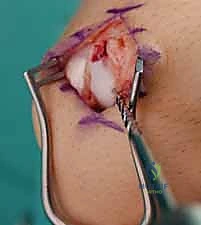
1. Lateral Approach to the Fibula
An incision is made directly over the posterolateral border of the fibula.
* Internervous Plane: Care is taken to avoid the superficial peroneal nerve anteriorly and the sural nerve posteriorly.
* Dissection: Full-thickness fasciocutaneous flaps are raised. The periosteum is incised longitudinally, preserving as much soft tissue attachment to the bone fragments as possible to maintain vascularity.
2. Medial Approach to the Medial Malleolus
A longitudinal or slightly curved incision is made centered over the medial malleolus.
* Structures at Risk: The great saphenous vein and the saphenous nerve run anterior to the medial malleolus and must be identified and protected.
* Dissection: The incision is carried down to the periosteum. The joint capsule is opened anteriorly to inspect the medial gutter and the talar dome for osteochondral lesions.
3. Posterolateral Approach to the Posterior Malleolus
Utilized when the patient is prone. The incision is made midway between the posterior border of the fibula and the lateral border of the Achilles tendon.
* Internervous Plane: The sural nerve is identified and protected laterally.
* Dissection: The fascia is incised, and the interval between the peroneal tendons (retracted laterally) and the flexor hallucis longus (FHL) muscle belly (retracted medially) is developed to expose the posterior tibia.
STEP-BY-STEP OPERATIVE TECHNIQUE (ORIF)
Fixation of the Lateral Malleolus (Fibula)
Restoring fibular length and rotation is the most critical step in ankle fracture surgery.
1. Reduction: The fracture site is debrided of hematoma. The fracture is reduced using point-to-point reduction forceps.
2. Lag Screw Fixation: For oblique or spiral fractures (e.g., SER patterns), a 3.5 mm cortical lag screw is placed perpendicular to the fracture plane to achieve interfragmentary compression.
3. Neutralization Plating: A one-third tubular plate or a pre-contoured locking plate is applied to the lateral or posterolateral surface of the fibula to neutralize torsional and bending forces.
4. Antiglide Plating: For short oblique fractures, a posterolateral antiglide plate can be utilized. This biomechanically superior construct resists the proximal migration of the distal fragment.
Fixation of the Medial Malleolus
- Joint Inspection: The medial clear space is inspected. Any inverted periosteum or deltoid ligament obstructing reduction is extracted.
- Reduction: The medial malleolus is reduced using a pointed reduction clamp. Anatomical reduction of the articular surface is verified visually and fluoroscopically.
- Fixation: Two parallel 4.0 mm partially threaded cancellous screws are typically placed from the tip of the malleolus directed superiorly and slightly laterally. For small or comminuted fragments, a tension band wire construct may be preferred to prevent fragmentation.
Fixation of the Posterior Malleolus
Historically, posterior malleolus fragments involving less than 25% of the articular surface were managed non-operatively. Modern evidence suggests that fixing even smaller fragments restores the PITFL, significantly enhancing syndesmotic stability.
1. Direct Reduction: Through a posterolateral approach, the fragment is directly visualized, reduced, and held with K-wires.
2. Plating: A posterior buttress plate (e.g., a 2.7 mm or 3.5 mm T-plate or one-third tubular plate) is applied in an anti-glide fashion. This is biomechanically superior to anterior-to-posterior (AP) lag screws.
Evaluation and Fixation of the Syndesmosis
Once the malleoli are fixed, the syndesmosis must be dynamically tested.
1. The Cotton Test: A bone hook is placed around the fibula, and a lateral pull is applied under fluoroscopy. Widening of the tibiofibular clear space indicates syndesmotic instability.
2. Fixation: If unstable, the syndesmosis is reduced with a large clamp (placed in neutral rotation and slight dorsiflexion). Fixation is achieved using either one or two 3.5 mm/4.5 mm cortical screws (placed across 3 or 4 cortices) or a dynamic suture-button construct.
Pitfall: Malreduction of the syndesmosis is a leading cause of poor outcomes. Always obtain a perfect mortise view and consider intraoperative CT or contralateral fluoroscopic comparison if there is any doubt regarding the reduction.
POSTOPERATIVE PROTOCOL AND REHABILITATION
Postoperative care must balance the need for tissue healing with the prevention of joint stiffness.
* Phase 1 (0-2 Weeks): The patient is placed in a well-padded short leg splint and remains strictly non-weight-bearing (NWB). Elevation and strict edema control are paramount.
* Phase 2 (2-6 Weeks): Sutures are removed at 2 weeks. The patient is transitioned to a controlled ankle motion (CAM) boot. Early active range of motion (ROM) exercises (dorsiflexion and plantarflexion) are initiated to nourish the articular cartilage and prevent capsular contracture. The patient remains NWB.
* Phase 3 (6-12 Weeks): Radiographs are obtained at 6 weeks to assess callus formation and hardware position. If clinical and radiographic union is progressing, the patient begins progressive weight-bearing in the CAM boot, transitioning to regular footwear by 8-10 weeks. Formal physical therapy focuses on proprioception, peroneal strengthening, and gait mechanics.
COMPLICATIONS
Despite meticulous surgical technique, complications can arise:
* Wound Dehiscence and Infection: The soft tissue envelope around the ankle is notoriously thin. Meticulous handling of tissues and delaying surgery until swelling subsides are critical preventative measures.
* Post-Traumatic Osteoarthritis: Directly correlated with the severity of the initial cartilage impact and the accuracy of the surgical reduction.
* Hardware Prominence: Lateral fibular plates frequently cause irritation, necessitating hardware removal in up to 15-20% of patients after complete bony union (typically >1 year post-op).
* Syndesmotic Malreduction: Leads to chronic pain and early arthrosis. Revision surgery may be required if identified early.
By adhering to strict biomechanical principles, respecting the soft tissue envelope, and executing precise osteosynthesis, the orthopedic surgeon can reliably restore the complex anatomy of the ankle, offering the patient the highest probability of a return to pre-injury function.
