Management of Ankle Fractures in Patients with Diabetes: A Comprehensive Surgical Guide
Key Takeaway
The management of ankle fractures in diabetic patients presents a profound orthopedic challenge due to compromised bone quality, peripheral neuropathy, and microvascular disease. While nonoperative treatment risks malunion and Charcot arthropathy, operative intervention carries a 43% complication rate. Successful outcomes demand meticulous soft-tissue handling, augmented rigid internal fixation—often utilizing multiple syndesmotic or trans-articular screws—and strictly prolonged postoperative immobilization to prevent catastrophic construct failure and neuroarthropathic collapse.
Comprehensive Introduction and Patho-Epidemiology
Although malleolar fractures are generally considered to be relatively benign and highly predictable injuries in the healthy, active population, their occurrence in patients with diabetes mellitus represents a profoundly limb-threatening pathology. The operative and nonoperative treatment of ankle fractures in the diabetic cohort is fraught with significant clinical challenges and is associated with a markedly elevated complication profile that demands a specialized, highly vigilant approach. These patients are frequently older and present with a devastating triad of systemic compromises: peripheral vascular disease (angiopathy), peripheral neuropathy, and compromised bone mineral density (diabetic osteopathy). This triad fundamentally alters the healing cascade, biomechanical stability, and immunological response of the lower extremity.
Historically, complication rates in the diabetic cohort have been reported to be as high as 43%, standing in stark contrast to the 15.5% complication rate observed in age-matched patients without diabetes. The spectrum of these complications is not merely an increase in minor morbidities; it encompasses catastrophic events including deep and superficial surgical site infections, catastrophic loss of fixation, malunion, nonunion, marginal wound necrosis, and ultimately, major lower extremity amputation. The "Diabetic Ankle" must never be treated as a standard ankle fracture. The presence of peripheral neuropathy fundamentally alters the biomechanical feedback loop, allowing patients to inadvertently overload and destroy standard fixation constructs due to an absence of protective pain sensation.
Despite these exorbitant risks, the modern paradigm of orthopedic care requires a highly nuanced, aggressive approach. While nonoperative treatment in diabetic patients has historically demonstrated an unacceptably high frequency of loss of reduction and malunion, these patients are often minimally symptomatic due to their profound neuropathy. However, inadequate immobilization or unstable alignment can rapidly precipitate Charcot neuroarthropathy, leading to architectural collapse of the midfoot and hindfoot. Therefore, if surgical treatment of the ankle fracture is biomechanically indicated, it should not be delayed or avoided simply because the patient carries a diagnosis of diabetes. The soft tissue compromise resulting from bony pressure of an unreduced fracture, combined with the inevitable malunion and Charcot collapse, carries a demonstrably higher amputation risk than a meticulously executed open reduction and internal fixation (ORIF).
The patho-epidemiology of the diabetic ankle fracture is driven by cellular-level dysfunctions. Hyperglycemia induces the formation of advanced glycation end-products (AGEs), which accumulate in the bone matrix and alter collagen cross-linking. This renders the bone highly brittle and prone to hardware pull-out, even in the absence of classic dual-energy x-ray absorptiometry (DEXA)-defined osteoporosis. Furthermore, microvascular ischemia impairs the delivery of fibroblasts, leukocytes, and essential nutrients to the fracture hematoma and surgical incision, severely blunting the host's ability to mount an appropriate healing or infection-fighting response. Understanding this underlying pathophysiology is paramount for the orthopedic surgeon to appropriately stratify risk and modify standard surgical techniques.
Detailed Surgical Anatomy and Biomechanics
A profound understanding of the surgical anatomy and altered biomechanics in the diabetic patient is essential for minimizing iatrogenic soft tissue complications and maximizing construct longevity. The vascular anatomy of the foot and ankle is conceptualized through the angiosome principle, which describes three-dimensional blocks of tissue supplied by specific source arteries. The anterior tibial artery supplies the anterior compartment and dorsum of the foot; the posterior tibial artery supplies the medial ankle and plantar aspect of the foot; and the peroneal artery supplies the lateral ankle and heel. In the diabetic patient, microvascular disease disproportionately affects the choke vessels that connect these angiosomes. Consequently, surgical incisions must be meticulously planned to stay within the boundaries of a single angiosome or directly over the source artery, as the collateral circulation is often insufficient to perfuse random-pattern flaps or wide skin bridges.
Osteologically, the ankle joint is a highly congruent mortise, relying on the precise anatomic relationship between the distal tibia, fibula, and talus. The syndesmotic ligamentous complex (anterior inferior tibiofibular ligament, posterior inferior tibiofibular ligament, interosseous membrane, and transverse ligament) maintains the integrity of the distal tibiofibular articulation. In a healthy patient, standard AO principles dictate that the syndesmosis should be stabilized dynamically, allowing for physiologic micromotion during dorsiflexion and plantarflexion. However, in the diabetic patient with loss of protective sensation (LOPS), this dynamic stabilization is a liability. Neuropathic patients lack the proprioceptive feedback required to modulate weight-bearing forces, leading to repetitive, supraphysiologic loading of the syndesmosis. If standard dynamic fixation is utilized, the repetitive micromotion inevitably leads to fatigue failure of the screws or catastrophic widening of the mortise.
The biomechanics of diabetic bone further complicate surgical intervention. The accumulation of AGEs leads to a paradoxical state where the bone may appear adequately dense on standard radiographs but is biomechanically brittle and lacks toughness. The pull-out strength of standard cortical and cancellous screws is significantly diminished. When a standard partially threaded cancellous screw is placed into the medial malleolus of a diabetic patient, the thread purchase is often inadequate to resist the shear forces generated during unperceived weight-bearing. This biomechanical reality necessitates a fundamental shift away from standard lag screw techniques toward load-bearing, angle-stable constructs that do not rely solely on bone-screw friction for stability.
Furthermore, the neuroarthropathic cascade (Charcot arthropathy) is a constant biomechanical threat. Any alteration in joint contact pressures—whether from a minor malreduction, a prominent screw head, or subtle syndesmotic widening—creates localized areas of extreme stress. In the insensate foot, these stress risers initiate a hyperemic inflammatory response. The resulting osteoclastic resorption further weakens the periarticular bone, leading to microfractures, joint subluxation, and eventual structural collapse. Therefore, the biomechanical goal in diabetic ankle fracture surgery is not merely anatomic reduction, but absolute, rigid, and unyielding stability that neutralizes all forces across the mortise until definitive osseous union is achieved.
Exhaustive Indications and Contraindications
The decision-making algorithm for the management of diabetic ankle fractures is complex and requires a careful balancing of the risks of surgical intervention against the near-certainty of catastrophic failure with inadequate nonoperative care. The historical dogma of "avoiding surgery at all costs" in the diabetic population has been largely abandoned in favor of a proactive, mechanically sound approach. However, patient selection remains the most critical determinant of success.
Operative management is strictly indicated for any displaced, unstable ankle fracture where anatomic reduction cannot be achieved or maintained via closed means. This includes bimalleolar and trimalleolar fractures, lateral malleolus fractures with dynamic or static widening of the medial clear space, and any fracture pattern involving disruption of the syndesmotic complex. Furthermore, if a closed reduction requires excessive molding or places the skin under tension, surgical stabilization is urgently indicated to prevent pressure necrosis of the compromised diabetic soft tissue envelope. The threshold for operative intervention should be lower in the diabetic patient with an unstable fracture, as the consequences of malunion—specifically the rapid onset of Charcot neuroarthropathy—are far more devastating than in the non-diabetic population.
Nonoperative management is reserved for a highly specific and narrow subset of clinical scenarios. It is indicated for strictly nondisplaced, stable fracture patterns (e.g., isolated Weber B fibula fractures with an intact deltoid ligament and no medial clear space widening on stress radiographs). It may also be considered in non-ambulatory patients, patients with severe, uncorrectable medical comorbidities precluding anesthesia, or those with an absolutely prohibitive soft tissue envelope (e.g., active, untreated osteomyelitis or massive soft tissue sloughing). When nonoperative management is elected, it mandates the use of a total contact cast (TCC) or a well-padded, rigid bivalved cast, combined with exceptionally close clinical and radiographic surveillance to detect early displacement.
Contraindications to immediate internal fixation must be respected to avoid catastrophic infectious complications. Absolute contraindications include active, uncontrolled deep infection at the surgical site, critically ischemic limbs with impending gangrene (requiring prior or concurrent vascular bypass or endovascular intervention), and medically unstable patients in extremis. Relative contraindications include severe, poorly controlled hyperglycemia (though acute trauma often transiently elevates glucose, making strict cutoffs difficult), massive fracture blisters overlying the planned surgical incisions, and severe patient non-compliance. In cases where internal fixation is contraindicated but stability is required, external fixation or salvage percutaneous techniques must be employed.
| Parameter | Indications for Operative Management (ORIF/Augmented Fixation) | Indications for Nonoperative Management (TCC/Casting) | Absolute Contraindications to Internal Fixation |
|---|---|---|---|
| Fracture Pattern | Displaced bimalleolar, trimalleolar, or syndesmotic equivalent injuries. | Strictly nondisplaced, stable Weber A or B fractures with intact medial structures. | N/A (Dictated by host factors rather than fracture pattern). |
| Soft Tissue Status | Intact skin, resolving edema, positive wrinkle sign, absent or clear fracture blisters. | Mild to moderate edema, intact skin, patient capable of tolerating a rigid cast. | Active deep infection, purulence, massive hemorrhagic blisters directly over incisions. |
| Vascular Status | Palpable pulses, ABI > 0.9, TcPO2 > 40 mmHg, biphasic Doppler signals. | Mild to moderate ischemia where surgical incisions would fail to heal. | Critical limb ischemia (ABI < 0.5, TcPO2 < 30 mmHg), impending gangrene. |
| Patient Profile | Ambulatory, independent or assisted, capable of adhering to prolonged NWB protocols. | Non-ambulatory, bedbound, or extreme medical comorbidities precluding anesthesia. | Medically unstable for anesthesia (e.g., acute myocardial infarction, severe sepsis). |
Pre-Operative Planning, Templating, and Patient Positioning
The preoperative evaluation of the diabetic patient with an ankle fracture must be exhaustive, focusing on the physiological envelope of the host as much as the osseous injury. The triad of neuropathy, angiopathy, and osteopathy must be systematically quantified. Neurologic status is assessed using a 5.07 Semmes-Weinstein monofilament to document the loss of protective sensation (LOPS). The presence of LOPS fundamentally dictates the surgical plan, requiring the surgeon to abandon standard fixation in favor of augmented, hyper-rigid constructs. Vascular assessment is equally critical; palpable pulses do not rule out microvascular ischemia. Non-invasive vascular studies, including Ankle-Brachial Indices (ABI) and Transcutaneous Oxygen Measurements (TcPO2), should be obtained if vascular compromise is suspected. A TcPO2 greater than 30-40 mmHg is generally required to support surgical wound healing. If the vascular supply is deemed inadequate, urgent consultation with vascular surgery for revascularization prior to definitive fixation is mandatory.
Glycemic control must be optimized, though the acute stress response to trauma often causes transient hyperglycemia. While an HbA1c level is useful for assessing chronic control and risk-stratifying for postoperative infections (with HbA1c > 8.0% correlating with higher complication rates), acute perioperative glucose management is more critical. Intravenous insulin protocols should be utilized to maintain perioperative blood glucose levels between 140 and 180 mg/dL, minimizing the risk of surgical site infections and optimizing leukocyte function.
Preoperative imaging should include orthogonal radiographs of the ankle, full-length tibia/fibula views to rule out proximal extension, and foot radiographs to assess for pre-existing Charcot changes or midfoot collapse. Computed Tomography (CT) scans are highly recommended, particularly for trimalleolar fractures or when comminution is suspected. CT provides invaluable information regarding the size of the posterior malleolar fragment, the presence of marginal impaction, and the true density of the cancellous bone, which aids in preoperative templating. Templating should anticipate the need for longer plates, multiple syndesmotic screws, and alternative fixation modalities (e.g., fibular nails) if the soft tissue envelope is marginal.
Patient positioning in the operating room must be executed with meticulous care to protect the insensate diabetic skin from pressure necrosis. The patient is typically positioned supine with a bump under the ipsilateral hip to internally rotate the leg, bringing the lateral malleolus anteriorly. All bony prominences (sacrum, contralateral heel, elbows) must be heavily padded. The use of a pneumatic tourniquet requires careful consideration. In patients with severe peripheral vascular disease or heavily calcified vessels, tourniquet inflation may cause plaque rupture, acute limb ischemia, or severe reperfusion injury. If a tourniquet is used, the duration should be minimized, and inflation pressures kept as low as possible. Many experienced surgeons prefer to operate without a tourniquet in severe diabetic vasculopaths, utilizing meticulous hemostasis and electrocautery to maintain visualization.
Step-by-Step Surgical Approach and Fixation Technique
The execution of surgical fixation in the diabetic ankle requires a departure from standard AO techniques. The surgeon must employ the "Rule of Double": double the fixation density and double the duration of postoperative immobilization. The primary goal is hyper-rigid stabilization with minimal soft tissue stripping, respecting the fragile microvascular supply of the diabetic periosteum.
Soft Tissue Handling and Incision Planning
Incision placement must respect the angiosomes of the foot and ankle. Full-thickness flaps, incorporating the skin, subcutaneous fat, and periosteum as a single layer, are mandatory. "No-touch" techniques should be employed for the skin edges. The surgeon must avoid the use of standard self-retaining retractors (such as Weitlaners), which cause localized pressure necrosis of the wound edges. Instead, manual retraction with blunt instruments or the use of K-wires placed into the bone to hold the flaps open (the "no-touch" technique) is preferred. Subcutaneous dissection should be minimized to preserve the delicate perforating vessels supplying the skin.
Fibular Reduction and Fixation
The lateral malleolus is approached via a direct lateral or slightly posterolateral incision, depending on the need to access the posterior malleolus. Following anatomic reduction of the fibula, a locking plate is applied. In diabetic patients, the plate should ideally span a longer segment of the fibula than in non-diabetic patients to distribute stress over a larger area of osteopenic bone. A minimum of four to five bicortical locking screws proximal to the fracture site is recommended. Standard one-third tubular plates are often biomechanically insufficient and should be substituted with pre-contoured, anatomically specific locking plates that offer higher bending stiffness and angular stability.
Medial Malleolar Fixation
The medial malleolus is a frequent site of catastrophic failure in diabetic patients. Standard partially threaded 4.0 mm cancellous screws often lack sufficient purchase in the osteopenic void of the diabetic medial talar dome and distal tibia. To prevent failure, the surgeon should consider using fully threaded cortical screws, which provide superior thread purchase in poor bone. Alternatively, tension band wiring or the application of a medial buttress plate (such as a contoured one-third tubular plate or a dedicated medial distal tibia plate) provides a mechanically superior anti-glide construct that neutralizes the shear forces across the vertical or transverse medial malleolar fracture line.
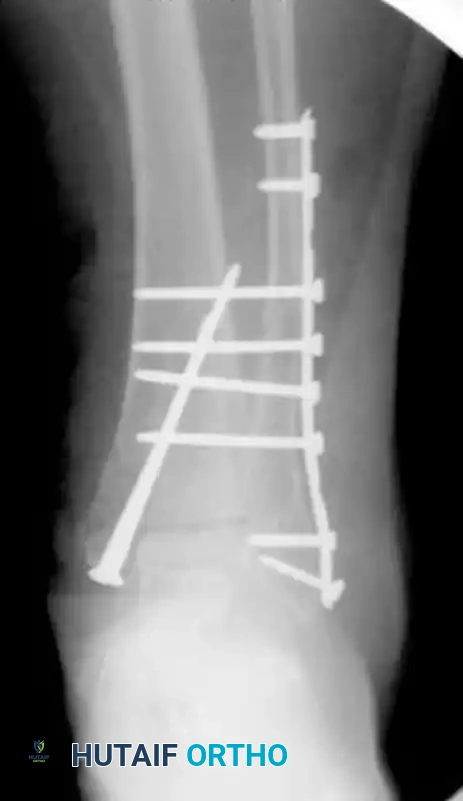
Construct Augmentation (The Transfibular/Transtibial Technique)
Consider the scenario of an elderly patient with osteoporosis, diabetes mellitus, and peripheral neuropathy who sustains an open comminuted bimalleolar ankle fracture-dislocation.
Following meticulous surgical débridement of the open wound, the fracture must be definitively treated with internal fixation. Because of the patient’s profoundly poor bone quality and lack of protective sensation, standard fixation is inadequate. To achieve the necessary stability, the construct is augmented. Multiple transfibular/transtibial screws are utilized to significantly increase fixation purchase, locking the fibula to the tibia and effectively neutralizing the syndesmosis and the distal tibiofibular articulation. This technique deliberately bypasses the standard dynamic syndesmotic principles in favor of absolute rigidity. Typically, three or four fully threaded 3.5 mm or 4.5 mm cortical screws are placed across three or four cortices, spanning from the fibular plate into the tibia. These screws are not intended for removal; they are left in place to provide permanent, rigid stabilization.
Alternative and Salvage Stabilization Techniques
In scenarios where the soft tissue envelope is catastrophically compromised, or where existing hardware/bone loss precludes standard ORIF, alternative stabilization methods must be employed. Intramedullary devices, such as fibular nails, offer a minimally invasive, load-sharing alternative that spares the lateral soft tissues while providing excellent bending stiffness. For severe salvage scenarios or acute Charcot reconstructions, Tibiotalocalcaneal (TTC) nails provide massive rigidity, though they sacrifice the subtalar and ankle joints. When external fixation is required due to massive blistering or active infection, a circular frame (Ilizarov or Taylor Spatial Frame) is preferred over uniplanar fixators. It is imperative to include the forefoot in the frame construct; neuropathic patients will rapidly develop an Achilles contracture, and pinning the forefoot prevents equinus posturing, which is vital should the frame become necessary for definitive, long-term management.
Complications, Incidence Rates, and Salvage Management
The management of complications in the diabetic ankle fracture requires aggressive, early intervention. The complication profile is vast, and the surgeon must maintain a high index of suspicion during every postoperative visit. The most common and devastating complications include surgical site infection (SSI), hardware failure, malunion/nonunion, and the onset of Charcot neuroarthropathy.
Surgical site infections and marginal wound necrosis occur at significantly higher rates in diabetic patients, driven by microvascular ischemia and impaired immune response. Superficial necrosis may present as eschar formation along the incision line. If the eschar is dry and stable, it can often be observed or managed with topical enzymatic debridement and oral antibiotics. However, deep infections involving the hardware are limb-threatening emergencies. Management requires aggressive, immediate surgical débridement in the operating room. If the hardware remains rigidly fixed to the bone and the fracture is not yet healed, the hardware may be temporarily retained to provide stability, supplemented with targeted, suppressive intravenous antibiotics and negative pressure wound therapy. If the hardware is loose, it must be completely explanted, the bone aggressively debrided of all necrotic tissue, and the ankle stabilized with a spanning external fixator until the infection is eradicated.
Loss of fixation and subsequent malunion or nonunion are direct consequences of neuropathic overloading and osteopenic bone. Neuropathic patients frequently ambulate on a fractured ankle despite strict non-weight-bearing instructions, leading to catastrophic screw pull-out, plate bending, or fracture displacement. Salvage of a failed construct is technically demanding. It typically requires revision ORIF utilizing extended TTC nailing, massive structural bone grafting, and heavy locking plates. In many cases, an attempt to reconstruct the joint is abandoned in favor of a definitive tibiotalocalcaneal arthrodesis to provide a stable, plantigrade, and braceable foot.
The most devastating non-infectious complication is the postoperative development of Charcot neuroarthropathy. This presents clinically as a profoundly swollen, warm, and erythematous foot and ankle, often mimicking a deep infection. Radiographically, it is characterized by rapidly progressive joint subluxation, periarticular fragmentation, and massive bone resorption. Immediate, strict offloading is required to halt the inflammatory cascade. Definitive management often necessitates complex midfoot or hindfoot arthrodesis, utilizing massive internal fixation or circular external fixation, once the acute inflammatory phase (Eichenholtz Stage I) has subsided and the disease enters the coalescent phase (Eichenholtz Stage II).
| Complication | Estimated Incidence in Diabetic Cohort | Pathophysiology / Risk Factors | Salvage Management Strategy |
|---|---|---|---|
| Superficial Wound Necrosis | 15% - 25% | Microvascular ischemia, excessive soft tissue stripping, tension on skin edges. | Local wound care, enzymatic debridement, oral antibiotics, delayed primary closure. |
| Deep Surgical Site Infection | 5% - 15% | Poor glycemic control (HbA1c > 8.0%), prolonged operative time, open fracture. | Aggressive I&D, hardware removal if loose, external fixation, IV antibiotics. |
| Hardware Failure / Nonunion | 10% - 20% | LOPS, premature weight-bearing, failure to utilize augmented fixation, osteopenia. | Revision ORIF with augmented constructs, TTC nailing, structural bone grafting, arthrodesis. |
| Charcot Neuroarthropathy | 5% - 10% | Pre-existing neuropathy, subtle malreduction, inadequate immobilization duration. | Immediate rigid offloading (TCC), eventual complex arthrodesis once acute phase resolves. |
| Major Amputation | 2% - 5% | Uncontrollable deep infection, massive Charcot collapse with unhealable ulceration. | Below-knee amputation (BKA) optimized for prosthetic fitting. |
Phased Post-Operative Rehabilitation Protocols
The postoperative rehabilitation of the diabetic ankle fracture deviates significantly from standard orthopedic protocols. The cornerstone of successful management is prolonged, rigid immobilization and a strictly delayed return to weight-bearing. The standard six-week radiographic healing timeline observed in healthy adults is entirely inapplicable to the neuropathic diabetic patient. The surgeon must adhere to a phased, highly conservative protocol to prevent late hardware failure and Charcot initiation.
Phase 1: Immediate Postoperative (Weeks 0-6)
Immediately following surgery, the patient is placed in a well-padded, rigid posterior splint or a bivalved cast. Strict Non-Weight-Bearing (NWB) status is absolutely mandatory. The patient must be educated extensively on the dangers of unperceived weight-bearing. Weekly or bi-weekly wound checks are required to monitor for early signs of marginal necrosis or infection. Sutures or staples are left in place significantly longer than in non-diabetic patients—typically 3 to 4 weeks—to prevent premature wound dehiscence, as diabetic collagen cross-linking and tensile strength recovery are markedly delayed.
Phase 2: Intermediate Healing (Weeks 6-12)
Once the incisions are fully healed and the sutures are removed, the patient is transitioned to a total contact cast (TCC) or a locked Charcot Restraint Orthotic Walker (CROW) boot. The TCC is the gold standard for offloading and protecting the insensate limb, as it intimately molds to the contours of the leg, distributing pressure evenly and preventing the shear forces that lead to ulceration. Strict NWB status is generally maintained for a minimum of 8 to 12 weeks postoperatively. Radiographic evidence of definitive bridging callus across at least three cortices must be clearly visualized before any weight-bearing is permitted. Clinical absence of pain is irrelevant in this population due to neuropathy; radiographic union is the sole determinant for advancing the protocol.
Phase 3: Gradual Return to Function (Months 3-6)
Progressive weight-bearing is initiated exclusively within a CROW boot or a custom-molded, rigid ankle-foot orthosis (AFO). The patient begins with partial weight-bearing with assistive devices (crutches or a walker) and slowly advances to full weight-bearing over a period of 4 to 6 weeks. During this phase, surveillance is critical. The patient must be monitored closely for signs of unilateral erythema, swelling, or warmth—the hallmark signs of an acute Charcot event. Temperature monitoring of the affected limb compared to the contralateral limb can be a useful adjunct; a temperature differential of greater than 2 degrees Celsius is highly suspicious for acute inflammation. If these signs occur, the patient must immediately revert to strict NWB status and total contact casting until the inflammation subsides.
Summary of Landmark Literature and Clinical Guidelines
The evolution of treatment protocols for diabetic ankle fractures is deeply rooted in several landmark clinical studies that have shifted the paradigm from nonoperative avoidance to aggressive, augmented surgical management. Understanding this literature is crucial for evidence-based practice and appropriate risk stratification.
Early historical literature painted a grim picture of operative intervention, citing catastrophic complication rates. However, recent literature provides a much more stratified and nuanced view of operative risks. A pivotal, paradigm-shifting study by Guo et al. compared patients with preoperatively neglected type 2 diabetes against a nondiabetic matched cohort. By employing meticulous soft tissue handling and rigid fixation principles, they found no significant increase in postoperative infection rates following the immediate operative stabilization of closed ankle fractures. This study underscored the principle that the soft tissue envelope, rather than the systemic disease alone, dictates the immediate infection risk.
Similarly, Jones et al. demonstrated that operatively treated ankle fractures in diabetic patients without severe end-organ comorbidities (such as end-stage renal disease or profound peripheral vascular disease) had complication rates comparable to nondiabetic patients. However, they critically noted that the presence of advanced diabetic comorbidities—and in particular, a history of Charcot arthropathy or profound LOPS—exponentially increased the likelihood of postoperative construct failure. This highlighted the need to treat "diabetes" not as a single binary variable, but as a spectrum of systemic compromise.
In a large, definitive series analyzing high-risk cohorts, Costigan et al. reported on 84 patients who underwent ORIF for acute closed ankle fractures. They identified open fractures, insulin dependence, and severe peripheral neuropathy as the primary, independent risk factors for construct failure and deep infection. Their work solidified the modern recommendation that patients exhibiting these specific risk factors require augmented fixation constructs (the "Rule of Double") rather than standard AO techniques. Furthermore, consensus guidelines published by Wukich et al. regarding the "Diabetic Ankle" emphasize that a swollen, warm ankle postoperatively in a diabetic patient must be considered Charcot neuroarthropathy or deep infection until definitively proven otherwise. The assumption of "standard postoperative edema" in this population is a frequent and devastating clinical error.
In conclusion, the management of ankle fractures in patients with diabetes requires a high index of suspicion for complications, a profound understanding of altered bone biomechanics, and a willingness to deviate significantly from standard fixation principles. By employing augmented fixation strategies—such as multiple transfibular screws and extended locking plates—respecting the fragile soft tissue envelope, and enforcing strictly prolonged postoperative immobilization, the orthopedic surgeon can successfully navigate these highly complex injuries, achieving stable osseous union and preserving the patient's limb and functional independence.
