Post-Traumatic Elbow Contracture: Diagnosis & Management of Heterotopic Ossification

Key Takeaway
Severe post-traumatic elbow stiffness often results from heterotopic ossification (HO), where bone forms in soft tissues after injury or surgery. Diagnosis involves detailed patient history, clinical examination, and advanced imaging like CT scans with 3D reconstructions to precisely map HO and plan effective surgical management, restoring essential range of motion.
You are presented with this 48-year-old patient eight months post-ORIF of a distal humerus fracture. He has a significantly restricted elbow arc and is complaining of ulnar nerve symptoms. Look at the provided radiograph. What are the critical findings, and how does this influence your management strategy?
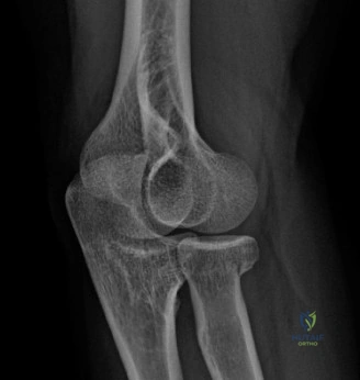
Candidate: The radiograph shows post-traumatic heterotopic ossification (HO) in the posterior-medial compartment, bridging the olecranon fossa. There is hardware present from the previous distal humerus fixation. The patient has a clear mechanical block to both flexion and extension. I would perform a CT scan to confirm maturity and plan the resection, and I am concerned about the ulnar nerve given his preoperative paresthesia.
Candidates often focus solely on the HO excision. They fail to mention the need to assess the hardware (is it prominent? does it need removal?), they skip the formal classification (e.g., Hastings and Graham), and they often forget to mention the mandatory prophylaxis against recurrence (NSAIDs vs. Radiation).
A high-scoring answer starts with the "3-S" approach: Status of the joint (mechanical block vs. articular cartilage health), Status of the hardware (is it causing impingement or does it need removal to access the HO?), and Status of the neurovascular bundle (specifically ulnar nerve vulnerability). The candidate must state: "I would classify this using the Hastings and Graham system, confirm maturity via CT scan, and counsel the patient on a structured post-operative protocol including pharmacological or radiation prophylaxis, as this is critical to avoid recurrence."
During your preoperative planning, you look at the 3D CT reconstruction. How does this specifically change your surgical approach compared to standard radiographs?
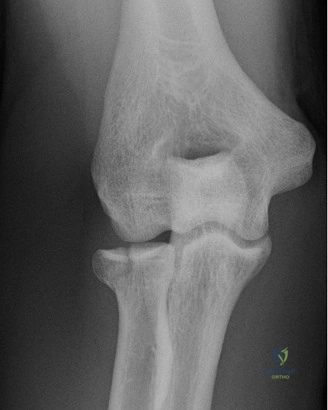
Candidate: The 3D CT allows me to map the exact location of the ossification. It helps me distinguish between extra-articular HO and intra-articular bony ankylosis, which changes the surgical effort. It also helps in planning the windows—medial or lateral—needed to reach specific deposits without violating stable ligamentous structures.
Failing to mention the "danger zones." Candidates who don't explicitly discuss how the 3D scan protects the medial collateral ligament (MCL) or the ulnar nerve by showing the exact extent of the bone are missing the clinical utility of the reconstruction.
The perfect answer highlights: 1. Spatial Mapping: Defining the relationship between the HO and the ulnar nerve path. 2. Hardware Interaction: Visualizing if hardware is embedded in the HO. 3. Ligament Preservation: Identifying the "safe" margins for excision to avoid iatrogenic valgus instability (MCL damage) or posterolateral rotatory instability (LUCL damage).
This image depicts the intraoperative setup. Why is the posterior approach with potential extension preferred, and what are the critical steps for the ulnar nerve in this specific patient?
Candidate: The posterior approach is preferred because it offers a universal "workhorse" view. I would perform a full neurolysis of the ulnar nerve from the arcade of Struthers to the FCB aponeurosis. Given his pre-existing symptoms, I would likely perform an anterior submuscular or subcutaneous transposition to prevent postoperative tension during flexion.
Neglecting the "skin" aspect. Candidates often forget to mention the need for full-thickness fasciocutaneous flaps to avoid wound complications—a high-risk area in previous surgical sites.
The examiner wants to hear: "The posterior approach allows for full-thickness flap elevation, providing access to both medial and lateral columns. Regarding the nerve, I would perform routine anterior transposition because the restoration of flexion will significantly increase the stretch on the nerve bed. I would also perform a formal neurolysis, ensuring the nerve is not tethered by the resected HO edges."
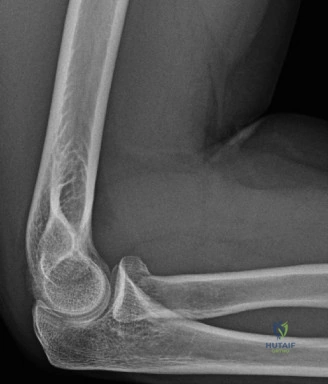
Look at this final radiograph post-excision. What are the most important postoperative considerations to prevent failure (recurrence of stiffness)?
Candidate: Early range of motion is key. I would use a static-progressive or dynamic splinting program, initiate physical therapy within 48 hours, and ensure the patient completes the prescribed course of Indomethacin or radiation therapy for HO prophylaxis.
Ignoring the "pain-motion" cycle. Many candidates fail to mention how they will manage post-op pain (e.g., nerve blocks) to allow the patient to actually perform the requested range of motion exercises.
A perfect answer structure: 1. Prophylaxis: Confirming compliance with NSAIDs or XRT. 2. Pain Control: Indwelling regional blocks to facilitate painless early ROM. 3. Splinting: A dynamic splinting regime that targets the patient's specific end-range deficits. 4. Expectation Management: Reminding the examiner that total range of motion will not match the contralateral side, and success is defined by achieving a functional arc.
