Operative Management of Elbow Pyarthrosis and Arthroscopic Lateral Compartment Pathology
Key Takeaway
Elbow pyarthrosis requires emergent surgical intervention to prevent irreversible articular cartilage destruction. While early infections may be managed via arthroscopic irrigation and débridement, extensive periarticular swelling necessitates an open approach. This guide details the indications, biomechanics, patient positioning, and step-by-step surgical techniques for elbow arthroscopy, including portal placement, joint decortication, and postoperative rehabilitation protocols essential for restoring joint function and eradicating infection.
Comprehensive Introduction and Patho-Epidemiology
Elbow pyarthrosis, commonly referred to as septic arthritis of the elbow, represents a profound orthopaedic emergency characterized by a destructive, purulent infection within the constrained articular space of the radiocapitellar and ulnohumeral joints. The rapid accumulation of purulent exudate initiates a catastrophic biochemical cascade. This exudate is densely populated with polymorphonuclear leukocytes (PMNs) that release host-derived matrix metalloproteinases (MMPs), elastases, and lysosomal enzymes. Concurrently, bacterial pathogens release exotoxins and stimulate the local production of pro-inflammatory cytokines, notably Interleukin-1 (IL-1) and Tumor Necrosis Factor-alpha (TNF-alpha). If left untreated, this hostile intra-articular environment leads to the irreversible degradation of glycosaminoglycans and the collagenous matrix of articular cartilage within 24 to 48 hours, ultimately resulting in severe secondary osteoarthritis, joint contracture, or systemic sepsis.
While the large weight-bearing joints such as the knee and hip are more frequently afflicted by septic arthritis, the elbow accounts for approximately 3% to 9% of all native joint pyarthrosis cases. The etiology is predominantly hematogenous seeding due to the highly vascularized nature of the synovial membrane, which lacks a limiting basement membrane, thereby allowing pathogens to easily translocate from the capillary bed into the joint space. However, direct inoculation from penetrating trauma, iatrogenic introduction via intra-articular corticosteroid injections, or contiguous spread from adjacent infectious foci—most notably olecranon bursitis—are also well-documented mechanisms. Patient populations at elevated risk include those with systemic immunosuppression, rheumatoid arthritis, diabetes mellitus, chronic kidney disease, and a history of intravenous drug use (IVDU).
The microbiological profile of elbow pyarthrosis is overwhelmingly dominated by Gram-positive organisms. Staphylococcus aureus remains the most frequently isolated pathogen, with Methicillin-resistant Staphylococcus aureus (MRSA) representing an increasing proportion of cases, particularly in urban centers and among the IVDU demographic. Streptococcus species constitute the second most common group. In neonates, elderly patients, and immunocompromised cohorts, Gram-negative bacilli (e.g., Escherichia coli, Pseudomonas aeruginosa) must be strongly considered. Furthermore, in sexually active young adults presenting with acute monoarticular or oligoarticular arthritis, Neisseria gonorrhoeae remains a critical differential diagnosis, often presenting alongside tenosynovitis and characteristic skin lesions.
The primary goals of surgical intervention in the setting of elbow pyarthrosis are multifaceted: the immediate and complete eradication of the offending organism, the mechanical decompression of the joint space to restore normal intra-articular pressure, the exhaustive removal of destructive inflammatory mediators and necrotic debris, and the preservation of the articular cartilage and complex joint biomechanics. Concurrently, the surgeon may encounter contiguous or pre-existing lateral compartment pathology, such as recalcitrant lateral epicondylitis (angiofibroblastic tendinosis of the extensor carpi radialis brevis). The arthroscopic management of such concomitant pathology during the same surgical setting requires a nuanced understanding of the lateral compartment's anatomy to prevent catastrophic iatrogenic instability.
Detailed Surgical Anatomy and Biomechanics
A profound, three-dimensional understanding of elbow anatomy is absolutely non-negotiable for the operating surgeon, particularly when navigating the lateral compartment, establishing arthroscopic portals, and performing aggressive synovectomies in the setting of distorted, edematous tissues. The elbow is a highly congruent, constrained hinge joint (ginglymus) with an additional pivot joint (trochoid) allowing for forearm rotation. The articular congruence provides primary stability, while the capsuloligamentous structures and dynamic muscle forces provide secondary and tertiary stability.
The lateral collateral ligament (LCL) complex is the primary static stabilizer against varus and posterolateral rotatory stress. It is a Y-shaped structure consisting of three distinct components: the radial collateral ligament (RCL), the lateral ulnar collateral ligament (LUCL), and the annular ligament. The RCL originates from the lateral epicondyle and inserts into the annular ligament, providing stability to the radial head. The annular ligament encircles the radial head, maintaining its articulation with the lesser sigmoid notch of the ulna. The LUCL is the most critical structure for posterolateral stability; it originates from the lateral epicondyle, blends seamlessly with the RCL and the capsule, and inserts distally on the supinator crest of the proximal ulna. It acts as an essential dynamic sling for the radial head. Iatrogenic transection of the LUCL during lateral compartment débridement, synovectomy, or extensor carpi radialis brevis (ECRB) release will inevitably result in posterolateral rotatory instability (PLRI), a devastating complication characterized by subluxation of the radial head posterior to the capitellum during combined axial load, valgus, and supination forces.
The extensor carpi radialis brevis (ECRB) plays a central role in lateral compartment pathology. It originates from the lateral epicondyle, positioned anterior and medial to the extensor digitorum communis (EDC) and immediately superficial to the LCL complex. The macroscopic appearance of the ECRB origin is clinically relevant during arthroscopy; its tendinous fibers are oriented longitudinally and appear distinct from the deeper, more obliquely oriented capsuloligamentous fibers. In cases where chronic inflammation, angiofibroblastic tendinosis, or contiguous infection involves the lateral compartment, arthroscopic débridement of the ECRB origin may be performed concomitantly with joint lavage. The surgeon must meticulously separate the pathological ECRB tissue from the underlying, healthy LUCL to avoid destabilizing the joint.
Navigating the neurovascular safe zones is the most critical aspect of elbow arthroscopy, particularly when the joint capsule is distended with purulence or contracted by chronic inflammation. The Ulnar Nerve is located in the cubital tunnel posterior to the medial epicondyle; it is at highest risk during the establishment of the proximal medial and anteromedial portals, necessitating a protective anterior trajectory of the trocar. The Radial Nerve crosses the radiocapitellar joint anteriorly, descending between the brachialis and brachioradialis muscles; it is highly vulnerable during anterior capsulectomy, synovectomy, and anterolateral portal placement. The Median Nerve and Brachial Artery are located medially in the anterior compartment, protected by the robust muscle belly of the brachialis; however, in a severely contracted or distorted elbow, the brachialis may be thinned, placing these critical structures at risk during anterior compartment instrumentation.
Exhaustive Indications and Contraindications
The surgical approach to elbow pyarthrosis is dictated by a multitude of factors, including the chronicity of the infection, the specific Gächter staging of the septic arthritis, the degree of periarticular soft tissue compromise, the presence of concomitant pathology, and the surgeon’s arthroscopic proficiency. Arthroscopic irrigation and débridement have emerged as the gold standard for early-stage pyarthrosis (Gächter stages I and II), offering the distinct advantages of minimal soft tissue morbidity, superior visualization of the anterior and posterior compartments, and the ability to perform a thorough, magnified synovectomy while preserving the stabilizing capsuloligamentous structures.
However, arthroscopy is not universally applicable and must be judiciously selected. When extensive periarticular swelling, severe cellulitis, and distention of anatomical landmarks have occurred, or when the infection has progressed to Gächter stages III or IV (characterized by extensive pannus formation, cartilage destruction, and subchondral bone involvement), an open arthrotomy is strongly preferable. Severe periarticular edema distorts the normal anatomical safe zones, significantly increasing the distance from the skin to the joint capsule and exponentially elevating the risk of iatrogenic injury to the radial, median, and ulnar nerves during blind or semi-blind portal placement. In such advanced, neglected cases, an open arthrotomy—typically via a lateral or posterior approach—ensures safe access, adequate decompression, and thorough mechanical débridement without jeopardizing neurovascular integrity.
The management of concomitant lateral compartment pathology, such as recalcitrant lateral epicondylitis requiring ECRB release, is indicated when the patient has a documented history of chronic lateral elbow pain that has failed exhaustive conservative management (e.g., physical therapy, bracing, biologic injections) for a minimum of six months, and this pathology is encountered during the arthroscopic evaluation for the acute pyarthrosis. The surgeon must weigh the benefits of a single-stage procedure against the risks of prolonging surgical time and potentially disseminating the infection into freshly decorticated bone or newly opened fascial planes.
Indications and Contraindications for Arthroscopic Management
| Parameter | Arthroscopic Management (Irrigation & Débridement) | Open Arthrotomy |
|---|---|---|
| Primary Indications | Acute pyarthrosis (<48-72 hours); Gächter Stage I-II; Concomitant ECRB tendinosis; Intact anatomical landmarks. | Delayed presentation; Gächter Stage III-IV; Severe periarticular edema; Osteomyelitis; Abscess extending into muscle compartments. |
| Relative Contraindications | Moderate soft tissue edema; Previous ulnar nerve transposition (alters medial safe zones); Limited surgeon experience. | Early, uncomplicated pyarthrosis (due to higher morbidity); Severe medical comorbidities precluding prolonged anesthesia. |
| Absolute Contraindications | Complete obliteration of anatomical landmarks; Overlying active cellulitis at portal sites; Extensive capsular necrosis. | Medically unstable patient (hemodynamic collapse requiring bedside aspiration/drainage instead). |
| Lateral Compartment (ECRB) | Concomitant chronic lateral epicondylitis failing >6 months of conservative therapy; clearly identifiable ECRB footprint. | Acute localized infection without chronic tendinosis; Inability to visualize the LUCL due to severe inflammation. |
Pre-Operative Planning, Templating, and Patient Positioning
Meticulous preoperative planning begins with a definitive diagnosis. A high index of suspicion must be maintained in any patient presenting with acute elbow pain, effusion, erythema, and severe restriction of both active and passive range of motion. Joint aspiration (arthrocentesis) prior to the administration of empirical antibiotics is the absolute gold standard for definitive diagnosis. The aspirate must be immediately sent for cell count with differential, Gram stain, aerobic and anaerobic cultures, and crystal analysis. A synovial fluid white blood cell (WBC) count exceeding 50,000 cells/mm³ with greater than 90% polymorphonuclear leukocytes is highly indicative of pyarthrosis, though lower counts do not definitively exclude infection, particularly in immunocompromised hosts or those with concurrent inflammatory arthropathies.
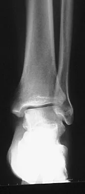
Imaging modalities play a crucial, albeit secondary, role in the acute setting. Standard anteroposterior and lateral radiographs of the elbow are mandatory to rule out concomitant fractures, foreign bodies, or signs of chronic osteomyelitis (e.g., periosteal reaction, osteolysis) and to establish a baseline for joint space narrowing. While Magnetic Resonance Imaging (MRI) is highly sensitive for detecting joint effusions, osteomyelitis, and soft tissue abscesses, its use should never delay emergent surgical decompression in a clinically obvious case of pyarthrosis. Preoperative templating in the setting of pyarthrosis focuses primarily on mapping the portal trajectories and anticipating the need for potential open conversion based on the degree of soft tissue swelling observed clinically.
Proper patient positioning is critical for successful elbow arthroscopy, ensuring adequate access to all compartments while rigorously protecting the patient from pressure-related neuropathies and positioning-related complications. The procedure is typically performed under general anesthesia. While regional blocks (e.g., supraclavicular or axillary) may be utilized for postoperative pain control, they are generally avoided or used with extreme caution in the setting of acute pyarthrosis, as they can mask the early clinical signs of postoperative neurological deficits or the development of compartment syndrome.
The patient is placed in either the lateral decubitus or supine suspended position, with the supine suspended position being favored by many arthroscopists for its ease of setup and excellent airway access. Ensure meticulous padding of the patient’s thorax, contralateral extremities, and all bony prominences to prevent decubitus ulcers and neuropraxia. The affected extremity is positioned with the ipsilateral shoulder abducted to 90 degrees, and the arm is supported with a precut foam holder or a specialized mechanical arm positioner. This allows the elbow to rest comfortably at 90 degrees of flexion, relaxing the anterior neurovascular structures and maximizing the intra-articular capacity. A non-sterile tourniquet is placed high on the brachium. Crucially, the surgeon must never use an Esmarch bandage for exsanguination in the setting of a florid pyarthrosis, as the compressive forces can drive purulent material proximally into the fascial planes of the arm or systemically. Instead, the arm is simply elevated for 3 to 5 minutes prior to tourniquet inflation (typically to 250 mm Hg). In cases of severe pyarthrosis with proximal cellulitis, tourniquet use may be entirely omitted to prevent proximal systemic seeding.
Step-by-Step Surgical Approach and Fixation Technique
Joint Distention and Initial Access
The fundamental key to safe portal placement in elbow arthroscopy is maximizing the intra-articular volume, which physically displaces the anterior neurovascular bundles (radial and median nerves, brachial artery) away from the anterior capsule. After meticulously marking the anatomical landmarks—the medial and lateral epicondyles, the radial head, the olecranon tip, and the course of the ulnar nerve—the joint is distended. Introduce an 18-gauge needle through the direct lateral portal, also known as the "soft spot." This portal is located in the center of the anconeus triangle, formed by the lateral epicondyle, the radial head, and the tip of the olecranon. Inject 20 to 30 mL of sterile normal saline. A spontaneous backflow of purulent fluid upon removal of the syringe confirms accurate intra-articular placement. It is imperative that fresh, intraoperative fluid and tissue cultures are obtained at this exact moment, prior to initiating continuous lavage, and that broad-spectrum intravenous antibiotics are instituted immediately thereafter by the anesthesia team.
Establishing the Proximal Medial Portal
The proximal medial (or superomedial) portal serves as the primary workhorse viewing portal for the anterior compartment of the elbow. It is located approximately 2 cm proximal to the medial epicondyle and 1 cm anterior to the palpable medial intermuscular septum. Make a superficial skin incision with a #11 blade, taking care to incise only the dermis. Use a blunt hemostat to spread the subcutaneous tissues down to the level of the joint capsule, ensuring no superficial cutaneous nerves (e.g., medial antebrachial cutaneous nerve) are injured. Introduce the blunt trocar and cannula anterior to the intermuscular septum. The surgeon must maintain constant tactile contact with the anterior aspect of the humerus as the trocar is directed distally and laterally toward the radial head. This specific trajectory ensures that the median nerve and brachial artery are pushed anteriorly and safely avoided. Once the capsule is breached, a 2.7-mm or 4.0-mm, 30-degree arthroscope is inserted to commence the diagnostic evaluation.
Synovectomy and Lavage
Once visualization is established from the medial side, an anterolateral portal is created under direct intra-articular vision using an outside-in spinal needle localization technique. The anterolateral portal is typically located 3 cm distal and 1 cm anterior to the lateral epicondyle. A motorized shaver and a radiofrequency ablation wand are introduced through this working portal. The surgeon must perform a systematic, aggressive, and exhaustive synovectomy. All fibrinous exudate, hypertrophic pannus, and necrotic cartilaginous debris must be evacuated. Copious irrigation—typically utilizing 6 to 9 liters of normal saline—is employed to mechanically wash out the bacterial load, destructive MMPs, and lysosomal enzymes. The posterior compartment is subsequently addressed using standard direct posterior and posterolateral portals, ensuring that the olecranon fossa, posterolateral gutter, and radiocapitellar joint are completely cleared of purulence.
Arthroscopic Management of Concomitant Lateral Pathology
In clinical scenarios where the surgeon encounters concomitant lateral compartment pathology, such as recalcitrant lateral epicondylitis (angiofibroblastic tendinosis of the ECRB), targeted débridement is indicated. After the pathological tissue is identified, establish the superolateral portal with an 18-gauge needle guided directly into the lesion. This portal is typically located 1 to 2 cm proximal and 1 cm anterior to the lateral epicondyle. Using a full-radius resector, excise the overlying capsule to clearly identify the undersurface of the ECRB tendon. The ECRB is visually distinct; its tendinous fibers orient longitudinally, contrasting sharply with the obliquely oriented capsular tissue. Using a combination of an arthroscopic curet and a motorized shaver, débride the pathological tendinous attachment of the ECRB and decorticate the lateral epicondyle. The goal is to comprehensively resect the degenerative, friable, gray tissue until healthy, bleeding margins are obtained. Decortication of the lateral epicondylar ridge can be performed with an arthroscopic burr or radiofrequency wand to stimulate a robust, vascularized healing response.
Protecting the LUCL: The Critical Step
During the release of the ECRB tendon and decortication of the lateral epicondyle, the surgeon must view the overlying muscle belly of the extensor musculature. The most critical step of this portion of the procedure is the rigorous protection of the lateral ulnar collateral ligament (LUCL). The surgeon must strictly limit the amount of posterior resection. The LUCL lies immediately posterior to the ECRB origin, precisely at the equator of the capitellum. Resection must never extend posterior to the midline of the radiocapitellar joint. A common and catastrophic pitfall is the overzealous use of the motorized burr on the lateral epicondyle, which can inadvertently catch, wrap, and avulse the LUCL fibers. To prevent this, the surgeon must always keep the burr blades facing anteriorly and superiorly, directing the suction and cutting action away from the ligamentous footprint. Violation of the LUCL will result in iatrogenic posterolateral rotatory instability, necessitating a complex, open ligamentous reconstruction using an autograft or allograft.
Drain Placement and Closure
Following the exhaustive irrigation and débridement of both the anterior and posterior compartments, surgical drains are routinely placed. A medium Hemovac or Jackson-Pratt drain is placed in the anterior compartment (exiting via the anterolateral portal) and the posterior compartment (exiting via the posterolateral portal) to prevent the postoperative re-accumulation of purulent fluid, hematoma, and inflammatory mediators. The remaining portals are either loosely approximated with simple non-absorbable sutures (e.g., 3-0 Nylon) or left entirely open to heal by secondary intention, depending heavily on the severity of the initial infection, the virulence of the organism, and the degree of soft tissue edema. A sterile, non-adherent dressing is applied, followed by a bulky compressive dressing and a posterior plaster splint to immobilize the joint in a resting position.
Complications, Incidence Rates, and Salvage Management
The operative management of elbow pyarthrosis, particularly when combined with lateral compartment débridement, carries a distinct and formidable complication profile. The complications can be broadly categorized into those related to the infection itself (persistence, recurrence, systemic sepsis), those related to the arthroscopic access (neurovascular injury, compartment syndrome), and those related to the structural intervention (iatrogenic instability, arthrofibrosis).
Neurological injury remains the most feared complication of elbow arthroscopy. The ulnar nerve is most frequently injured during the establishment of medial portals, particularly if the trocar is directed too posteriorly or if the patient has a subluxating ulnar nerve. The radial nerve is at risk during anterolateral portal placement and aggressive anterior capsulectomy. Most nerve injuries are transient neuropraxias resulting from fluid extravasation and compression, resolving within weeks to months. However, direct transection requires immediate microsurgical repair.
Iatrogenic Posterolateral Rotatory Instability (PLRI) is a devastating structural complication resulting from the inadvertent resection or thermal ablation of the LUCL during ECRB release or lateral synovectomy. Patients will present postoperatively with lateral elbow pain, a sensation of the elbow "giving way" when pushing out of a chair, and a positive pivot-shift test. Salvage management requires an open LUCL reconstruction, typically utilizing a palmaris longus or gracilis autograft, once the intra-articular infection has been definitively eradicated.
Arthrofibrosis is the most common functional complication following elbow pyarthrosis. The intense inflammatory response, combined with postoperative immobilization, leads to rapid capsular contracture and loss of the functional arc of motion. Prevention through early, aggressive rehabilitation is paramount. If profound stiffness persists beyond 6 months post-infection eradication, an open or arthroscopic capsular release (arthrolysis) may be indicated.
Complications, Incidence, and Salvage Strategies
| Complication | Estimated Incidence | Mechanism / Etiology | Salvage Management / Treatment |
|---|---|---|---|
| Recurrent/Persistent Infection | 10% - 25% | Inadequate initial débridement; Highly virulent organism (e.g., MRSA); Biofilm formation. | Serial arthroscopic or open washouts every 48-72 hours; Prolonged IV antibiotics; Infectious Disease consult. |
| Transient Neuropraxia | 2% - 10% | Fluid extravasation causing compression; Tourniquet palsy; Retractor stretch. | Observation; Gabapentinoids; Typically resolves spontaneously within 3-6 months. EMG at 6 weeks if no improvement. |
| Nerve Transection (Radial/Ulnar) | < 1% | Direct laceration by trocar, scalpel, or motorized shaver during portal placement or capsulectomy. | Immediate or early microsurgical epineural repair; Possible nerve grafting or tendon transfers for late presentations. |
| Iatrogenic PLRI | 1% - 3% | Inadvertent resection of the LUCL during ECRB release or aggressive lateral synovectomy. | Open LUCL reconstruction (autograft/allograft) delayed until complete eradication of the joint infection is confirmed. |
| Arthrofibrosis / Contracture | 30% - 50% | Capsular fibrosis secondary to intense inflammation and prolonged postoperative immobilization. | Aggressive physical therapy; Static progressive splinting; Late arthroscopic or open capsular release (arthrolysis). |
| Compartment Syndrome | < 1% | Massive fluid extravasation into the forearm or arm fascial compartments during prolonged arthroscopy. | Emergent open fasciotomies of the affected compartments; Delayed primary closure or skin grafting. |
Phased Post-Operative Rehabilitation Protocols
The postoperative rehabilitation protocol following the operative management of elbow pyarthrosis is a delicate, constantly evolving balancing act. The surgeon and physical therapist must simultaneously manage the absolute necessity of soft tissue rest to facilitate infection eradication and wound healing, while aggressively addressing the joint's profound propensity for rapid arthrofibrosis and contracture.
Immediate Postoperative Phase (0-48 Hours)
In the immediate postoperative period, the primary focus is on infection control, pain management, and edema reduction. The arm is placed in a bulky compressive dressing and a posterior plaster splint or sling, with the elbow immobilized at 90 degrees of flexion and the forearm in neutral rotation. This position minimizes tension on the anterior capsule and provides optimal patient comfort. Intravenous antibiotics, meticulously tailored to the intraoperative culture sensitivities, are continued without interruption. An infectious disease consultation is highly recommended to manage the specific antibiotic selection, dosing, monitoring of renal/hepatic function, and the eventual transition to a prolonged course of oral or outpatient intravenous antibiosis (e.g., via a PICC line). The intra-articular drains are typically removed at 48 hours, provided the output is minimal and non-purulent. If the clinical signs of infection are receding—evidenced by a down-trending C-Reactive Protein (CRP) and Erythrocyte Sedimentation Rate (ESR), resolving local erythema, and decreased resting pain—the splint is discontinued, and the patient transitions to the next phase.
Intermediate Phase and Secondary Interventions (3 Days to 3 Weeks)
The clinical trajectory must be monitored with extreme vigilance during the first week. If the patient exhibits persistent fevers, escalating pain, or if the inflammatory markers fail to decrease, the infection is deemed uncontrolled, and the patient must be returned to the operating room for a repeat irrigation and débridement. Serial washouts (every 48 to 72 hours) may be required in cases involving highly virulent pathogens or delayed initial presentation. Once the infection is clinically eradicated and the portal sites are stabilizing, gentle active and active-assisted range-of-motion (ROM) exercises are initiated. The immediate goal is to restore the functional arc of motion, defined as 30 to 130 degrees of flexion and 50 degrees of both pronation and supination. Passive stretching is generally avoided in this early phase to prevent exacerbating capsular inflammation and heterotopic ossification.
Late Rehabilitation and Strengthening Phase (4 Weeks and Beyond)
As the joint capsule heals and the infection is definitively cleared, the focus shifts to terminal range of motion and muscular strengthening. If a concomitant ECRB release and lateral epicondyle decortication were performed, the patient must avoid resisted wrist extension and forceful gripping for the first 4 to 6 weeks to allow the extensor origin sufficient time to scar and heal to the decorticated bone. Around the 6-week mark, the patient progresses to progressive resistance exercises, beginning with isometric wrist extension and advancing to eccentric strengthening exercises, mimicking standard lateral epicondylitis rehabilitation protocols. Static progressive splinting (e.g., turnbuckle splints) may be introduced if the patient plateaus in their ROM progression and demonstrates a persistent flexion or extension contracture.
Summary of Landmark Literature and Clinical Guidelines
The evolution of operative management for elbow pyarthrosis has been heavily influenced by a transition from open, highly morbid arthrotomies to minimally invasive arthroscopic techniques. Landmark literature in the late 1990s and early 2000s, spearheaded by pioneers in elbow arthroscopy, demonstrated that arthroscopic irrigation and débridement yielded equivalent, if not superior, rates of infection eradication compared to open procedures, with significantly reduced postoperative pain, faster recovery of range of motion, and shorter hospital stays.
The staging system proposed by Gächter for septic arthritis of the knee has been widely adapted and validated for the elbow. The literature consistently supports that Gächter Stages I (opacity of fluid, redness of synovial membrane) and II (severe inflammation, fibrinous deposits, no cartilage damage) are highly amenable to arthroscopic management. Conversely, studies evaluating outcomes in Stages III (thickening of synovial membrane, compartment formation, early cartilage damage) and IV (aggressive pannus, extensive cartilage destruction) advocate for open arthrotomy due to the high failure rate of arthroscopy in adequately clearing loculated abscesses and necrotic bone.
Regarding lateral compartment pathology, the anatomical studies by O'Driscoll et al. remain the definitive cornerstone for understanding the LUCL and the pathophysiology of posterolateral rotatory instability. Their work underscores the absolute necessity of preserving the LUCL during any lateral compartment intervention. Furthermore, clinical trials evaluating the arthroscopic release of the ECRB for recalcitrant lateral epicondylitis, such as those by Baker and colleagues, have demonstrated excellent long-term outcomes, provided the LUCL is protected and the degenerative tissue is adequately resected. Current clinical guidelines from major orthopaedic and infectious disease societies recommend a multidisciplinary approach to pyarthrosis, mandating immediate surgical decompression followed by a targeted, culture-directed antibiotic regimen lasting a minimum of 4 to 6 weeks, with the precise duration dictated by the pathogen's virulence and the host's immune status.
