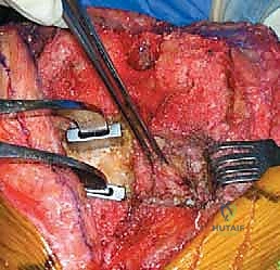
Orthopedic Sports Medic Review | Dr Hutaif Sports Medic -...
Key Takeaway
This topic focuses on Orthopedic MCQS online sports Medicine, Orthopedic care involves diagnosing and treating diverse conditions, including chronic knee instability with osteoarthritis, often requiring high tibial osteotomy with decreased tibial slope. It also addresses acute injuries in a 14-year-old, such as concussions, emphasizing a graduated return to activity. Post-surgical complications like those following total shoulder arthroplasty necessitate specific management plans for b a yearold patients.
Orthopedic Sports Medic Review | Dr Hutaif Sports Medic -...
Comprehensive 100-Question Exam
00:00
Start Quiz
Question 1
A 25-year-old professional soccer player undergoes an anterior cruciate ligament (ACL) reconstruction. Preoperative MRI revealed a suspicious fluid signal posterior to the medial meniscus. Intraoperatively, the surgeon suspects a 'ramp lesion'. Which of the following arthroscopic approaches provides the most optimal visualization for accurate diagnosis and repair of this specific lesion?
Explanation
Question 2
A 22-year-old rugby player presents with recurrent anterior shoulder instability. A 3D CT scan reveals 25% anterior glenoid bone loss. The surgeon plans to perform a Latarjet procedure. Which of the following biomechanical mechanisms is considered the primary stabilizer provided by this procedure when the shoulder is in the abducted and externally rotated position?
Explanation
Question 3
During a double-bundle posterior cruciate ligament (PCL) reconstruction, understanding the reciprocal tension pattern of the native PCL bundles is critical. Which of the following accurately describes the biomechanical behavior of the native PCL bundles during knee motion?
Explanation
Question 4
A 19-year-old female presents with a history of recurrent lateral patellar dislocations. Non-operative management has failed. Advanced imaging demonstrates an intact patellofemoral cartilage profile but reveals an elevated tibial tubercle-trochlear groove (TT-TG) distance. Above what specific TT-TG threshold is a medializing tibial tubercle osteotomy generally indicated?
Explanation
Question 5
A 28-year-old male volleyball player presents with insidious onset of vague posterior shoulder pain and isolated weakness in external rotation. An MRI reveals a paralabral cyst extending posteriorly. Compression of the involved neural structure at which of the following anatomic locations is the most likely cause of his specific physical exam findings?
Explanation
Question 6
A 30-year-old weightlifter feels a sudden 'pop' and tearing sensation in his anterior axillary fold while performing a heavy bench press. He is diagnosed with a pectoralis major tendon rupture. Which of the following accurately describes the anatomic relationship of the pectoralis major tendon at its insertion site?
Explanation
Question 7
A 45-year-old male undergoes a single-incision anterior approach for a distal biceps tendon rupture repair. Postoperatively, he complains of numbness and paresthesia along the radial aspect of his forearm. Which of the following nerves is most commonly at risk and likely injured due to superficial retraction during this specific surgical approach?
Explanation
Question 8
A 55-year-old female reports a sudden, sharp pain in the back of her knee while descending stairs. MRI of the knee demonstrates a classic 'ghost sign'. This specific radiographic sign is most indicative of which of the following pathologies?
Explanation
Question 9
A 23-year-old collegiate hockey player complains of insidious anterior groin pain that worsens with deep flexion. Physical examination reveals limited internal rotation in flexion, and a positive anterior impingement test. Radiographs show an alpha angle of 68 degrees. In Cam-type femoroacetabular impingement (FAI), the offending osseous deformity is most frequently located at which aspect of the proximal femur?
Explanation
Question 10
A 30-year-old male presents to the clinic after a hyperextension knee injury. During physical examination, the Dial test reveals 15 degrees of increased external rotation at 30 degrees of knee flexion compared to the contralateral side. However, at 90 degrees of knee flexion, the external rotation is symmetric bilaterally. This clinical finding isolated to 30 degrees of flexion primarily implicates injury to which of the following structures?
Explanation
Question 11
A 42-year-old construction worker sustains a severe direct blow to the superior aspect of his shoulder, resulting in an unstable, completely displaced Type V acromioclavicular (AC) joint separation. The surgeon plans an anatomic reconstruction of the coracoclavicular (CC) ligaments. What is the correct anatomical orientation of the native CC ligaments?
Explanation
Question 12
A 35-year-old competitive water skier sustains an acute, complete, three-tendon proximal hamstring avulsion with 4 cm of retraction. Surgical repair is indicated. Which of the following peripheral nerves is anatomically situated closest to the ischial tuberosity footprint and is at the highest risk of iatrogenic injury during deep surgical dissection?
Explanation
Question 13
A 40-year-old male weekend warrior sustains an acute midsubstance Achilles tendon rupture. He elects to undergo a percutaneous repair to minimize wound complications. The surgeon must be meticulously aware of the sural nerve trajectory during lateral suture placement. What is the typical anatomic course of the sural nerve relative to the Achilles tendon?
Explanation
Question 14
A 25-year-old professional baseball pitcher presents with posterior shoulder pain. Workup reveals 'internal impingement'. This pathologic process involves the articular surface of the rotator cuff becoming pinched between the greater tuberosity and the posterosuperior glenoid. During which specific phase of the throwing motion does this impingement primarily occur?
Explanation
Question 15
A 60-year-old male manual laborer presents with chronic shoulder weakness and pseudoparalysis of forward elevation. MRI demonstrates a massive, retracted, and irreparable posterosuperior rotator cuff tear (supraspinatus and infraspinatus) with advanced fatty infiltration (Goutallier stage 4), but the subscapularis is fully intact. There is no evidence of glenohumeral osteoarthritis. Which of the following is the most appropriate tendon transfer to restore active external rotation and elevation?
Explanation
Question 16
A surgeon is performing a medial patellofemoral ligament (MPFL) reconstruction on an 18-year-old female. Precise anatomic location of the femoral attachment is critical to avoid graft anisometry. Radiographically identified by Schöttle's point, where is the true anatomic femoral footprint of the MPFL located?
Explanation
Question 17
A 13-year-old male athlete presents with vague anterior knee pain and catching. Radiographs and subsequent MRI confirm the presence of Osteochondritis Dissecans (OCD) with intact overlying articular cartilage. Statistically, what is the most common anatomic location for an OCD lesion in the knee?
Explanation
Question 18
A 21-year-old collegiate baseball pitcher presents with medial elbow pain and diminished throwing velocity. He demonstrates a positive moving valgus stress test, and MRI confirms a full-thickness tear of the ulnar collateral ligament (UCL). Which distinct anatomical structure forms the primary restraint to valgus stress at the elbow?
Explanation
Question 19
A 20-year-old elite track athlete sustains an acute fracture at the metaphyseal-diaphyseal junction of the 5th metatarsal (Jones fracture) during a sprint. Given her status as a high-level competitive athlete, what is the gold standard treatment to minimize the risk of nonunion and expedite return to play?
Explanation
Question 20
A 9-year-old boy (Tanner Stage 1) sustains a complete anterior cruciate ligament (ACL) rupture while skiing. He has wide-open physes and significant remaining growth potential. The parents opt for surgical intervention due to severe recurrent instability. Which surgical technique is most strongly recommended to minimize the risk of iatrogenic leg length discrepancy or angular deformity?
Explanation
Question 21
A 25-year-old football player sustains a knee hyperextension injury. On physical examination, the dial test reveals a 15-degree increase in external rotation at 30 degrees of knee flexion compared to the contralateral side. However, at 90 degrees of knee flexion, external rotation is symmetric bilaterally. What is the most likely diagnosis?
Explanation
Question 22
A 22-year-old rugby player presents with recurrent anterior shoulder dislocations. Preoperative CT imaging reveals 25% anterior glenoid bone loss and an engaging Hill-Sachs lesion. Which of the following is the most appropriate surgical treatment?
Explanation
Question 23
An 11-year-old boy (Tanner stage 1) sustains a midsubstance ACL tear. Radiographs confirm widely open physes. The family elects for surgical management to prevent further meniscal damage. Which of the following surgical options is most appropriate?
Explanation
Question 24
A 26-year-old hockey player presents with chronic anterior groin pain exacerbated by hip flexion and internal rotation. Radiographs reveal an alpha angle of 70 degrees, a normal center-edge angle (CEA), and a negative cross-over sign. What is the primary pathomechanism of this patient's condition?
Explanation
Question 25
What is the primary underlying biomechanical cause of posteromedial impingement (valgus extension overload syndrome) in the elbow of a throwing athlete?
Explanation
Question 26
A 19-year-old female presents with recurrent lateral patellar dislocations. Evaluation reveals a tibial tubercle-trochlear groove (TT-TG) distance of 22 mm and a Caton-Deschamps index of 1.0. The trochlea shows mild dysplasia. What is the most appropriate surgical management?
Explanation
Question 27
A 14-year-old gymnast complains of lateral elbow pain and catching. MRI of the elbow shows an osteochondritis dissecans (OCD) lesion of the capitellum with intact articular cartilage, but there is a rim of T2-hyperintense fluid behind the lesion. What is the best initial surgical management?
Explanation
Question 28
A 45-year-old female felt a 'pop' in the back of her knee while squatting. MRI demonstrates a complete posterior root tear of the medial meniscus with 4 mm of meniscal extrusion, but no significant osteoarthritis. Mechanical alignment is neutral. What is the recommended treatment?
Explanation
Question 29
Regarding the coracoclavicular (CC) ligaments in the setting of an acromioclavicular (AC) joint reconstruction, which of the following statements is true regarding their anatomic orientation and biomechanical function?
Explanation
Question 30
As the Achilles tendon descends toward its insertion on the calcaneus, how do the constituent fibers of the gastrocnemius and soleus typically rotate?
Explanation
Question 31
A 40-year-old water skier suffers a forced hyperflexion injury of the hip with an extended knee, resulting in a proximal hamstring avulsion. Which of the following muscles form the conjoined tendon of the proximal hamstring complex at the ischial tuberosity?
Explanation
Question 32
During the surgical repair of a complete pectoralis major rupture in a weightlifter, the surgeon must anatomically restore the footprint. What is the anatomic relationship of the sternal head footprint relative to the clavicular head footprint on the humerus?
Explanation
Question 33
A 28-year-old volleyball player complains of vague posterior shoulder pain and numbness over the lateral deltoid. MRI confirms Quadrilateral Space Syndrome causing compression of the axillary nerve and posterior humeral circumflex artery. Which of the following structures forms the inferior border of this anatomic space?
Explanation
Question 34
A 'ramp lesion' of the knee is frequently encountered during anterior cruciate ligament (ACL) reconstruction. This pathology specifically refers to a tear located in which of the following anatomic zones?
Explanation
Question 35
A 22-year-old collegiate baseball pitcher presents with posterior shoulder pain during the late cocking phase of throwing. Physical examination reveals a 20-degree glenohumeral internal rotation deficit (GIRD). Which of the following MRI findings is most characteristic of 'internal impingement' in this patient?
Explanation
Question 36
An athlete sustains a syndesmotic ('high') ankle sprain. According to classic biomechanical studies, which ligament is typically the first to fail during the external rotation mechanism of injury?
Explanation
Question 37
A 45-year-old construction worker presents with a symptomatic Type II SLAP tear. According to recent literature, compared to arthroscopic SLAP repair, primary biceps tenodesis in patients in this age demographic is associated with:
Explanation
Question 38
During a single-bundle posterior cruciate ligament (PCL) reconstruction, the femoral tunnel is positioned to anatomically reconstruct the dominant bundle. Which specific bundle is reconstructed, and at what degree of knee flexion should the graft typically be tensioned?
Explanation
Question 39
In the evaluation of anterior shoulder instability, the 'glenoid track' concept is used to determine the risk of engagement. A Hill-Sachs lesion is considered 'off-track' (and thus at high risk of engagement) if:
Explanation
Question 40
The anterior bundle of the medial ulnar collateral ligament (UCL) of the elbow is the primary restraint to valgus stress. Which specific sub-portion of the anterior bundle is most taut in full elbow extension?
Explanation
Question 41
A 45-year-old active male sustains a medial meniscus posterior root tear. Biomechanically, what is the consequence of leaving this tear unrepaired compared to a completely meniscectomized knee?
Explanation
Question 42
During an ACL reconstruction, the surgeon utilizes a bone-patellar tendon-bone (BPTB) autograft. Compared to hamstring autografts, BPTB grafts are historically associated with a higher incidence of which postoperative complication?
Explanation
Question 43
A 19-year-old female gymnast presents with atraumatic multidirectional shoulder instability. Following 6 months of physical therapy, she continues to subluxate inferiorly. If surgical intervention is performed, which capsular structure must be primarily addressed to correct the inferior instability?
Explanation
Question 44
A 62-year-old male with a massive, irreparable posterosuperior rotator cuff tear and no glenohumeral arthritis undergoes superior capsular reconstruction (SCR). What is the primary biomechanical goal of this procedure?
Explanation
Question 45
An MPFL reconstruction is planned for a 17-year-old female with recurrent lateral patellar dislocations. To prevent postoperative patellofemoral over-constraint, where must the femoral tunnel be positioned relative to radiographic landmarks (Schöttle's point)?
Explanation
Question 46
During an anatomic posterolateral corner (PLC) reconstruction, accurate placement of the fibular collateral ligament (FCL) graft is critical. Where is the native FCL femoral attachment located relative to the popliteus sulcus?
Explanation
Question 47
A 26-year-old baseball pitcher presents with posterior shoulder pain and a "dead arm" sensation during the late cocking phase of throwing. He exhibits a 25-degree glenohumeral internal rotation deficit (GIRD). What is the primary pathomechanical cause of his suspected labral pathology?
Explanation
Question 48
During hip arthroscopy for mixed femoroacetabular impingement (FAI), excessive bony resection of a cam lesion extending too far posterosuperiorly on the femoral head-neck junction risks injury to the terminal branches of which artery?
Explanation
Question 49
A 30-year-old runner with a focal 3 cm² full-thickness chondral defect on the medial femoral condyle undergoes Matrix-induced autologous chondrocyte implantation (MACI). What is the primary histological goal of the repair tissue generated by MACI compared to microfracture?
Explanation
Question 50
A 42-year-old male opts for non-operative management with early functional rehabilitation for an acute Achilles tendon rupture. Which of the following is the most significant advantage of this approach compared to traditional open surgical repair?
Explanation
Question 51
A 25-year-old sustains a Type III acromioclavicular (AC) joint separation. Biomechanically, which ligamentous complex serves as the primary restraint to superior translation of the distal clavicle?
Explanation
Question 52
A lateral extra-articular tenodesis (LET) is performed to augment an ACL reconstruction in an athlete with a high-grade pivot shift. Biomechanically, LET or anterolateral ligament (ALL) reconstruction primarily limits which coupled motion?
Explanation
Question 53
A 35-year-old weightlifter undergoes an acute distal biceps tendon repair using a single-incision anterior approach. Which nerve is at the highest risk of iatrogenic injury due to lateral retraction during this exposure?
Explanation
Question 54
During an ulnar collateral ligament (UCL) reconstruction utilizing a docking technique, what is the primary biomechanical rationale for precise graft placement on the medial ulna?
Explanation
Question 55
A 24-year-old volleyball player is scheduled for surgical debridement of recalcitrant proximal patellar tendinopathy (Jumper's knee). Where is the classic pathological lesion located in this condition?
Explanation
Question 56
A 45-year-old female presents with acute posterior knee pain after deep flexion. MRI shows a medial meniscus posterior root tear with 3 mm of extrusion. What is the primary biomechanical consequence of leaving this unrepaired?
Explanation
Question 57
A 20-year-old rugby player has recurrent anterior shoulder instability. CT shows 15% glenoid bone loss and a Hill-Sachs lesion. Based on the 'glenoid track' concept, an 'off-track' Hill-Sachs lesion is defined by which of the following?
Explanation
Question 58
During medial patellofemoral ligament (MPFL) reconstruction, accurate femoral tunnel placement is crucial. Using fluoroscopy, where is the anatomic femoral origin of the MPFL located relative to Schöttle's point?
Explanation
Question 59
A 28-year-old male sustains a KD-III-M knee dislocation. He presents with a foot drop and inability to evert the foot. Electromyography (EMG) at 6 weeks shows no motor unit potentials in the tibialis anterior. What is the most appropriate management regarding the nerve injury?
Explanation
Question 60
A 30-year-old male hockey player undergoes hip arthroscopy for symptomatic femoroacetabular impingement (FAI). An isolated cam lesion is resected. Which anatomic landmark marks the most common location of a cam deformity on the femoral head-neck junction?
Explanation
Question 61
A 19-year-old gymnast requires ACL reconstruction. The surgeon considers a bone-patellar tendon-bone (BPTB) autograft. Which of the following is the most widely recognized long-term complication associated with this specific graft choice compared to hamstring autograft?
Explanation
Question 62
A 65-year-old male presents with pseudoparalysis of the shoulder and an irreparable massive posterosuperior rotator cuff tear. Radiographs show Hamada Grade 4 changes. Which is the most appropriate surgical treatment?
Explanation
Question 63
A 29-year-old bodybuilder feels a pop in his anterior axilla while bench pressing. Examination reveals a loss of the anterior axillary fold. MRI confirms a rupture of the pectoralis major at the sternocostal head insertion. Where does the sternocostal head insert relative to the clavicular head on the humerus?
Explanation
Question 64
A 14-year-old male presents with knee pain. MRI reveals a 2x2 cm osteochondritis dissecans (OCD) lesion on the lateral aspect of the medial femoral condyle. Which MRI finding is most indicative of lesion instability requiring surgical fixation rather than non-operative management?
Explanation
Question 65
A 24-year-old professional baseball pitcher presents with vague anterior shoulder pain and a 'dead arm' sensation. Examination reveals Glenohumeral Internal Rotation Deficit (GIRD) of 25 degrees. During the late cocking phase of throwing, which mechanism is primarily responsible for a Type II SLAP tear?
Explanation
Question 66
A 22-year-old football player sustains a syndesmotic ankle sprain. Intraoperative evaluation using the Cotton test indicates instability. The surgeon opts for suture button fixation over syndesmotic screws. What is the primary biomechanical advantage of dynamic suture button fixation?
Explanation
Question 67
A 21-year-old collegiate pitcher undergoes ulnar collateral ligament (UCL) reconstruction using a palmaris longus autograft. Which bundle of the native UCL is the primary restraint to valgus stress between 30 and 120 degrees of elbow flexion, and thus the target for reconstruction?
Explanation
Question 68
A 26-year-old active female has a symptomatic full-thickness chondral defect on her medial femoral condyle measuring 4.5 cm^2. She has failed conservative therapy. Which of the following surgical interventions is most appropriate for a defect of this size?
Explanation
Question 69
A 45-year-old female presents with acute posterior knee pain after a deep squat. MRI reveals a complete radial tear at the posterior horn of the medial meniscus, 2 mm from its root attachment. What is the expected biomechanical consequence if this lesion is left untreated?
Explanation
Question 70
During reconstruction of the medial patellofemoral ligament (MPFL), identifying the correct femoral footprint is critical for graft isometry. According to Schottle's radiographic landmarks, where is the optimal femoral attachment located on a strictly lateral radiograph?
Explanation
Question 71
In evaluating a patient with suspected glenohumeral instability, which ligamentous structure serves as the primary restraint to inferior translation of the humerus when the shoulder is abducted to 90 degrees?
Explanation
Question 72
A 22-year-old collegiate baseball pitcher complains of medial elbow pain during the late cocking phase of throwing. What is the primary restraint to valgus stress at the elbow during this specific phase of the throwing motion?
Explanation
Question 73
A 28-year-old male undergoes hip arthroscopy for symptomatic femoroacetabular impingement (FAI). He has a large cam lesion requiring osteochondroplasty. To minimize the risk of a postoperative iatrogenic femoral neck fracture, the maximum recommended depth of the resection should not exceed what percentage of the femoral neck diameter?
Explanation
Question 74
A 65-year-old patient presents with an irreparable massive rotator cuff tear, pseudoparalysis of the shoulder (active elevation less than 90 degrees), and an intact deltoid. Radiographs show Hamada grade 3 changes. What is the most appropriate definitive surgical management?
Explanation
Question 75
A 25-year-old football player sustains a contact injury to his knee. Clinical examination reveals increased external rotation of the tibia at 30 degrees of knee flexion compared to the contralateral side, but equal external rotation at 90 degrees. Which structure is most likely injured?
Explanation
Question 76
A 12-year-old gymnast is diagnosed with an osteochondritis dissecans (OCD) lesion on the lateral aspect of the medial femoral condyle. Her physes are wide open. MRI shows a 1.5 cm lesion with no high T2 signal behind the fragment. What is the most appropriate initial management?
Explanation
None
