العربية
Part of the Master Guide

Updated Orthopedic Review | Dr Hutaif General Orthopedi -...

Orthopedic Sport Review | Dr Hutaif Sports Medicine Rev -...

23 Apr 2026 60 min read 109 Views
Orthopedic Sports MCQs: Expert Discussion for Every Question

Key Takeaway

In this comprehensive guide, we discuss everything you need to know about ORTHOPEDIC MCQS ONLINE 013 SPORT. This orthopedic interactive module offers a sports medicine self-assessment with multiple-choice questions covering various clinical scenarios, such as shoulder dislocation and knee injuries. Each question includes a detailed explanation and a comprehensive discussion for questions, helping users understand appropriate diagnoses and treatment strategies in sports medicine and orthopedics.

Orthopedic Sport Review | Dr Hutaif Sports Medicine Rev -...

Comprehensive 100-Question Exam


00:00

Start Quiz

Question 1

Anatomic ACL reconstruction relies on identifying the femoral footprint. The lateral intercondylar ridge (Resident's ridge) serves as a key surgical landmark.

Where is the native ACL femoral footprint located relative to this ridge when the knee is viewed at 90 degrees of flexion?


Explanation

The native ACL footprint is posterior to the lateral intercondylar ridge (Resident's ridge) and anterior to the lateral bifurcate ridge (which separates the AM and PL bundles) when the knee is viewed in 90 degrees of flexion. Placing the tunnel anterior to Resident's ridge results in a non-anatomic, vertical graft that fails to control rotational stability.

Question 2

A 24-year-old athlete undergoes an isolated Posterior Cruciate Ligament (PCL) reconstruction using a double-bundle technique.

To accurately recreate the biomechanics of the native PCL, at what degree of knee flexion should the anterolateral (AL) and posteromedial (PM) bundles be tensioned and fixed, respectively?


Explanation

The anterolateral (AL) bundle is the larger bundle of the PCL and is tightest in flexion; it is traditionally tensioned and fixed at 90 degrees of flexion. The posteromedial (PM) bundle is tightest in extension and is tensioned and fixed at 0 to 30 degrees of flexion (near extension).

Question 3

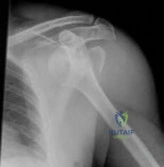
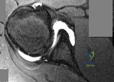
During a Latarjet procedure for recurrent anterior shoulder instability with significant glenoid bone loss, the coracoid process is transferred to the anterior glenoid.

Which nerve is at greatest risk of iatrogenic injury during the medial retraction of the conjoined tendon and subsequent screw fixation?


Explanation

The musculocutaneous nerve typically enters the coracobrachialis muscle 3-8 cm distal to the tip of the coracoid. Retraction of the conjoined tendon medially during the Latarjet procedure places significant traction on this nerve, making it the most commonly injured neurologic structure during this operation.

Question 4

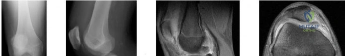
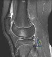
A 45-year-old male sustains a posterior root tear of the medial meniscus during a deep squat.

Biomechanically, what is the consequence of this specific injury on the tibiofemoral joint contact pressures if left untreated?


Explanation

A medial meniscus posterior root tear disrupts the hoop stresses of the meniscus. Biomechanical studies have demonstrated that this extrusion results in medial compartment contact pressures and contact areas that are functionally equivalent to those seen after a total medial meniscectomy, leading to rapid chondrolysis.

Question 5

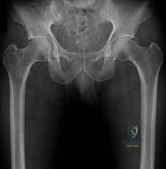
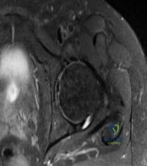
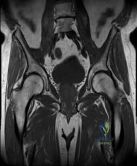
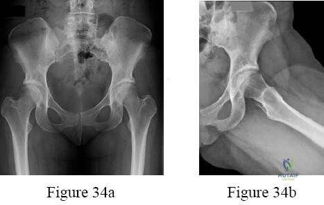
A 22-year-old hockey player presents with anterior groin pain exacerbated by hip flexion and internal rotation. Imaging reveals Cam-type femoroacetabular impingement (FAI).

During hip arthroscopy, which of the following intra-articular pathologic findings is most commonly associated with isolated Cam impingement?


Explanation

Cam impingement is caused by an aspherical femoral head-neck junction creating sheer forces on the anterosuperior acetabular rim during flexion and internal rotation. This reliably leads to chondral delamination (the 'wave sign') and tearing of the labrum at the chondrolabral junction in the anterosuperior quadrant.

Question 6

During a Medial Patellofemoral Ligament (MPFL) reconstruction for recurrent patellar instability, the surgeon inadvertently places the femoral tunnel proximal to the anatomic insertion site.

What is the expected biomechanical consequence during knee range of motion?


Explanation

If the femoral tunnel in an MPFL reconstruction is placed too proximal, the distance between the patellar and femoral attachments increases as the knee flexes. This causes the graft to become excessively tight in flexion (leading to loss of flexion and medial patellofemoral overload) and relatively loose in extension.

Question 7

A 30-year-old runner has an isolated, full-thickness 3.5 cm^2 chondral defect on the medial femoral condyle. The surgeon considers Matrix-induced Autologous Chondrocyte Implantation (MACI).

Which of the following represents an absolute or strong relative contraindication to MACI for this patient?


Explanation

MACI is indicated for symptomatic, unipolar, full-thickness cartilage defects in the knee in young, active patients. Bipolar ('kissing') lesions, uncorrected malalignment, uncorrected ligamentous instability, and advanced osteoarthritis are significant contraindications due to unacceptably high failure rates.

Question 8

A 20-year-old collegiate baseball pitcher undergoes an ulnar collateral ligament (UCL) reconstruction utilizing the Docking technique.

Which bundle of the native UCL is the primary restraint to valgus stress at 90 degrees of elbow flexion and is the primary structure surgically reconstructed?


Explanation

The anterior bundle of the UCL is the primary restraint to valgus stress. It is divided into anterior and posterior bands. The anterior band is the primary restraint to valgus stress at 30, 60, and 90 degrees of elbow flexion, whereas the posterior band is most tense at 120 degrees of flexion. The anterior band is the principal structure reconstructed in 'Tommy John' surgery.

Question 9

A 35-year-old overhead athlete presents with posterior shoulder pain and profound weakness in external rotation. An MRI reveals a paralabral cyst in the spinoglenoid notch.

Which physical exam finding and associated intra-articular pathology is most likely present?


Explanation

A cyst at the spinoglenoid notch typically compresses the suprascapular nerve after it has innervated the supraspinatus, leading to isolated infraspinatus denervation and isolated external rotation weakness. Spinoglenoid cysts are highly associated with posterior labral tears, through which synovial fluid escapes via a one-way valve mechanism.

Question 10

A 28-year-old football player sustains a multiligamentous knee injury. Physical exam reveals a positive posterior drawer test and increased external tibial rotation at 30 degrees of knee flexion compared to the contralateral side. However, at 90 degrees of knee flexion, the external tibial rotation is symmetric bilaterally. What is the most likely injury pattern?


Explanation

The Dial test measures external tibial rotation at 30 and 90 degrees of flexion. An increase of >10 degrees of external rotation at 30 degrees only indicates an isolated PLC injury. An increase at both 30 and 90 degrees indicates a combined PLC and PCL injury.

Question 11

In the surgical management of a Type V acromioclavicular (AC) joint dislocation, reconstructing the coracoclavicular (CC) ligaments is prioritized.

What is the precise anatomic orientation and insertion of the native conoid and trapezoid ligaments on the clavicle?


Explanation

The coracoclavicular (CC) ligaments consist of the conoid and trapezoid. The conoid ligament inserts on the conoid tubercle of the clavicle, which is located posteromedial to the trapezoid insertion. The trapezoid ligament inserts more anterolaterally. The conoid is the primary restraint to superior translation.

Question 12

A 35-year-old male sustains an acute, complete rupture of the Achilles tendon.

He opts for non-operative management utilizing a functional rehabilitation protocol. Compared to surgical repair, which of the following is true regarding his clinical outcomes according to recent level-1 evidence?


Explanation

High-quality RCTs (e.g., Willits et al.) demonstrate that when a functional rehabilitation protocol (early weight-bearing and mobilization) is utilized, the re-rupture rate for non-operative management is statistically similar to surgical repair, while entirely avoiding surgical wound complications and infections.

Question 13

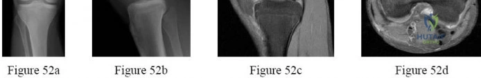
A 14-year-old female gymnast complains of lateral elbow pain, clicking, and a loss of 15 degrees of extension. Radiographs and MRI demonstrate a 12mm osteochondral defect of the capitellum with subchondral fluid and a loose cartilaginous flap.

What is the most appropriate surgical intervention?


Explanation

Osteochondritis dissecans (OCD) of the capitellum affects young throwing athletes and gymnasts. Indications for surgery include unstable lesions (fluid behind the fragment, cartilaginous flap, loose bodies) or failure of non-operative management. For fragmented, unsalvageable lesions, arthroscopic debridement and marrow stimulation (microfracture) is indicated. In situ drilling is reserved for intact lesions.

Question 14

A 22-year-old female with an isolated ACL deficiency and genu varum is scheduled for an ACL reconstruction and an opening-wedge high tibial osteotomy (HTO). What is the potential biomechanical consequence of a standard medial opening-wedge HTO on the sagittal plane of the knee, and how does it affect the ACL graft?


Explanation

A standard medial opening-wedge HTO tends to inadvertently increase the posterior tibial slope because the anterior aspect of the proximal tibia is narrower than the posterior aspect. An increased posterior slope exacerbates anterior tibial translation during weight-bearing, placing higher stress on an ACL graft. Surgeons must intentionally adjust the gap to avoid this in ACL-deficient knees.

Question 15

In overhead throwing athletes, a Type II SLAP tear is often attributed to the 'peel-back' mechanism.

During which phase of the throwing motion does the maximal peel-back force occur on the superior labrum?


Explanation

The 'peel-back' mechanism occurs in the late cocking phase of throwing when the shoulder is in maximum abduction and external rotation. In this position, the biceps vector shifts posteriorly, creating a torsional force at the biceps anchor that twists and peels back the superior labrum from the glenoid rim.

Question 16

During the surgical evaluation of an acute ankle injury, the 'Cotton test' is performed to assess the integrity of the syndesmosis. Which specific anatomic structure is considered the primary restraint to anterior subluxation of the distal fibula and is typically the first to tear in an external rotation syndesmotic injury?


Explanation

Syndesmotic ankle sprains usually occur via external rotation forces. The anterior inferior tibiofibular ligament (AITFL) is typically the first ligament to tear, followed by the interosseous membrane/ligament, and finally the PITFL. The AITFL is the primary restraint to anterior translation and external rotation of the fibula.

Question 17

A 9-year-old Tanner stage 1 male sustains a complete, mid-substance ACL tear.

A physeal-sparing extra-articular reconstruction is planned. Which structure is traditionally utilized and routed over the 'over-the-top' position to reconstruct the ACL without violating the open physes?


Explanation

In very young children with wide-open physes (Tanner stage 1), extra-articular physeal-sparing techniques are preferred to prevent growth arrest. The classic physeal-sparing technique (modified MacIntosh or Micheli procedure) utilizes a strip of the Iliotibial (IT) band routed through the 'over-the-top' position on the femur and under the intermeniscal ligament, entirely avoiding the physes.

Question 18

A 65-year-old manual laborer presents with a massive, irreparable posterosuperior rotator cuff tear (supraspinatus and infraspinatus) with an intact subscapularis and functional deltoid. He lacks active external rotation (positive hornblower's sign). Which tendon transfer is most biomechanically appropriate to restore external rotation in this patient?


Explanation

For irreparable posterosuperior cuff tears with profound external rotation weakness, the lower trapezius transfer has a line of pull that closely replicates the native infraspinatus and allows for in-phase firing, making it biomechanically superior for restoring active external rotation. The latissimus dorsi is an internal rotator and requires out-of-phase retraining.

Question 19

In the Schenck classification of knee dislocations (KD), a KD III-L injury indicates disruption of the ACL, PCL, and the Posterolateral Corner (PLC).

This specific injury pattern carries the highest risk of injury to which of the following neurovascular structures?


Explanation

While popliteal artery injuries are a classic complication of knee dislocations, injuries to the common peroneal nerve are most highly associated with damage to the posterolateral corner (PLC). A KD III-L has the highest incidence of common peroneal nerve palsy due to traction on the nerve as the joint severely opens laterally.

Question 20

A 28-year-old competitive weightlifter feels a 'pop' in his anterior axilla while performing a heavy bench press. MRI confirms a complete rupture of the pectoralis major tendon.

At the humeral insertion site, what is the spatial relationship of the sternocostal head relative to the clavicular head?


Explanation

The pectoralis major consists of the clavicular and sternocostal heads. As the muscle fibers course laterally toward the humerus, the sternocostal head twists 180 degrees on itself, resulting in its insertion being posterior (deep) and proximal to the clavicular head insertion. This twisting puts the inferior fibers under maximum tension during the eccentric phase of a bench press, making them the most vulnerable to rupture.

Question 21

A 23-year-old professional soccer player presents with anterior knee pain and swelling. MRI reveals a 4.5 cm² full-thickness osteochondral defect with a 6 mm deep subchondral bone cyst on the weight-bearing surface of the medial femoral condyle. He has previously undergone a failed microfracture procedure. What is the most appropriate definitive surgical treatment?


Explanation

Fresh osteochondral allograft (OCA) transplantation is the treatment of choice for large (>2-3 cm²) osteochondral defects, especially when associated with subchondral bone loss or cysts. OATS is typically reserved for smaller defects (<2 cm²) due to donor site morbidity. MACI is excellent for large (>2 cm²) purely chondral defects but does not address significant subchondral bone loss unless performed as a 'sandwich' technique with bone grafting, making OCA the more direct and preferred single-stage option for large bony defects.

Question 22

A 45-year-old construction worker complains of deep shoulder pain that worsens with overhead lifting. Physical examination reveals a positive O'Brien's test and pain with resisted forearm supination. MRI demonstrates a Type II Superior Labrum Anterior to Posterior (SLAP) tear. Given his age and occupation, what is the most appropriate surgical management if conservative therapy fails?


Explanation

In older patients (typically >35-40 years old) or manual laborers, biceps tenodesis provides superior, more reliable outcomes and faster return to work compared to SLAP repair. SLAP repairs in this demographic are associated with higher rates of postoperative stiffness, persistent pain, and revision surgery. Biceps tenotomy is an option for elderly or low-demand patients but can cause cramping and a Popeye deformity in heavy laborers.

Question 23

During the evaluation of a patient with a multiligamentous knee injury, the examiner performs the Dial test. The test demonstrates 15 degrees of increased external rotation of the tibia relative to the femur at 30 degrees of knee flexion compared to the contralateral side. At 90 degrees of knee flexion, the external rotation is symmetric bilaterally. This clinical finding most likely indicates an isolated injury to which of the following?


Explanation

The Dial test evaluates external rotation of the tibia. Asymmetry of >10-15 degrees compared to the normal knee indicates injury. An increase in external rotation at 30 degrees of flexion, but symmetric at 90 degrees, indicates an isolated injury to the posterolateral corner (PLC). If the increase is present at both 30 and 90 degrees, it indicates a combined injury to the PLC and the PCL.

Question 24

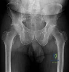
A 28-year-old professional hockey player presents with chronic groin pain exacerbated by hip flexion and internal rotation. Radiographs demonstrate a prominent bony bump at the anterosuperior femoral head-neck junction with an alpha angle of 65 degrees. He is diagnosed with Femoroacetabular Impingement (FAI). Which of the following conditions is a known risk factor for the development of this specific type of lesion?


Explanation

The patient has a Cam lesion, characterized by an abnormal femoral head-neck offset (alpha angle >50-55 degrees). A mild or subclinical Slipped Capital Femoral Epiphysis (SCFE) leads to an anterior and superior metaphyseal prominence, a well-known cause of secondary Cam-type FAI. Acetabular retroversion and coxa profunda are associated with Pincer-type FAI (acetabular overcoverage).

Question 25

A 19-year-old collegiate baseball pitcher complains of medial elbow pain during the late cocking and early acceleration phases of throwing. The 'moving valgus stress test' is strongly positive. If surgical reconstruction is indicated, which bundle of the ulnar collateral ligament (UCL) must be primarily reconstructed, and at what degree of flexion is it the primary restraint to valgus stress?


Explanation

The anterior bundle of the UCL is the primary restraint to valgus stress of the elbow between 30 and 120 degrees of flexion. It is the bundle that is reconstructed in 'Tommy John' surgery. The radiocapitellar joint is an important secondary restraint to valgus stress.

Question 26

A 16-year-old female presents with recurrent patellar dislocations. Imaging reveals a ruptured Medial Patellofemoral Ligament (MPFL). If the surgeon plans a reconstruction, the anatomic femoral attachment of the MPFL (Schöttle point) should be located in relation to which osseous landmarks on a true lateral radiograph?


Explanation

The Schöttle point represents the radiographic femoral origin of the MPFL. On a true lateral radiograph, it is situated 1 mm anterior to the posterior femoral cortical line, 2.5 mm distal to the posterior origin of the medial femoral condyle, and proximal to the level of the posterior aspect of Blumensaat's line. Clinically, it lies in the saddle between the adductor tubercle and the medial epicondyle.

Question 27

A 32-year-old weightlifter feels a sudden pop in his anterior chest wall while performing a heavy bench press. He presents with a loss of the anterior axillary fold and ecchymosis extending down his arm. An MRI confirms a complete rupture of the pectoralis major tendon. Which of the following best describes the pathoanatomy and usual location of this specific injury?


Explanation

Pectoralis major ruptures most commonly occur during eccentric loading, such as the downward phase of a bench press. The most common site of failure is an avulsion of the sternal head from its insertion on the lateral lip of the bicipital groove of the humerus. The sternal head inserts deep and proximal to the clavicular head.

Question 28

A 55-year-old patient undergoes an arthroscopic partial meniscectomy for an isolated posterior horn tear of the medial meniscus. Intraoperatively, the tear is identified as a complete radial tear located 2 mm from the posterior bony attachment. Which of the following correctly describes the biomechanical consequence of leaving this root tear un-repaired?


Explanation

A complete medial meniscus posterior root tear disrupts the circumferential hoop fibers, completely un-tethering the meniscus. Biomechanical studies have shown this renders the meniscus non-functional, equating the contact pressures in the medial compartment to those seen following a total medial meniscectomy. This often leads to rapid articular cartilage wear and spontaneous osteonecrosis or insufficiency fractures if not repaired.

Question 29

A 24-year-old professional football player suffers an acute ankle injury after his foot is planted and externally rotated while a defender falls on his leg. Physical examination reveals a positive squeeze test and tenderness extending 6 cm proximal to the ankle joint over the anterior tibiofibular ligament. What is the most reliable intraoperative dynamic test to confirm syndesmotic instability?


Explanation

The patient has a syndesmotic 'high ankle' sprain. The Cotton test is performed by grasping the heel and applying a lateral translational force while observing the ankle mortise under fluoroscopy. Widening of the medial clear space (>4-5 mm) or tibiofibular clear space confirms syndesmotic instability requiring operative stabilization. An external rotation stress test under fluoroscopy is also highly reliable.

Question 30

A 13-year-old gymnast complains of chronic, insidious onset lateral elbow pain, stiffness, and clicking. Examination reveals a 15-degree flexion contracture. Radiographs show a radiolucent lesion on the anterolateral aspect of the capitellum. MRI confirms Osteochondritis Dissecans (OCD) with an intact articular surface. What differentiates capitellar OCD from Panner's disease?


Explanation

Panner's disease is an osteochondrosis of the entire capitellar ossific nucleus, typically occurring in children aged 7-10 years, and is self-limiting. Capitellar OCD is a focal osteochondral defect, typically seen in older adolescent athletes (11-15 years) involved in repetitive valgus loading (gymnasts, throwers), and carries a higher risk of loose body formation and long-term sequelae.

Question 31

A 60-year-old female presents with lateral hip pain that radiates down her lateral thigh. She reports pain when rising from a seated position and lying on the affected side. On exam, she has a positive Trendelenburg sign and weakness in hip abduction. Trochanteric bursitis treatments have failed. MRI reveals a full-thickness tear of the gluteus medius tendon. At which anatomic footprint does this tendon most commonly tear?


Explanation

The gluteus medius inserts primarily on the lateral and superoposterior facets of the greater trochanter. Tears of the 'rotator cuff of the hip' most commonly involve the gluteus medius at the lateral facet of the greater trochanter. The gluteus minimus inserts on the anterior facet.

Question 32

A 22-year-old professional baseball pitcher presents with posterior shoulder pain. On exam, he has a 25-degree deficit in glenohumeral internal rotation (GIRD) compared to his non-throwing arm, but total arc of motion is equal bilaterally. When his shoulder is placed in 90 degrees of abduction and maximal external rotation, he complains of deep posterior pain. What is the classic pathoanatomic finding on arthroscopy for 'Internal Impingement' in this population?


Explanation

Internal impingement classically occurs in overhead athletes during the late cocking phase of throwing (abduction/external rotation). It involves the pathological contact (impingement) of the articular-sided fibers of the posterosuperior rotator cuff (supraspinatus and infraspinatus) against the posterosuperior glenoid labrum, often exacerbated by posterior capsular contracture (GIRD) and anterior capsular laxity.

Question 33

A 26-year-old male is involved in a high-speed motorcycle accident. In the ER, his knee is grossly unstable in multiple planes, and he has a documented Knee Dislocation (Schenck KD-III). His Ankle-Brachial Index (ABI) is measured at 0.85. What is the most appropriate next step in management?


Explanation

In the setting of a knee dislocation, an ABI < 0.9 is highly concerning for a popliteal artery injury. The definitive next step for a subnormal ABI or asymmetric pulses (without hard signs of ischemia requiring immediate OR exploration) is a CT angiogram (CTA) to rule out an intimal flap or vascular occlusion. Hard signs of ischemia (absent pulses, expanding hematoma, active pulsatile bleeding) warrant immediate surgical exploration.

Question 34

A 34-year-old recreational weightlifter presents with severe shoulder pain and inability to actively internally rotate the shoulder after a forceful extension injury. On exam, he has increased passive external rotation compared to the normal side and a positive 'lift-off' test. An MRI confirms an isolated subscapularis tendon rupture. Which accompanying pathology is most frequently associated with a complete rupture of the upper subscapularis?


Explanation

The upper fibers of the subscapularis provide the medial stabilizing sling for the long head of the biceps tendon at the bicipital groove. A tear of the upper subscapularis often disrupts this transverse humeral ligament/medial sling complex, leading to medial subluxation or frank dislocation of the long head of the biceps tendon into the joint.

Question 35

A 29-year-old professional volleyball player complains of isolated, painless weakness of the throwing arm. On physical examination, she demonstrates marked weakness in active external rotation with the arm at the side, but normal internal rotation, normal abduction, and no sensory deficits. An MRI is performed. What is the most likely pathological finding?


Explanation

Isolated weakness of the infraspinatus (external rotation) without supraspinatus involvement (abduction) suggests compression of the suprascapular nerve at the spinoglenoid notch. This is classically caused by a paralabral cyst associated with a posterior labral tear in overhead athletes. Compression at the suprascapular notch would affect both the supraspinatus and infraspinatus.

Question 36

A 19-year-old dancer presents with a painful snapping sensation in her anterior hip when extending her hip from a flexed, abducted, and externally rotated position. An ultrasound demonstrates the iliopsoas tendon snapping over a bony prominence. What is the most common anatomic structure over which the iliopsoas snaps in 'Internal Snapping Hip' (Coxa Saltans Interna)?


Explanation

Internal snapping hip (coxa saltans interna) is caused by the iliopsoas tendon catching or snapping over the iliopectineal eminence or the anterior capsule/femoral head. External snapping hip involves the iliotibial (IT) band snapping over the greater trochanter.

Question 37

A 35-year-old recreational basketball player sustains an acute Achilles tendon rupture. He is discussing operative vs. non-operative management. According to recent high-level evidence, if an early functional rehabilitation protocol is utilized, how do the outcomes of non-operative management compare to operative repair?


Explanation

Recent Level I studies (e.g., Willits et al.) have demonstrated that when acute Achilles tendon ruptures are managed with early functional weight-bearing and range-of-motion rehabilitation protocols, there is no clinically significant difference in re-rupture rates between operative and non-operative groups. However, operative management consistently carries a higher risk of surgical wound complications and infections.

Question 38

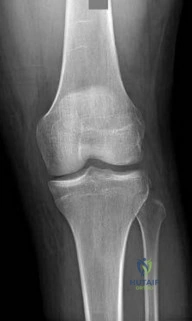
A 14-year-old male presents with non-specific knee pain. Imaging reveals Osteochondritis Dissecans (OCD) of the knee. What is the most common anatomical location for an OCD lesion in the knee?


Explanation

The classic and most common location (accounting for roughly 70-80% of cases) for an OCD lesion in the knee is the lateral aspect of the medial femoral condyle (LAME - Lateral Aspect Medial Epicondyle/Condyle). This is thought to be related to repeated impingement from the tibial spine or localized vascular insufficiency.

Question 39

A 25-year-old rugby player sustains an acute anterior shoulder dislocation. During the reduction in the emergency department, the physician notes weakness in shoulder abduction and decreased sensation over the lateral deltoid. Which of the following describes the most likely associated nerve injury and its typical prognosis?


Explanation

The axillary nerve is the most commonly injured nerve during an anterior glenohumeral dislocation. It presents with weakness in the deltoid and teres minor, and sensory loss over the 'regimental badge' area (lateral deltoid). Fortunately, the injury is almost always a neuropraxia (stretching) that resolves spontaneously over weeks to months, and observation is the standard of care.

Question 40

A 40-year-old male falls directly onto his shoulder point. Radiographs show an acromioclavicular (AC) joint separation with 150% superior displacement of the clavicle relative to the acromion. There is no disruption of the deltotrapezial fascia. Based on the Rockwood classification, what type of injury is this, and what is the generally recommended treatment for this specific grade?


Explanation

A Rockwood Type V AC joint injury involves disruption of the AC and coracoclavicular (CC) ligaments, with superior displacement of the clavicle >100% to 300% of the normal contralateral side. The deltotrapezial fascia is severely stripped. Unlike Type III injuries, which are controversially managed but often treated non-operatively, Type V injuries generally require operative reconstruction to restore shoulder biomechanics and relieve skin tension.

Question 41

A 25-year-old male sustains a dashboard injury during a motor vehicle collision. Physical examination reveals a grade III posterior sag sign. A Posterior Cruciate Ligament (PCL) reconstruction is planned. What distinct biomechanical advantage does the tibial inlay technique offer over the traditional transtibial technique for PCL reconstruction?


Explanation

The primary biomechanical advantage of the tibial inlay technique over the transtibial technique in PCL reconstruction is the elimination of the 'killer turn.' In a transtibial reconstruction, the graft must make a sharp acute angle as it exits the posterior tibial tunnel to reach the femoral footprint. This acute angle can lead to graft abrasion, attenuation, and eventual failure over time. The tibial inlay technique involves an open posterior approach where a bone block is fixed directly to the posterior tibial footprint, avoiding this sharp turn. While both techniques are utilized, avoiding the killer turn is the specific rationale for the inlay procedure.

Question 42

A 22-year-old collegiate soccer player is evaluated for a knee injury. The dial test demonstrates 15 degrees of increased external rotation at 30 degrees of knee flexion compared to the uninjured contralateral side, but symmetric external rotation at 90 degrees of knee flexion. Which of the following is the most likely isolated injured structure?


Explanation

The dial test is utilized to differentiate between isolated posterolateral corner (PLC) injuries and combined PCL/PLC injuries. Increased external rotation of greater than 10 degrees (compared to the normal knee) at 30 degrees of flexion, with normal rotation at 90 degrees, indicates an isolated PLC injury. If the external rotation is increased at both 30 and 90 degrees, it suggests a combined PLC and PCL injury, as the PCL is a secondary restraint to external rotation at 90 degrees.

Question 43

A 16-year-old female presents with recurrent lateral patellar dislocations. Surgical stabilization with Medial Patellofemoral Ligament (MPFL) reconstruction is indicated. Based on the classic anatomical layers of the medial knee described by Warren and Marshall, the native MPFL is located in which layer?


Explanation

According to the classic anatomical description by Warren and Marshall, the medial side of the knee is divided into three layers. Layer I is the superficial layer (sartorius fascia). Layer II contains the superficial medial collateral ligament (sMCL), medial patellofemoral ligament (MPFL), and the posterior oblique ligament (POL). Layer III is the deep layer, containing the joint capsule and deep MCL. Therefore, the MPFL is located in Layer II.

Question 44

A 20-year-old collegiate baseball pitcher undergoes an ulnar collateral ligament (UCL) reconstruction using the docking technique. What is the primary anatomical landmark for the accurate placement of the ulnar bone tunnel?


Explanation

The anterior bundle of the ulnar collateral ligament (UCL) is the primary restraint to valgus stress at the elbow. It originates on the anterior inferior surface of the medial epicondyle and inserts on the sublime tubercle of the proximal ulna (located at the anteromedial aspect of the coronoid). Therefore, the sublime tubercle is the critical anatomical landmark for ulnar tunnel placement during UCL reconstruction.

Question 45

A 13-year-old male gymnast presents with a 4-month history of lateral elbow pain, locking, and catching. MRI demonstrates an osteochondritis dissecans (OCD) lesion of the capitellum with T2 fluid signal interposing between the osteochondral fragment and the underlying bone. What is the most appropriate next step in management?


Explanation

The patient has an unstable OCD lesion of the capitellum, as indicated by the presence of locking/catching and the MRI finding of T2 fluid signal behind the fragment (which signifies instability). While stable lesions in patients with open physes can be treated nonoperatively (rest), unstable lesions require surgical intervention. Arthroscopic evaluation to assess stability, followed by drilling (to promote healing) and internal fixation of the salvageable fragment, is the standard of care. Open osteochondral allograft is reserved for large, unsalvageable defects that have failed primary fixation.

Question 46

A 22-year-old professional baseball pitcher presents with posterior shoulder pain. Physical examination demonstrates Glenohumeral Internal Rotation Deficit (GIRD) with internal rotation decreased by 25 degrees and the total arc of motion decreased by 15 degrees compared to the non-throwing shoulder. What is the primary pathophysiological driver of this true pathologic GIRD?


Explanation

In overhead throwing athletes, a loss of internal rotation (GIRD) is common. Physiologic GIRD is characterized by a loss of internal rotation matched by an equal gain in external rotation, resulting in a symmetric total arc of motion; this is primarily due to osseous adaptation (humeral retrotorsion). However, 'pathologic GIRD' is defined by a loss of internal rotation that exceeds the gain in external rotation, resulting in a decreased total arc of motion (>5 degrees difference). This pathologic state is primarily driven by posterior capsular contracture due to repetitive eccentric loading during the deceleration phase of throwing.

Question 47

During surgical repair of an acute, complete pectoralis major rupture in a weightlifter, the surgeon must accurately identify and reattach the tendinous footprint. Which of the following statements correctly describes the anatomical relationship of the pectoralis major insertion on the humerus?


Explanation

The pectoralis major tendon undergoes a 180-degree twist as it approaches its insertion on the lateral lip of the bicipital groove. Because of this twist, the inferior (sternocostal) fibers insert superiorly (proximally) and deep (posteriorly) to the superior (clavicular) fibers. The sternal head is the most commonly ruptured portion during eccentric loading activities like the bench press.

Question 48

A 25-year-old hockey player presents with chronic groin pain exacerbated by hip flexion, adduction, and internal rotation. Radiographs reveal an alpha angle of 65 degrees. He is diagnosed with Femoroacetabular Impingement (FAI) and undergoes hip arthroscopy for cam lesion resection. A cam lesion most commonly occurs at which location on the proximal femur?


Explanation

Cam-type femoroacetabular impingement (FAI) is caused by an aspherical femoral head or a decreased head-neck offset, leading to abutment against the acetabular rim. This prominent bone (cam lesion) most commonly forms at the anterolateral head-neck junction. An alpha angle > 55 degrees on a modified Dunn or frog-leg lateral radiograph is diagnostic of a cam lesion.

Question 49

A 28-year-old professional soccer player presents with chronic lower abdominal and proximal medial thigh pain unresponsive to 6 months of nonoperative management. MRI reveals a core muscle injury (athletic pubalgia). The primary pathology typically involves a disruption or imbalance at the confluence of which two structures?


Explanation

Athletic pubalgia, or 'sports hernia,' represents a core muscle injury involving the pubic aponeurosis. The primary anatomic pathology is a disruption or imbalance of the opposing forces at the pubic symphysis, specifically the insertion of the rectus abdominis (which pulls superiorly) and the origin of the adductor longus (which pulls inferiorly). Injury to the conjoint tendon of these structures leads to the classic severe lower abdominal and groin pain.

Question 50

A 21-year-old cross-country runner presents with bilateral exercise-induced anterolateral leg pain that resolves 30 minutes after stopping activity. Chronic exertional compartment syndrome (CECS) is suspected. According to the Pedowitz criteria, which of the following intramuscular compartment pressure measurements is diagnostic of CECS?


Explanation

The Pedowitz criteria are the gold standard for diagnosing chronic exertional compartment syndrome (CECS). Diagnosis requires ONE of the following pressure readings: a resting baseline pressure ≥ 15 mm Hg, a 1-minute post-exercise pressure ≥ 30 mm Hg, or a 5-minute post-exercise pressure ≥ 20 mm Hg. Therefore, a resting baseline ≥ 15 mm Hg is the correct diagnostic option.

Question 51

A 45-year-old recreational overhead athlete is diagnosed with an isolated Type II Superior Labrum Anterior and Posterior (SLAP) tear. According to recent literature, what is the expected clinical advantage of primary biceps tenodesis over SLAP repair in this specific patient demographic?


Explanation

Management of Type II SLAP tears is age-dependent. In patients over 35-40 years of age, SLAP repair is associated with higher rates of postoperative stiffness, lower satisfaction, and higher reoperation rates compared to primary biceps tenodesis. Biceps tenodesis effectively removes the pathological pull of the long head of the biceps on the labrum and provides more predictable pain relief and lower reoperation rates in older athletes.

Question 52

A 50-year-old female marathon runner complains of recalcitrant lateral hip pain. Physical examination demonstrates a positive Trendelenburg sign. MRI confirms a full-thickness tear of the gluteus medius tendon. During an open repair, the surgeon isolates the primary footprint of the gluteus medius. This tendon inserts onto which specific facet(s) of the greater trochanter?


Explanation

The gluteus medius, often referred to as the 'rotator cuff of the hip,' has a broad insertion on the greater trochanter. Its primary footprint is located on the lateral and superoposterior facets of the greater trochanter. The gluteus minimus inserts more anteriorly on the anterior facet. The gluteus maximus inserts on the gluteal tuberosity of the femur and the iliotibial band.

Question 53

A 19-year-old ballet dancer complains of a painful popping sensation deep in her anterior hip when she extends her hip from a flexed, abducted, and externally rotated position. Dynamic ultrasound confirms internal coxa saltans. Which anatomical structures are mechanically interacting to cause this snapping?


Explanation

Internal snapping hip (internal coxa saltans) is caused by the iliopsoas tendon snapping over the iliopectineal eminence or the femoral head, typically when the hip is moved from a flexed, abducted, externally rotated position into extension and internal rotation. External snapping hip involves the iliotibial band or gluteus maximus snapping over the greater trochanter.

Question 54

A 40-year-old male sustains a severe hyperflexion injury to his knee and is diagnosed with a posterior medial meniscal root tear. Which of the following statements best describes the in vivo biomechanical consequence of this specific injury?


Explanation

A complete radial tear at the meniscal root functionally detaches the meniscus from its bony tibial anchor. This prevents the conversion of axial loads into circumferential hoop stresses, leading to a complete loss of meniscal hoop stresses. Biomechanically, a medial meniscal root tear is equivalent to a total medial meniscectomy, leading to significantly increased peak contact pressures in the medial compartment and rapid progression of osteoarthritis if left untreated.

Question 55

A 25-year-old professional basketball player presents with persistent medial knee pain. MRI reveals an isolated, unipolar, full-thickness (Outerbridge Grade IV) chondral defect on the weight-bearing surface of the medial femoral condyle, measuring 4.5 cm². The subchondral bone is completely intact without cysts or edema. He has failed nonoperative treatment. Which cartilage restoration procedure is most appropriate?


Explanation

The treatment of focal chondral defects depends on the size of the lesion and the status of the subchondral bone. Microfracture and OATS are generally reserved for smaller lesions (< 2.0 to 2.5 cm²). For a large isolated chondral defect (4.5 cm²) with intact subchondral bone, an autologous chondrocyte implantation (such as MACI) is indicated. If there was significant subchondral bone involvement (e.g., deep cystic changes), an osteochondral allograft would be the preferred choice.

Question 56

During an anatomic reconstruction of the coracoclavicular (CC) ligaments for a high-grade acromioclavicular (AC) joint separation, the surgeon must drill clavicular and coracoid tunnels to recreate the conoid and trapezoid ligaments. Which of the following statements accurately describes their native anatomical relationship?


Explanation

The coracoclavicular (CC) ligaments consist of the conoid and trapezoid. Anatomically, the conoid is located medial and posterior to the trapezoid. The conoid is shaped like a cone and is the primary restraint to superior translation of the clavicle. The trapezoid is anterolateral and primarily resists axial compression of the clavicle.

Question 57

A 35-year-old water skier suffers a forceful hip flexion injury with the knee extended, resulting in a 3 cm retracted avulsion of the proximal hamstring complex. During open surgical repair, the surgeon identifies the ischial tuberosity footprint. Which of the following accurately describes the anatomical footprint of the proximal hamstring complex?


Explanation

The proximal hamstring complex originates on the ischial tuberosity. The footprint is divided into two distinct areas. The semimembranosus originates laterally and anteriorly (superiorly) and has a more crescentic footprint. The conjoint tendon, composed of the long head of the biceps femoris and the semitendinosus, originates medially and posteriorly (inferiorly). The short head of the biceps originates from the linea aspera of the femur, not the ischium. The sciatic nerve lies lateral to the ischial tuberosity.

Question 58

A 12-year-old baseball pitcher presents with progressive medial elbow pain and decreased pitch velocity. Radiographs reveal widening and irregularity of the medial epicondyle apophysis. He is diagnosed with 'Little Leaguer's Elbow' (medial epicondyle apophysitis). What is the primary biomechanical force responsible for this condition?


Explanation

During the late cocking and early acceleration phases of throwing, tremendous valgus torque is placed on the elbow. In adults, this force is primarily resisted by the ulnar collateral ligament (UCL). In skeletally immature athletes, the apophyseal growth plate is the weakest link, leading to tension overload and subsequent medial epicondyle apophysitis, commonly known as Little Leaguer's Elbow. It is a tension failure, whereas the lateral side undergoes compression (capitellar OCD).

Question 59

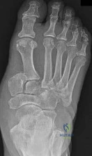
A 21-year-old collegiate basketball player sustains an acute Zone II fifth metatarsal base fracture (Jones fracture). To minimize the risk of nonunion and expedite return to play, intramedullary screw fixation is planned. Which of the following vascular structures is most uniquely at risk of disruption by this specific fracture pattern, predisposing to nonunion?


Explanation

A true Jones fracture occurs at the metaphyseal-diaphyseal junction of the fifth metatarsal (Zone II). This specific area represents a vascular watershed region. The blood supply to the proximal diaphysis is provided by a nutrient artery that enters the medial cortex at the mid-diaphysis and flows retrogradely. A fracture in Zone II disrupts this retrograde intraosseous blood supply, significantly increasing the risk of delayed union or nonunion.

Question 60

A 19-year-old football player sustains a severe blunt trauma to his anterior thigh from a helmet collision. Three weeks later, he presents with significantly restricted knee flexion and a firm, painful mass in his quadriceps. Radiographs demonstrate peripheral calcification within the muscle belly. What is the most appropriate initial management for this condition?


Explanation

The patient has developed myositis ossificans traumatica following a severe quadriceps contusion. The hallmark radiographic finding at 3-4 weeks is peripheral maturation/calcification with a radiolucent center. Initial management is strict nonoperative care, including rest, gentle active (not passive) range of motion, and NSAIDs (like Indomethacin) to help halt further heterotopic bone formation. Aggressive passive stretching or massage can exacerbate the condition. Surgical excision is absolutely contraindicated in the acute/immature phase due to an extremely high risk of recurrence; it is only considered if the mass remains symptomatic after full maturation (typically 6-12 months).

Question 61

A 24-year-old hockey player presents with anterior groin pain exacerbated by hip flexion and internal rotation. Radiographs demonstrate a prominent alpha angle.

Which of the following is the most likely location of the associated labral or chondral pathology in this condition?


Explanation

Cam impingement typically causes chondral delamination and labral tears in the anterosuperior quadrant of the acetabulum (clock face 1 to 3 o'clock). The abnormal morphology of the femoral head-neck junction is forced into the joint during flexion and internal rotation.

Question 62

A 25-year-old football player sustains a high-energy knee dislocation (KD-III).

Following closed reduction in the emergency department, his Ankle-Brachial Index (ABI) is measured at 0.85. He has palpable distal pulses. What is the most appropriate next step in management?


Explanation

In the setting of a knee dislocation, an Ankle-Brachial Index (ABI) less than 0.9 is an indication for advanced imaging, preferably CT angiography, to evaluate for popliteal artery injury. Immediate surgical exploration is indicated only if there are 'hard' signs of vascular injury (e.g., absent pulses after reduction, expanding hematoma, pulsatile bleeding).

Question 63

A 22-year-old collegiate baseball pitcher complains of posterior shoulder pain during the late cocking phase of throwing. He exhibits increased external rotation and decreased internal rotation (GIRD) compared to the contralateral side. What is the primary pathophysiologic mechanism of this condition?


Explanation

This patient has internal impingement, common in overhead athletes. It occurs during extreme abduction and external rotation (late cocking phase), leading to pathologic contact between the articular surface of the posterior rotator cuff (supraspinatus/infraspinatus) and the posterosuperior glenoid and labrum.

Question 64

During an ulnar collateral ligament (UCL) reconstruction using the docking technique, the surgeon decides to transpose the ulnar nerve subcutaneously. Which of the following is the primary risk factor for postoperative ulnar neuropathy in this specific scenario?


Explanation

When performing an anterior transposition of the ulnar nerve (subcutaneous or submuscular), it is critical to resect the medial intermuscular septum to prevent kinking, tethering, or compression of the nerve as it transitions into the anterior compartment of the elbow.

Question 65

A 26-year-old professional soccer player suffers an external rotation injury to his ankle. Radiographs show no fracture.

An MRI demonstrates a complete tear of the anterior inferior tibiofibular ligament (AITFL) and interosseous membrane extending 4 cm proximal to the joint line, with an intact deltoid ligament. Dynamic stress views show no widening of the medial clear space. What is the most appropriate management?


Explanation

Stable syndesmotic injuries (characterized by an intact deltoid ligament and no medial clear space widening on stress views) can be treated nonoperatively, even with proximal extension of the interosseous membrane tear. Operative intervention is reserved for unstable injuries.

Question 66

A 21-year-old athlete undergoes an anatomic posterolateral corner (PLC) reconstruction. The fibular collateral ligament (FCL) graft is being secured on the femur. What are the correct anatomic landmarks for the femoral footprint of the FCL?


Explanation

On the lateral femoral condyle, the anatomic footprint of the fibular collateral ligament (FCL) is situated approximately 18.5 mm proximal and posterior to the popliteus tendon insertion.

Question 67

A 45-year-old female marathon runner complains of recalcitrant lateral hip pain. Examination shows a positive Trendelenburg sign and pain with resisted hip abduction. MRI reveals a full-thickness tear of the gluteus medius at its insertion. To which facet(s) of the greater trochanter does the main tendon of the gluteus medius primarily attach?


Explanation

The gluteus medius inserts primarily onto the lateral and superoposterior facets of the greater trochanter. The gluteus minimus inserts more anteriorly onto the anterior facet.

Question 68

A 35-year-old weightlifter feels a "pop" in his anterior shoulder during a heavy bench press. He now has increased passive external rotation and profound weakness in internal rotation.

He tests positive on the bear hug test. Which of the following associated injuries is most likely present given this pathology?


Explanation

Acute subscapularis tears, particularly traumatic ruptures in younger patients, are highly associated with damage to the biceps pulley (superior glenohumeral ligament and coracohumeral ligament). This leads to medial subluxation or dislocation of the long head of the biceps tendon out of the bicipital groove.

Question 69

A 19-year-old soccer player has a symptomatic 2.5 cm^2 full-thickness chondral defect on the weight-bearing surface of the medial femoral condyle. He has failed conservative management. What is the most appropriate surgical treatment that provides hyaline-like cartilage repair for a defect of this size?


Explanation

For a 2.5 cm^2 defect (>2 cm^2), microfracture is relatively contraindicated (fails to provide durable hyaline-like cartilage) and OATS carries high donor site morbidity. Autologous chondrocyte implantation (ACI) provides hyaline-like cartilage and is the standard of care for symptomatic defects between 2 and 10 cm^2 in young patients.

Question 70

A 22-year-old college basketball player sustains a fracture at the metaphyseal-diaphyseal junction of the fifth metatarsal.

He is in mid-season and wishes to return to play as soon as possible. What is the recommended treatment?


Explanation

In elite athletes, acute true Jones fractures (Zone 2 metaphyseal-diaphyseal junction) are generally treated with intramedullary screw fixation to decrease the time to return to play and reduce the high risk of nonunion associated with conservative management in this population.

Question 71

A 28-year-old professional volleyball player presents with vague posterior shoulder pain and weakness in external rotation. Examination reveals isolated atrophy of the infraspinatus muscle.

Where is the most likely site of neural compression?


Explanation

The suprascapular nerve innervates both the supraspinatus and infraspinatus. Compression at the suprascapular notch affects both muscles. Compression further distal at the spinoglenoid notch (often due to a paralabral cyst associated with a posterior labral tear) selectively denervates the infraspinatus, sparing the supraspinatus.

Question 72

A 14-year-old male presents with knee pain and catching. Radiographs demonstrate a classical osteochondritis dissecans (OCD) lesion.

What is the most common anatomical location for this lesion in the knee?


Explanation

The most common location for osteochondritis dissecans (OCD) of the knee is the lateral aspect of the medial femoral condyle (accounting for 70-80% of all knee OCD lesions), classically remembered by the mnemonic LAME (Lateral Aspect Medial Epicondyle/condyle).

Question 73

A 42-year-old competitive weightlifter undergoes an anterior single-incision repair for an acute complete distal biceps tendon rupture.

Postoperatively, he complains of significant numbness over the lateral aspect of his forearm. Which nerve is most likely injured?


Explanation

The lateral antebrachial cutaneous nerve (LABC) is the terminal sensory branch of the musculocutaneous nerve. It exits the deep fascia lateral to the biceps tendon. It is the most commonly injured nerve during a single-anterior-incision distal biceps repair due to aggressive retraction, leading to lateral forearm paresthesias.

Question 74

During an arthroscopic posterior cruciate ligament (PCL) reconstruction using a single-bundle technique, the tibial tunnel is prepared.

Which of the following describes the correct anatomic trajectory of the PCL tibial guide pin to optimize footprint coverage while avoiding neurovascular injury?


Explanation

The anatomic tibial footprint of the PCL is located in the posterior aspect of the tibia (PCL facet). The guide pin should be placed approximately 7 mm anterior to the posterior tibial cortex. This ensures a complete cortical rim remains for tunnel integrity and minimizes the risk of posterior pin penetration injuring the popliteal neurovascular bundle.

Question 75

A 60-year-old male undergoes arthroscopic rotator cuff repair.

To optimize tendon-to-bone healing, the surgeon decorticates the greater trochanter footprint. What is the primary histological mechanism of healing at the tendon-bone interface following this repair?


Explanation

Following surgical rotator cuff repair, the healing process does not reliably regenerate the native four-zone transitional anatomy (tendon, uncalcified fibrocartilage, calcified fibrocartilage, bone). Instead, it heals primarily by fibrovascular scar tissue formation, which is structurally and biomechanically weaker than the native insertion.

Question 76

A 24-year-old rugby player falls with his foot plantar flexed and another player lands on his heel.

Radiographs suggest a subtle widening between the first and second rays. MRI confirms a complete tear of the Lisfranc ligament. He undergoes open reduction and internal fixation. Which specific articulation does the primary Lisfranc ligament span?


Explanation

The Lisfranc ligament is a strong interosseous ligament that runs obliquely from the lateral aspect of the medial cuneiform to the medial aspect of the base of the second metatarsal. It is the primary stabilizer of the midfoot.

Question 77

A 16-year-old female dancer experiences her first episode of acute lateral patellar dislocation. Radiographs show no osteochondral loose bodies. MRI confirms an isolated full-thickness tear of the medial patellofemoral ligament (MPFL).

What is the most common site of MPFL injury in an acute lateral patellar dislocation?


Explanation

In the setting of an acute primary lateral patellar dislocation, the medial patellofemoral ligament (MPFL) most commonly tears at its femoral origin, which is located in the saddle region between the adductor tubercle and the medial epicondyle.

Question 78

A 45-year-old construction worker with chronic anterior shoulder pain undergoes an open subpectoral biceps tenodesis.

During the surgical approach, retractors are placed deep to the short head of the biceps and coracobrachialis (conjoined tendon). Which nerve is at greatest risk of injury with overly aggressive medial retraction in this specific area?


Explanation

The musculocutaneous nerve branches from the lateral cord and typically enters the coracobrachialis muscle approximately 5-8 cm distal to the coracoid process. Aggressive medial retraction of the conjoined tendon during a subpectoral biceps tenodesis places this nerve at significant risk of traction injury.

Question 79

A 20-year-old female ballet dancer presents with a palpable and audible "snap" over her deep anterior groin when she extends her hip from a flexed, abducted, and externally rotated position.

Dynamic ultrasound evaluation demonstrates a tendon snapping over a bony prominence. What is the most likely diagnosis?


Explanation

Internal coxa saltans (snapping hip syndrome) is caused by the iliopsoas tendon snapping over the iliopectineal eminence or the anterior femoral head during extension of the hip from a flexed, abducted, and externally rotated position. External coxa saltans involves the IT band snapping over the greater trochanter.

Question 80

A 30-year-old runner complains of lateral knee pain that is particularly worse when running downhill.

Examination reveals tenderness exquisitely localized over the lateral femoral epicondyle. Which clinical test is most specific for confirming the diagnosis of Iliotibial Band Friction Syndrome?


Explanation

The Noble compression test is specifically designed to elicit the pain of Iliotibial (IT) Band Friction Syndrome. Pain is typically reproduced at approximately 30 degrees of knee flexion as the IT band snaps over the lateral femoral epicondyle while manual pressure is applied. Ober's test evaluates for IT band tightness but is not specific for recreating the pain of friction syndrome.

Question 81

A 45-year-old male feels a pop in his posterior knee while squatting. MRI shows a medial meniscus posterior root tear. What is the primary biomechanical consequence of leaving this specific lesion untreated?


Explanation

A posterior root tear of the medial meniscus disrupts hoop stresses, leading to meniscal extrusion. Biomechanically, this failure is equivalent to a total meniscectomy, causing significantly increased peak contact pressures and rapid articular cartilage degeneration.

Question 82

During an arthroscopic anterior cruciate ligament (ACL) reconstruction, the femoral tunnel is inadvertently placed too anteriorly within the intercondylar notch. What is the most likely clinical consequence during postoperative rehabilitation?


Explanation

An anteriorly placed femoral tunnel causes the ACL graft to tighten excessively as the knee transitions into flexion. This over-tensioning captures the joint, leading to a restricted arc of motion and a significant loss of full knee flexion.

Question 83

A 25-year-old football player presents with an acute knee injury. The Dial test demonstrates 15 degrees of increased external rotation at 30 degrees of knee flexion compared to the contralateral side, but symmetric external rotation at 90 degrees of flexion. What is the most likely injury pattern?


Explanation

Increased external tibial rotation at 30 degrees of flexion, which normalizes at 90 degrees, is the hallmark of an isolated posterolateral corner (PLC) injury. If external rotation is increased at both 30 and 90 degrees, it strongly suggests a combined PLC and PCL injury.

Question 84

A 22-year-old rugby player suffers recurrent anterior shoulder instability. 3D CT reconstruction demonstrates 12% anterior glenoid bone loss and a deep, engaging Hill-Sachs lesion. Which of the following is the most appropriate surgical management?


Explanation

An engaging Hill-Sachs lesion combined with subcritical glenoid bone loss (<20-25%) is optimally managed with an arthroscopic Bankart repair and a Remplissage procedure. Remplissage prevents engagement by tenodesing the infraspinatus and posterior capsule into the Hill-Sachs defect.

Question 85

When performing a medial patellofemoral ligament (MPFL) reconstruction, accurate placement of the femoral attachment is critical to prevent graft anisometry. Based on Schöttle's anatomic landmarks, where is the correct femoral footprint of the MPFL located?


Explanation

The native MPFL femoral footprint is anatomically located in the saddle-like groove between the adductor tubercle (proximally) and the medial epicondyle (distally). Non-anatomic placement, particularly too proximal, leads to graft tightening in knee flexion and patellofemoral overload.

Question 86

A 19-year-old collegiate baseball pitcher undergoes a modified Jobe ulnar collateral ligament (UCL) reconstruction using a palmaris longus autograft. Which of the following represents the most common postoperative complication associated with this specific surgical technique?


Explanation

Ulnar neuropathy is the most frequent complication following UCL reconstruction, particularly with techniques like the modified Jobe that involve routine ulnar nerve transposition. Modern techniques, such as the docking procedure, minimize nerve handling and have significantly reduced this risk.

None

Clinic OS
Medically Verified Content by
Prof. Clinic OS
Consultant Orthopedic & Spine Surgeon
Chapter Index