العربية
Part of the Master Guide

Updated Orthopedic Review | Dr Hutaif General Orthopedi -...

Orthopedic Upper Limb Review | Dr Hutaif General Orthop -...

23 Apr 2026 36 min read 109 Views
Illustration of osteonecrosis of the humeral - Dr. Mohammed Hutaif

Key Takeaway

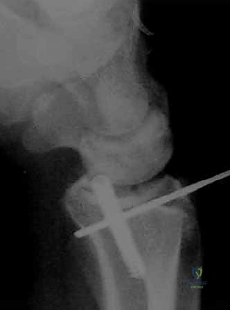
For anyone wondering about ONLINE ORTHOPEDIC MCQS UPPER LIMB08, Osteonecrosis of the humeral head refers to the death of bone tissue in the upper arm bone, often leading to severe shoulder pain and motion loss. Management, similar to advanced osteoarthritis, frequently involves total shoulder arthroplasty. Successful long-term results depend on factors like proper prosthetic placement and tuberosity osteosynthesis, crucial aspects highlighted in preventing complications and improving patient outcomes after shoulder reconstruction.

Orthopedic Upper Limb Review | Dr Hutaif General Orthop -...

Comprehensive 100-Question Exam


00:00

Start Quiz

Question 1

A 35-year-old male sustains a fall onto an outstretched hand, resulting in a terrible triad injury of the elbow. During surgical management, after repair of the coronoid fracture, fixation of the radial head, and repair of the lateral ulnar collateral ligament (LUCL), the elbow demonstrates persistent subluxation when extended past 30 degrees. What is the most appropriate next step in management?


Explanation

The standard algorithm for treating a terrible triad injury involves repairing structures from deep to superficial (or inside-out), typically addressing the coronoid, then radial head, then LUCL. If the elbow remains unstable in extension after these lateral structures and anterior structures are stabilized, the medial collateral ligament (MCL) should be repaired or a hinged external fixator applied.

Question 2

Which of the following best describes the vascular supply to the scaphoid and its clinical implication?


Explanation

The scaphoid receives its primary blood supply from branches of the radial artery that enter at the distal pole and flow retrogradely to the proximal pole. Because of this retrograde perfusion, proximal pole fractures have a high risk of avascular necrosis and nonunion.

Question 3

According to Hertel's radiographic criteria, which of the following fracture characteristics is the most accurate predictor of humeral head ischemia in proximal humerus fractures?


Explanation

Hertel et al. described predictors of ischemia in proximal humerus fractures. The combination of a disrupted medial hinge (>2 mm) and a short calcar segment (<8 mm attached to the articular segment) has the highest positive predictive value (97%) for subsequent humeral head ischemia.

Question 4

A 55-year-old male presents with chronic wrist pain and a known history of a scapholunate ligament tear. Radiographs demonstrate advanced osteoarthritis specifically involving the radioscaphoid joint and the capitolunate joint, with complete sparing of the radiolunate joint. What stage of Scapholunate Advanced Collapse (SLAC) does this represent?


Explanation

SLAC wrist arthritis progresses predictably: Stage I involves only the radial styloid-scaphoid articulation; Stage II involves the entire radioscaphoid joint; Stage III involves proximal migration of the capitate with capitolunate arthritis. The radiolunate joint is characteristically spared due to the conformal nature of the fossa and intact radiolunate ligaments.

Question 5

A 28-year-old male sustains a closed mid-shaft humeral fracture resulting in an immediate complete radial nerve palsy. He is treated non-operatively with a functional brace. At 12 weeks post-injury, the fracture shows early signs of union, but there is no clinical or electromyographic (EMG) evidence of radial nerve recovery. What is the most appropriate next step in management?


Explanation

Immediate radial nerve palsy associated with a closed humeral shaft fracture is typically treated expectantly. However, if there is no clinical or EMG evidence of recovery by 12 to 16 weeks (3-4 months), surgical exploration of the radial nerve is indicated. Continuing observation beyond this window risks irreversible motor endplate loss.

Question 6

In the context of a thumb ulnar collateral ligament (UCL) rupture (Skier's thumb), a Stener lesion is anatomically defined as the interposition of which structure between the torn ends of the UCL?


Explanation

A Stener lesion occurs when the torn ulnar collateral ligament (UCL) of the thumb metacarpophalangeal joint flips proximally and comes to rest superficial to the adductor pollicis aponeurosis. This interposition prevents primary ligamentous healing and is a strong indication for surgical repair.

Question 7

Which of the following best describes the biomechanical advantage achieved by a reverse total shoulder arthroplasty in a patient with rotator cuff tear arthropathy?


Explanation

A reverse total shoulder arthroplasty medializes and inferiorizes the center of rotation of the glenohumeral joint compared to native anatomy. This recruits more of the deltoid muscle fibers and significantly increases the moment arm and resting tension of the deltoid, allowing it to elevate the arm without a functioning rotator cuff.

Question 8

A patient sustains a laceration to the volar aspect of the hand, severing the flexor digitorum profundus (FDP) and flexor digitorum superficialis (FDS) of the middle finger. The injury is located between the A1 pulley and the insertion of the FDS on the middle phalanx. This injury corresponds to which flexor tendon zone?


Explanation

Zone II (historically called 'no man's land') extends from the proximal aspect of the A1 pulley (at the distal palmar crease) to the insertion of the flexor digitorum superficialis (FDS) on the middle phalanx. Both FDS and FDP tendons lie within the tight fibro-osseous sheath in this zone.

Question 9

Which of the following muscles is typically the most severely affected by ischemia in volar compartment syndrome of the forearm due to its deeper location and vascular anatomy?


Explanation

The flexor digitorum profundus (FDP) and flexor pollicis longus (FPL) are located in the deep volar compartment of the forearm. Due to their deep position immediately adjacent to the radius and ulna, and their epimysial blood supply, they are typically the most severely affected muscles in forearm compartment syndrome (leading to Volkmann's ischemic contracture).

Question 10

According to the Bado classification, a Monteggia fracture-dislocation characterized by a fracture of the ulnar diaphysis with posterior angulation and a posterior dislocation of the radial head is classified as:


Explanation

In the Bado classification for Monteggia lesions: Type I is an anterior dislocation of the radial head with anterior apex ulnar fracture; Type II is a posterior dislocation of the radial head with posterior apex ulnar fracture; Type III is lateral dislocation of the radial head with an ulnar metaphyseal fracture; Type IV is anterior dislocation with fractures of both the radius and ulna shafts.

Question 11

A 65-year-old female presents with sudden inability to extend her thumb interphalangeal joint 6 weeks after non-operative management of a non-displaced distal radius fracture. What is the most widely accepted etiology of this complication?


Explanation

Extensor pollicis longus (EPL) rupture is a classic complication following minimally displaced or non-displaced distal radius fractures. The primary etiology is a combination of mechanical attrition over the fracture callus/Lister's tubercle and ischemia due to increased pressure within the tight, intact third extensor compartment.

Question 12

In the setting of recurrent anterior shoulder instability, an engaging Hill-Sachs lesion is defined as an osseous defect that:


Explanation

An engaging Hill-Sachs lesion is a posterosuperior humeral head defect that dynamically engages (levers over or drops into) the anterior glenoid rim when the shoulder is placed in a functional position of abduction and external rotation, contributing to recurrent instability.

Question 13

A 32-year-old laborer presents with progressive dorsal wrist pain. Radiographs reveal sclerosis and fragmentation of the lunate with negative ulnar variance. According to the Lichtman classification, lunate collapse with fixed scaphoid rotation (signet ring sign) but no radiocarpal or midcarpal arthritis is classified as:


Explanation

Kienböck's disease Lichtman classification: Stage I has normal plain films but MRI changes; Stage II has lunate sclerosis without collapse; Stage IIIA has lunate collapse without fixed scaphoid rotation; Stage IIIB has lunate collapse with fixed scaphoid rotation; Stage IV has associated carpal arthritis.

Question 14

A 28-year-old male bodybuilder presents with severe pain and ecchymosis over his anterior axillary fold after bench pressing a heavy weight. Examination reveals loss of the anterior axillary fold contour and weakness in internal rotation. In this demographic, the most common site of a pectoralis major rupture occurs:


Explanation

In weightlifters performing activities with high eccentric loads (e.g., bench press), the vast majority of pectoralis major ruptures occur at the tendinous insertion onto the lateral lip of the bicipital groove of the humerus, predominantly involving the sternocostal head.

Question 15

A 6-year-old boy falls from monkey bars and sustains a widely displaced posterolateral extension-type supracondylar humerus fracture. Which nerve is most commonly injured in this specific fracture displacement pattern?


Explanation

In extension-type supracondylar humerus fractures, the direction of distal fragment displacement determines the structures at risk. Posterolateral displacement stretches the median nerve and specifically the anterior interosseous nerve (AIN) over the proximal metaphyseal spike. Posteromedial displacement places the radial nerve at risk.

Question 16

A 45-year-old female presents with vague volar forearm pain and paresthesias in the thumb, index, and middle fingers. Symptoms worsen with resisted forearm pronation. Examination reveals diminished sensation over the thenar eminence but no weakness of the flexor pollicis longus. What is the most likely diagnosis?


Explanation

Pronator syndrome is a proximal compression neuropathy of the median nerve. Numbness over the thenar eminence helps distinguish it from carpal tunnel syndrome, as the palmar cutaneous branch of the median nerve branches proximal to the carpal tunnel. Exacerbation with resisted pronation is characteristic. AIN syndrome is a pure motor neuropathy.

Question 17

A 42-year-old male undergoes surgical repair of a distal biceps tendon rupture via a single anterior incision. Post-operatively, he exhibits an inability to actively extend his metacarpophalangeal (MCP) joints and thumb interphalangeal joint, though tenodesis effect is intact. His wrist extension demonstrates radial deviation. Sensation is fully intact. Which nerve was most likely injured during the procedure?


Explanation

The posterior interosseous nerve (PIN) is at risk during an anterior single-incision approach to the distal biceps, particularly with overzealous lateral retraction. A PIN palsy results in loss of active extension of the digits and thumb. Wrist extension results in radial deviation because the extensor carpi radialis longus (innervated by the radial nerve proper) remains functional while the extensor carpi ulnaris (PIN innervated) is paralyzed. Sensation remains intact.

Question 18

During an in situ ulnar nerve decompression for cubital tunnel syndrome, a tight fascial band is encountered just distal to the medial epicondyle, bridging the humeral and ulnar heads of the flexor carpi ulnaris (FCU). What is the specific anatomical name of this structure?


Explanation

Osborne's ligament (or the cubital tunnel retinaculum) forms the roof of the cubital tunnel, bridging the humeral and ulnar heads of the flexor carpi ulnaris (FCU) just distal to the medial epicondyle. The Arcade of Struthers is a fascial band located approximately 8 cm proximal to the medial epicondyle.

Question 19

In Dupuytren's contracture, the neurovascular bundle is often displaced centrally and superficially by the spiral cord. The spiral cord is formed by the pathological alteration of several normal fascial structures. Which of the following normally occurring structures is a component of the spiral cord complex?


Explanation

The spiral cord in Dupuytren's disease displaces the neurovascular bundle medially/centrally and superficially, making it highly vulnerable during surgical fasciectomy. It is formed by the diseased amalgamation of four normal structures: the pretendinous band, the spiral band, the lateral digital sheet, and Grayson's ligament. Cleland's ligament is dorsal to the bundle and is typically spared.

Question 20

A patient presents with a chronic Boutonniere deformity consisting of proximal interphalangeal (PIP) joint flexion and distal interphalangeal (DIP) joint hyperextension. This deformity primarily results from the rupture or attenuation of which extensor mechanism structure, followed by volar subluxation of another?


Explanation

A Boutonniere deformity initiates with the disruption or attenuation of the central slip of the extensor mechanism at its insertion on the middle phalanx. This allows the lateral bands to subluxate volarly past the axis of rotation of the PIP joint, causing them to act as PIP flexors while maintaining their strong continuous extensor pull on the distal phalanx.

Question 21

A 60-year-old female undergoes volar locked plating for a distal radius fracture. Six months later, she presents with an inability to actively flex the interphalangeal joint of her thumb. Which radiographic finding is most predictive of this specific complication?


Explanation

Flexor pollicis longus (FPL) tendon rupture is a known complication of volar plating of the distal radius. Plates placed distal to the watershed line (Soong grade 2) have the highest risk of attritional FPL rupture due to tendon impingement.

Question 22

In a 25-year-old active male with a closed midshaft clavicle fracture, which of the following radiographic characteristics is the strongest independent predictor of nonunion if treated non-operatively?


Explanation

Complete fracture displacement with greater than 2 cm of shortening is a strong independent predictor of nonunion and poor functional outcomes in midshaft clavicle fractures. This finding strongly supports the indication for surgical fixation.

Question 23

A 6-year-old boy sustains a Bado Type I Monteggia fracture-dislocation. Following closed reduction and casting of the ulnar shaft fracture, radiographs reveal persistent anterior subluxation of the radial head. What is the most likely anatomic block to radial head reduction?


Explanation

In pediatric Bado Type I Monteggia injuries, failure to achieve or maintain radial head reduction after anatomic ulnar realignment is most commonly due to the interposition of the torn annular ligament or joint capsule. Open reduction of the radiocapitellar joint is required to extract the interposed tissue.

Question 24

A 45-year-old manual laborer presents with chronic wrist pain and is diagnosed with Scapholunate Advanced Collapse (SLAC). According to the predictable pattern of SLAC wrist arthropathy, which carpal articulation is characteristically spared even in advanced stages?


Explanation

In a SLAC wrist, the radiolunate joint is characteristically spared from degenerative changes. This occurs due to the congruent spherical anatomy of the lunate fossa and the stabilizing presence of the intact short radiolunate ligament.

Question 25

During a lateral deltoid-splitting approach for open reduction and internal fixation of a proximal humerus fracture, the surgeon must identify and protect the axillary nerve. At what average distance distal to the lateral edge of the acromion does the axillary nerve cross the humerus?


Explanation

The axillary nerve courses transversally across the surgical neck of the humerus at an average distance of 5 to 7 cm distal to the lateral border of the acromion. The deltoid split should not extend beyond 5 cm to avoid iatrogenic denervation of the anterior deltoid.

Question 26

A 30-year-old mechanic undergoes a Zone II flexor digitorum profundus (FDP) and superficialis (FDS) repair of the index finger. To optimize tendon gliding and minimize rupture risk, which of the following intraoperative pulley management strategies is most appropriate if the bulky repair catches during active flexion?


Explanation

Modern flexor tendon repair protocols demonstrate that venting up to 50% of the A2 pulley or completely venting the A4 pulley is safe if a bulky repair catches during excursion. This allows for smooth gliding and early active motion without causing clinically significant bowstringing.

Question 27

A 72-year-old female with pseudoparalysis secondary to severe rotator cuff tear arthropathy undergoes a reverse total shoulder arthroplasty (RTSA). According to Grammont's biomechanical principles, how does this prosthesis design improve the functional capacity of the deltoid muscle?


Explanation

Grammont's principles for RTSA involve medializing and distalizing the joint's center of rotation. This configuration increases the deltoid moment arm and tension, recruiting more deltoid fibers to elevate the arm in the absence of a functional rotator cuff.

Question 28

A 42-year-old male undergoes a single-incision anterior approach for a distal biceps tendon repair using cortical button fixation. Postoperatively, he notes weakness in extending his thumb and fingers, though wrist extension is preserved with a radial deviation bias. Which nerve was most likely injured during the procedure?


Explanation

The posterior interosseous nerve (PIN) is highly at risk during the single-incision anterior approach to the distal biceps, particularly from excessive lateral retraction or blind drilling of the posterior radial cortex. PIN injury results in weakened digit and thumb extension, while radial wrist extension (ECRL) remains preserved.

Question 29

A 5-year-old child sustains a Gartland type III supracondylar humerus fracture. On presentation, the hand is pink but the radial pulse is absent. Following emergent closed reduction and percutaneous pinning, the fracture is anatomically reduced, but the radial pulse remains absent. The hand remains warm with a capillary refill of less than 2 seconds. What is the most appropriate next step?


Explanation

In a well-perfused 'pink, pulseless' hand following anatomic reduction and pinning of a pediatric supracondylar humerus fracture, the standard of care is close clinical observation. Collateral circulation is typically adequate to perfuse the limb, and the radial pulse usually returns within days to weeks.

Question 30

A 55-year-old female presents with a highly comminuted, intra-articular distal radius fracture with significant volar displacement of the carpus alongside the volar fracture fragment. Which of the following is the most appropriate biomechanical principle for plate fixation of this injury pattern?


Explanation

A volar shear fracture (Barton's fracture) represents an unstable pattern where the carpus subluxates with the volar articular fragment. A volar plate acts as a buttress to counteract the shear forces, preventing volar subluxation of the carpus.

Question 31

A 24-year-old cyclist falls and sustains a completely displaced midshaft clavicle fracture. Which of the following radiographic parameters is the strongest absolute indication for open reduction and internal fixation (ORIF) to prevent symptomatic malunion?


Explanation

In midshaft clavicle fractures, shortening greater than 2 cm is highly associated with symptomatic malunion, persistent pain, and shoulder weakness. ORIF is indicated to restore resting length and optimize shoulder mechanics.

Question 32

A 40-year-old manual laborer with a chronic scapholunate ligament tear develops Scapholunate Advanced Collapse (SLAC). Radiographs reveal arthritis limited to the radioscaphoid joint, while the radiolunate joint is spared. Which ligament is primarily responsible for preserving the radiolunate articulation in the typical SLAC wrist progression?


Explanation

The short radiolunate ligament provides robust stabilization to the lunate, maintaining its relationship with the lunate fossa even as the scaphoid rotates into flexion. This anatomic feature spares the radiolunate joint from degenerative changes until late stages of SLAC.

Question 33

A 45-year-old male undergoes surgical repair of a distal biceps tendon rupture using a single-anterior-incision technique with cortical button fixation. Postoperatively, he complains of numbness over the lateral aspect of his forearm. Injury to which of the following nerves is the most common complication of this specific surgical approach?


Explanation

The lateral antebrachial cutaneous nerve (LABC) is highly susceptible to traction or iatrogenic injury during the single-anterior-incision approach to the distal biceps. In contrast, posterior interosseous nerve (PIN) injury or radioulnar synostosis is classically more associated with the two-incision technique.

Question 34

A 32-year-old male sustains a Bado Type I Monteggia fracture-dislocation. Following closed reduction of the ulnar fracture and radial head, the patient exhibits an inability to extend his thumb and fingers at the metacarpophalangeal joints, but wrist extension is preserved with radial deviation. Which nerve is most likely injured?


Explanation

The posterior interosseous nerve (PIN) is vulnerable to traction injury during anterior dislocation of the radial head. Preservation of wrist extension with radial deviation occurs because the extensor carpi radialis longus (ECRL) is innervated by the radial nerve proper proximal to the PIN branch.

Question 35

In a patient undergoing a Reverse Total Shoulder Arthroplasty (RTSA) for cuff tear arthropathy, how does the prosthesis alter the normal shoulder biomechanics to compensate for the deficient rotator cuff?


Explanation

RTSA medializes and distalizes the center of rotation of the glenohumeral joint. This significantly increases the moment arm and resting tension of the deltoid muscle, allowing it to elevate the arm without a functioning rotator cuff.

Question 36

A 21-year-old rugby player presents with recurrent anterior shoulder instability. A 3D CT scan reveals 26% anterior glenoid bone loss. Which of the following procedures is the most appropriate definitive management to prevent recurrent instability?


Explanation

In cases of anterior shoulder instability with critical glenoid bone loss (typically >20-25%), soft tissue stabilization alone has unacceptably high failure rates. The Latarjet procedure restores the bony arc and provides a dynamic sling effect via the conjoint tendon.

Question 37

A 65-year-old female presents with pseudoparalysis of her right shoulder, preserved passive motion, and severe glenohumeral arthritis. MRI shows a massive, retracted, and fatty-infiltrated rotator cuff tear. What is the most appropriate surgical treatment?


Explanation

Reverse total shoulder arthroplasty is the gold standard for rotator cuff tear arthropathy with pseudoparalysis. It restores overhead elevation by utilizing the deltoid muscle and medializing the center of rotation. Anatomic TSA is contraindicated due to the deficient rotator cuff causing eccentric glenoid wear.

Question 38

A 25-year-old cyclist falls directly onto his shoulder. Radiographs reveal a completely displaced midshaft clavicle fracture with 2.5 cm of shortening and no cortical contact. Which of the following is the most established biomechanical and clinical advantage of open reduction internal fixation (ORIF) compared to non-operative management in this patient?


Explanation

Displaced midshaft clavicle fractures with shortening greater than 2 cm have a high rate of nonunion and symptomatic malunion if treated non-operatively. ORIF significantly decreases the nonunion rate and improves early functional outcomes in this specific patient population.

Question 39

Following open reduction and internal fixation of a 3-part proximal humerus fracture with a locking plate, a patient complains of persistent pain and stiffness. What is the most common hardware-related complication leading to reoperation in this scenario?


Explanation

Intra-articular screw penetration is the most common hardware-related complication after locked plating of proximal humerus fractures, often due to avascular necrosis or fracture settling. Meticulous fluoroscopic evaluation and avoiding excessively long screws can help prevent this.

Question 40

A 40-year-old male sustains a closed fracture of the distal third of the humeral shaft. On presentation, he has a normal neurovascular exam. Following a closed reduction and application of a coaptation splint, he is noted to have a new complete radial nerve palsy. What is the most appropriate next step in management?


Explanation

A secondary (post-reduction) radial nerve palsy in the setting of a closed humeral shaft fracture, particularly a Holstein-Lewis type, is an absolute indication for immediate surgical exploration. The nerve may be iatrogenically entrapped in the fracture site during the reduction maneuver.

Question 41

A 30-year-old male sustains a highly comminuted radial head fracture, a tear of the interosseous membrane, and distal radioulnar joint (DRUJ) disruption. To prevent longitudinal instability of the forearm, which of the following interventions is critical in addition to stabilizing the DRUJ?


Explanation

This triad of injuries describes an Essex-Lopresti fracture-dislocation. Restoring radiocapitellar contact with a radial head arthroplasty is essential to prevent proximal migration of the radius, which would lead to chronic DRUJ pain and ulnocarpal impingement.

Question 42

A 45-year-old male falls from a ladder and sustains an isolated proximal ulna shaft fracture with an associated anterior dislocation of the radial head (Bado Type I Monteggia injury). During surgery, the ulna is anatomically reduced and plated, but the radial head remains persistently dislocated. What is the most likely interposing structure preventing reduction?


Explanation

In Monteggia fractures, anatomic restoration of ulnar length and alignment typically spontaneously reduces the radial head. If the radial head fails to reduce, the annular ligament or joint capsule is the most common interposing structure, requiring open reduction.

Question 43

A 28-year-old male sustains a high-energy wrist injury. Radiographs show a volar dislocation of the lunate, while the rest of the carpus remains aligned with the radius. The patient exhibits numbness in the thumb, index, and middle fingers. What is the appropriate initial management?


Explanation

This is a Stage IV perilunate (lunate) dislocation, which frequently causes acute carpal tunnel syndrome due to the volarly displaced lunate. Urgent closed reduction is required; if median nerve symptoms do not resolve immediately, an emergent carpal tunnel release and open reduction are indicated.

Question 44

A 55-year-old female undergoes volar locking plate fixation for a displaced distal radius fracture. Six months postoperatively, she suddenly loses the ability to actively flex her thumb interphalangeal joint. This complication is most directly related to plate placement in relation to which anatomic landmark?


Explanation

Placement of a volar plate distal to the watershed line of the distal radius places the flexor pollicis longus (FPL) tendon at high risk for attrition and rupture. The watershed line is a critical radiographic and surgical landmark used to prevent prominent hardware.

Question 45

Following a Zone II flexor tendon repair of the index finger, a standard rehabilitation protocol emphasizes early active mobilization. Which biomechanical factor in the surgical repair provides the necessary strength to safely permit early active motion without rupture?


Explanation

The ultimate tensile strength of a flexor tendon repair is directly proportional to the number of core suture strands crossing the repair site. A 4-strand or 6-strand core repair provides sufficient strength to withstand the forces of early active motion protocols.

Question 46

During surgical decompression for cubital tunnel syndrome, the ulnar nerve is traced distally into the forearm. Compression at this level is most commonly caused by the aponeurotic band between the two heads of the flexor carpi ulnaris (FCU). What is the name of this structure?


Explanation

Osborne's ligament (or Osborne's fascia) forms the roof of the cubital tunnel, bridging the olecranon and the medial epicondyle, and continues distally between the two heads of the FCU. The Arcade of Struthers is a distinct structure located proximal to the medial epicondyle.

Question 47

A 35-year-old male presents with chronic wrist pain. Radiographs reveal a scaphoid nonunion with radioscaphoid arthritis, but the capitolunate and radiolunate joints are completely spared. What is the most appropriate surgical treatment for this Stage II Scaphoid Nonunion Advanced Collapse (SNAC)?


Explanation

Stage II SNAC wrist involves arthritis between the distal radius and the scaphoid, sparing the midcarpal and radiolunate joints. Both scaphoid excision with four-corner fusion and proximal row carpectomy (PRC) are well-established, motion-preserving salvage procedures for this stage.

Question 48

A 42-year-old diabetic patient presents with a swollen, erythematous index finger. Of Kanavel's four cardinal signs for infectious pyogenic flexor tenosynovitis, which is considered the earliest and most sensitive clinical indicator?


Explanation

Pain with passive extension is the earliest and most sensitive of Kanavel's signs for pyogenic flexor tenosynovitis. This stretch places direct tension on the inflamed synovial sheath, eliciting severe pain before the other localized signs fully manifest.

Question 49

A patient presents with a brachial plexus injury resulting in Horner syndrome. Which root is most likely avulsed, and what is the prognosis for spontaneous recovery?


Explanation

Horner syndrome indicates a proximal preganglionic T1 root avulsion due to disruption of the sympathetic chain. Preganglionic injuries have a poor prognosis for spontaneous recovery and cannot be repaired directly with nerve grafting.

Question 50

In evaluating a patient with recurrent anterior shoulder instability, what is the defining characteristic of an "off-track" Hill-Sachs lesion?


Explanation

An "off-track" Hill-Sachs lesion engages the anterior glenoid rim because its medial margin extends further medially than the width of the intact glenoid track. This biomechanical mismatch typically requires a remplissage or bone block procedure.

Question 51

A 25-year-old male sustains a closed midshaft humeral fracture with an immediate radial nerve palsy. Closed reduction is performed, and post-reduction examination reveals a worsening of the radial nerve palsy. What is the most appropriate next step?


Explanation

Worsening or new-onset radial nerve palsy following closed reduction of a humeral shaft fracture is an absolute indication for immediate surgical exploration. This is because the nerve may be iatrogenically entrapped between the fracture fragments.

Question 52

Which intrinsic ligament of the wrist is the primary stabilizer of the scapholunate articulation, and what is its strongest component?


Explanation

The primary stabilizer of the scapholunate joint is the scapholunate interosseous ligament (SLIL). The dorsal component of the SLIL is the thickest and strongest, providing the most mechanical stability against dissociation.

Question 53

A 45-year-old manual laborer presents with chronic wrist pain. X-rays show Scapholunate Advanced Collapse (SLAC) stage II. Which articulation is classically spared in SLAC arthritis?


Explanation

In SLAC wrist, the radiolunate joint is characteristically spared due to the concentric shape of the lunate fossa and the absence of cartilage-shearing forces. Degeneration sequentially progresses from the radial styloid to the entire radioscaphoid joint, and then to the capitolunate joint.

Question 54

During a surgical approach to the anterior elbow (Henry approach), which interval is utilized to access the proximal radius, and which nerve must be protected?


Explanation

The anterior (Henry) approach to the proximal radius utilizes the internervous plane between the brachioradialis (innervated by the radial nerve) and the pronator teres (median nerve). The radial nerve and its branches (particularly the PIN) must be carefully protected during deep dissection.

Question 55

In the management of a closed midshaft clavicle fracture in an active adult, which of the following is a widely accepted relative indication for acute open reduction and internal fixation?


Explanation

Shortening of greater than 2 cm in a midshaft clavicle fracture is a widely accepted relative indication for acute surgical fixation in active adults. Severe shortening alters shoulder mechanics, weakens abduction, and increases the risk of nonunion.

Question 56

A 16-year-old male sustains a posterior sternoclavicular joint dislocation. He presents with mild dyspnea and dysphagia. Which of the following is the most appropriate initial management step in the emergency department?


Explanation

Posterior sternoclavicular dislocations can cause life-threatening compression of mediastinal structures, including the trachea and major vessels. An urgent CT of the chest with IV contrast is mandatory to evaluate for vascular or airway compromise before attempting reduction.

Question 57

In a patient with isolated palsy of the anterior interosseous nerve (AIN), which of the following clinical findings is expected?


Explanation

The AIN is a pure motor branch of the median nerve innervating the flexor pollicis longus, the flexor digitorum profundus to the index and middle fingers, and the pronator quadratus. An AIN palsy results in the inability to make an "OK" sign due to loss of index FDP and thumb FPL function.

Question 58

What is the primary restraint to valgus stress at the elbow during 30 to 120 degrees of flexion?


Explanation

The anterior bundle of the medial ulnar collateral ligament (MUCL) is the primary restraint to valgus stress at the elbow throughout the functional arc of flexion. The radial head acts as an important secondary bony restraint.

Question 59

A patient with long-standing rheumatoid arthritis presents with a sudden inability to extend the small and ring fingers at the MCP joints. What is the most likely etiology?


Explanation

Vaughan-Jackson syndrome describes the sequential attritional rupture of extensor tendons from ulnar to radial (starting with EDM and EDC to the small and ring fingers). This is caused by a dorsally prominent and unstable distal ulna, classically seen in rheumatoid arthritis.

Question 60

A 65-year-old female presents with a 4-part proximal humerus fracture. In planning for a reverse total shoulder arthroplasty (rTSA) for this injury, anatomical repair and healing of the tuberosities are essential for which of the following?


Explanation

In a reverse total shoulder arthroplasty, the deltoid primarily powers forward elevation, but active external rotation requires an intact teres minor and infraspinatus. Healing of the greater tuberosity is therefore critical to restore active external rotation and improve overall functional outcomes.

Question 61

Which of the following defines the "Camper chiasm" in flexor tendon anatomy of the hand?


Explanation

Camper's chiasm is the structural decussation of the flexor digitorum superficialis (FDS) tendon slips. It allows the deeper flexor digitorum profundus (FDP) tendon to pass through and become superficial before inserting on the distal phalanx.

Question 62

A 28-year-old male presents with a painful, swollen index finger held in slight flexion. There is tenderness along the entire flexor tendon sheath and severe pain with passive extension. What is the most common causative organism for this condition if acquired via a penetrating injury?


Explanation

The patient exhibits Kanavel's signs, indicating acute pyogenic flexor tenosynovitis. The most common causative organism overall, particularly following a penetrating injury, is Staphylococcus aureus.

None

Clinic OS
Medically Verified Content by
Prof. Clinic OS
Consultant Orthopedic & Spine Surgeon
Chapter Index