Complex Post-Traumatic Ankle Arthritis: Deformity, Malunion & Nonunion Management
Key Takeaway
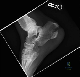
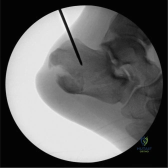
Severe post-traumatic ankle arthritis stemming from an old fracture often presents with chronic pain, limited ROM, and deformity. Imaging reveals tricompartmental joint space narrowing, significant osteophyte formation, fibular nonunion, malunion of malleoli, chronic syndesmosis diastasis, and hindfoot varus. Pre-operative CT templating is crucial for complex deformity correction.
A 65-year-old male presents with chronic end-stage right ankle pain, 33 years post-injury. He has a fixed varus deformity and a symptomatic lateral malleolar nonunion. Based on the radiographs provided, describe your assessment of the mechanical axis and how the fibular pathology influences your surgical decision-making.
Candidate: The patient has post-traumatic arthritis with a varus tilt and a lateral malleolar nonunion. Because the lateral support is gone, the talus has shifted laterally and tilted into varus. I would suggest an ankle arthrodesis, as the deformity is fixed and there is significant bone loss and nonunion. I would use the fibula as a bone graft to help the fusion.
Failing to emphasize the biomechanical consequences. Candidates often miss the "1mm lateral shift = 42% contact area loss" concept. They also often fail to mention the necessity of restoring the mechanical axis (0-5 degrees of valgus) to prevent long-term failure and adjacent segment disease.
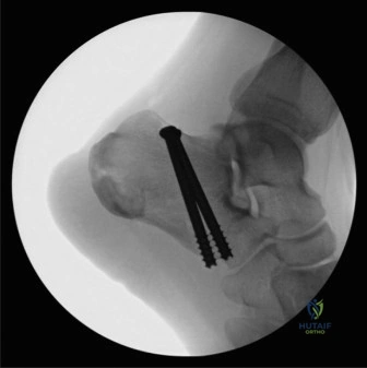
The candidate should identify the "lateral buttress" failure. State that the fibular nonunion and shortening lead to lateral talar shift, causing a dramatic reduction in tibiotalar contact area. The varus deformity creates medial overload. Therefore, the goal is: 1) Resect the nonunion to allow for correction of the varus. 2) Perform an ankle arthrodesis. 3) Utilize the excised fibula as an autologous structural onlay graft to restore lateral column support and biological bridging.
During your preoperative planning, you note the patient is a smoker and has Type 2 Diabetes. Why is this specifically concerning in the context of this planned procedure, and how do you manage it?
Candidate: Smoking and diabetes both significantly increase the risk of nonunion and surgical site infection (SSI). I would ask him to stop smoking and check his HbA1c to ensure it is under 7.5%. I might also use bone stimulators postoperatively.
Being passive. Simply "advising" the patient to stop smoking is insufficient. A high-scoring candidate discusses the objective verification (e.g., serum cotinine) and the multidisciplinary approach to metabolic control.
Acknowledge that nicotine causes vasoconstriction and inhibits osteogenesis, while hyperglycemia impairs neutrophil function and wound healing. The answer must be structured: 1) Objective smoking cessation verified by cotinine testing. 2) Optimization of HbA1c < 7.5%. 3) Preoperative infectious disease/endocrinology optimization. 4) A low threshold for prophylactic plastic surgery involvement for wound closure if the envelope is compromised.
You have decided on a transfibular approach. Detail your intraoperative setup and the specific surgical sequence to ensure a successful fusion.
Candidate: I would place the patient in the lateral decubitus position. I'll make a lateral incision, remove the distal fibula, and clear out the nonunion. I'll then remove the articular cartilage from the tibia and talus until I see bleeding bone. I'll align the foot in neutral dorsiflexion, a few degrees of valgus, and 5-10 degrees of external rotation, then fix it with screws.
Forgetting the soft tissue release. In a chronic varus deformity, the deltoid ligament is often contracted. Simply cutting the bone and fixating it will result in "varus malunion" if you don't release the medial tether.
Structure the answer: 1) Positioning (Lateral decubitus). 2) Approach (Internervous plane). 3) Fibular Management (Excision/decortication). 4) Articular Preparation (Denuding cartilage, "fish-scaling" subchondral bone). 5) Deformity Correction (Crucial: Medial deltoid release to correct varus). 6) Alignment (0° dorsiflexion, 5° valgus, 5-10° ER). 7) Fixation (Compression screws + fibular onlay strut).
