Operative Management of Hand Arthritis: Osteoarthritis, Rheumatoid Arthritis, and SLE
Key Takeaway
Hand osteoarthritis and systemic arthropathies like rheumatoid arthritis and systemic lupus erythematosus present complex reconstructive challenges. This guide details the biomechanics, clinical presentation, and step-by-step surgical management of these conditions. From trapeziometacarpal arthroplasty to soft-tissue realignments for lupus-induced ligamentous laxity, mastering these techniques is essential for restoring hand function, correcting severe deformities, and alleviating debilitating pain in the arthritic hand.
Comprehensive Introduction and Patho-Epidemiology
The human hand represents an evolutionary marvel of biomechanical engineering, providing both the power for forceful grip and the precision for intricate manipulation. Consequently, it is highly susceptible to a variety of degenerative and systemic arthropathies, each presenting with distinct pathoanatomy, biomechanical alterations, and reconstructive challenges. While osteoarthritis (OA) is driven by mechanical wear, cartilage degradation, and osteophyte formation, systemic conditions such as rheumatoid arthritis (RA) and systemic lupus erythematosus (SLE) are characterized by complex inflammatory cascades that target the synovium, tendons, and capsuloligamentous structures. For the reconstructive hand surgeon, understanding the nuanced differences between these entities is not merely an academic exercise, but the foundation of operative decision-making. Distinguishing between the fixed, erosive joint destruction of RA and the reducible, ligamentous laxity of SLE dictates whether a joint-sacrificing or joint-preserving procedure is indicated.
Osteoarthritis is the most ubiquitous of the rheumatic diseases affecting the upper extremity, with a striking predilection for postmenopausal women. Epidemiological data suggest that radiographic evidence of hand OA is present in over 70% of individuals above the age of 65. Unlike weight-bearing joints where OA is often secondary to trauma or malalignment, primary hand OA has a strong genetic predisposition, frequently manifesting in a polyarticular pattern involving the distal interphalangeal (DIP), proximal interphalangeal (PIP), and trapeziometacarpal (TMC) joints. The pathophysiology is primarily mechanical, characterized by progressive cartilage fragmentation, subchondral sclerosis, and marginal spur formation. Unlike rheumatoid arthritis, OA is less frequently associated with spontaneous tendon ruptures or severe inflammatory tenosynovitis, though these pathologies can occasionally coexist in the setting of severe osteophytic impingement.
Rheumatoid arthritis, conversely, is a systemic autoimmune disease characterized by a proliferative, erosive synovitis (pannus). The global prevalence of RA is approximately 0.5% to 1%, with a female-to-male ratio of 3:1. The pannus directly invades and destroys articular cartilage and subchondral bone while simultaneously attenuating capsuloligamentous structures through enzymatic degradation. This dual assault leads to complex, multi-planar, and rapidly fixed deformities. The hand and wrist are involved in up to 90% of RA patients, often serving as the initial site of clinical presentation. The advent of disease-modifying antirheumatic drugs (DMARDs) and biologic agents has dramatically altered the natural history of RA, reducing the incidence of end-stage deformities; however, the reconstructive surgeon still frequently encounters patients with medically refractory disease or those who present late with established joint destruction.
Systemic lupus erythematosus (SLE) introduces yet another distinct pathophysiological paradigm. SLE is a diffuse connective tissue disease capable of affecting multiple organ systems, with musculoskeletal involvement occurring in up to 90% of patients. Unlike RA, which targets the synovium, the inflammatory process in SLE primarily attacks the tendons, joint capsules, and ligaments. The classic hand presentation begins with profound ligamentous laxity at the metacarpophalangeal (MCP) and PIP joints. Because the primary pathology is capsuloligamentous attenuation rather than proliferative erosive synovitis, the articular cartilage remains remarkably well-preserved until very late in the disease process. This results in severe, yet passively reducible, deformities—a condition classically termed Jaccoud's arthropathy. Recognizing this reducible nature is paramount, as joint-preserving soft-tissue realignments are prioritized over arthrodesis or arthroplasty in the lupus hand.
Detailed Surgical Anatomy and Biomechanics
A profound mastery of hand anatomy and biomechanics is a prerequisite for executing successful reconstructive procedures. The trapeziometacarpal (TMC) joint is a biconcave-biconvex saddle joint that allows for an exceptional range of motion, including flexion, extension, abduction, adduction, and opposition. This mobility, however, comes at the cost of intrinsic osseous stability. Stability is therefore highly reliant on a complex network of ligaments. Historically, the anterior oblique ligament (AOL), also known as the volar beak ligament, was considered the primary stabilizer against dorsal subluxation of the first metacarpal. However, contemporary biomechanical studies have demonstrated that the dorsoradial ligament complex is equally, if not more, critical in preventing dorsal translation during pinch and grasp activities. During key pinch, the joint reaction forces across the TMC joint are magnified up to 12 times the force applied at the thumb tip, explaining the high propensity for degenerative failure at this specific articulation.
The extensor mechanism of the digits is an intricate, highly balanced system that is frequently disrupted in systemic arthropathies. The extrinsic extensor tendon (extensor digitorum communis) trifurcates over the proximal phalanx. The central slip inserts into the dorsal base of the middle phalanx, extending the PIP joint. The lateral slips diverge, join the intrinsic tendons (lumbricals and interossei) to form the lateral bands, and converge distally to insert on the dorsal base of the distal phalanx as the terminal tendon, extending the DIP joint. The delicate balance between these structures is maintained by the transverse retinacular ligament, which prevents dorsal subluxation of the lateral bands, and the triangular ligament, which prevents their volar subluxation. In rheumatoid arthritis, attenuation of these stabilizing ligaments leads to predictable collapse patterns, namely the swan-neck and boutonnière deformities.
In the rheumatoid hand, the metacarpophalangeal (MCP) joint is particularly vulnerable to volar and ulnar subluxation. The normal MCP joint is an ellipsoid articulation stabilized by the collateral ligaments, the volar plate, and the sagittal bands of the extensor hood. The rheumatoid pannus distends the joint capsule, stretching the collateral ligaments and the radial sagittal band. The natural ulnar-directed forces of daily grasp, combined with the unresisted pull of the ulnar intrinsic muscles (abductor digiti minimi and volar interossei), lead to ulnar drift. Concurrently, the extensor tendons subluxate into the ulnar valleys between the metacarpal heads, exacerbating the deformity and creating a vicious cycle of progressive subluxation and loss of active extension.
The vascular anatomy of the hand is of critical importance, particularly in patients with SLE who frequently suffer from severe Raynaud phenomenon. The superficial and deep palmar arches supply the common digital arteries, which bifurcate into the proper digital arteries. These vessels are highly reactive to sympathetic tone. In the lupus patient, an exaggerated vasospastic response to cold or emotional stress can lead to profound ischemia. The sympathetic nerve fibers travel within the adventitia of these digital arteries. Understanding this microanatomy is essential for performing a periarterial digital sympathectomy, a limb-salvage procedure designed to strip the adventitia and disrupt the vasospastic reflex arc, thereby restoring perfusion to the threatened digit.
Exhaustive Indications and Contraindications
Surgical intervention for hand arthropathies is generally reserved for patients who have failed exhaustive conservative management, including customized orthotic splinting, activity modification, nonsteroidal anti-inflammatory drugs (NSAIDs), and judicious intra-articular corticosteroid injections. The decision to proceed with surgery must carefully weigh the patient's functional demands, pain levels, and the specific pathoanatomy of their disease. In systemic diseases like RA and SLE, coordination with the treating rheumatologist is mandatory to optimize perioperative immunomodulation and assess the overall systemic burden of the disease.
For osteoarthritis of the trapeziometacarpal joint, the Eaton-Littler classification guides surgical decision-making. Stage I involves mild widening of the joint space (synovitis) without cartilage loss. Stage II exhibits slight joint space narrowing and osteophytes smaller than 2 mm. Stage III is characterized by significant joint space narrowing, subchondral sclerosis, and osteophytes larger than 2 mm. Stage IV involves pan-trapezial arthritis, with degenerative changes extending to the scaphotrapezial (STT) joint. Trapeziectomy with or without ligament reconstruction is typically indicated for symptomatic Stage III and IV disease. Arthrodesis of the TMC joint may be considered in young, high-demand manual laborers who require absolute stability at the expense of mobility, though it carries a risk of nonunion and adjacent joint degeneration.
In rheumatoid arthritis, the indications for surgery are dictated by the severity of joint destruction and the presence of tendon ruptures. Prophylactic tenosynovectomy is indicated when persistent tenosynovitis threatens tendon integrity, particularly at the dorsal wrist (Vaughan-Jackson syndrome) or volar wrist (Mannerfelt-Norman syndrome). For the MCP joints, silicone interpositional arthroplasty is indicated for fixed, painful, and subluxated joints with severe cartilage destruction (Larsen grade IV or V). For the PIP and DIP joints, arthrodesis is often the most reliable procedure to restore a stable pinch and grasp, particularly when bone stock is insufficient for arthroplasty or when the soft-tissue envelope is severely compromised.
Systemic lupus erythematosus requires a fundamentally different surgical algorithm. Because the articular cartilage is preserved (Jaccoud's arthropathy), joint-sacrificing procedures like arthrodesis or arthroplasty are absolutely contraindicated in the early and middle stages of the disease. Indications for surgery include severe, progressive soft-tissue deformities that significantly impair function, such as reducible ulnar drift or swan-neck deformities. Soft-tissue realignment, intrinsic release, and capsulodesis are the procedures of choice. Additionally, severe, medically refractory Raynaud phenomenon with impending digital ulceration or necrosis is a strict indication for digital sympathectomy.
Indications and Contraindications Table
| Procedure | Primary Indications | Absolute Contraindications | Relative Contraindications |
|---|---|---|---|
| TMC LRTI (Trapeziectomy) | Eaton Stage III/IV OA; severe pain refractory to conservative care; preserved MCP joint function. | Active local or systemic infection; severe medical comorbidities precluding anesthesia. | Age < 40 (consider osteotomy or arthrodesis); heavy manual laborer. |
| MCP Silicone Arthroplasty | Fixed, erosive RA (Larsen IV/V); severe ulnar drift with joint destruction; pain and loss of function. | Active infection; inadequate bone stock for stems; functioning, reducible joint. | Poor skin envelope; active, uncontrolled systemic disease flare. |
| PIP/DIP Arthrodesis | Severe, painful OA/RA with instability; fixed Boutonnière/Swan-neck deformities. | Active infection; lack of adjacent joint mobility (can lead to completely stiff finger). | Heavy smoking (nonunion risk); severe osteoporosis. |
| Digital Sympathectomy | SLE/Scleroderma with medically refractory Raynaud's; impending digital necrosis/ulceration. | Viable alternative medical therapies exist; complete arterial occlusion (requires bypass/grafting). | Vasculitis flare (requires medical optimization first). |
| Soft-Tissue Realignment | Reducible Jaccoud's arthropathy (SLE); early RA without joint erosion. | Fixed joint contractures; severe articular cartilage destruction (Larsen IV/V). | Patient inability to comply with complex postoperative rehab. |
Pre-Operative Planning, Templating, and Patient Positioning
Meticulous pre-operative planning is the cornerstone of successful hand reconstruction. For osteoarthritic conditions, high-quality, specialized radiographs are essential. The Robert's view (a hyper-pronated anteroposterior view of the thumb) provides a true AP projection of the TMC joint, allowing for accurate assessment of joint space narrowing, osteophyte formation, and subluxation. Stress views, obtained by having the patient press the tips of the thumbs together, can unmask dynamic instability. The surgeon must also critically evaluate the adjacent joints. Hyperextension of the thumb MCP joint greater than 30 degrees must be addressed concurrently during TMC arthroplasty (via volar capsulodesis or EPB transfer) to prevent a postoperative zigzag collapse deformity.
For rheumatoid and SLE patients, standard posteroanterior, lateral, and oblique radiographs of the hand and wrist are required. In RA, the surgeon must look for periarticular osteopenia, marginal erosions, and joint space loss. Templating is critical when planning for silicone MCP arthroplasty. The surgeon must measure the medullary canals of the metacarpals and proximal phalanges to ensure appropriate implant sizing, as oversized stems can cause iatrogenic fractures, while undersized stems lead to instability and early implant failure. Furthermore, cervical spine radiographs (flexion-extension views) are absolutely mandatory in all RA patients prior to surgery to rule out atlantoaxial subluxation, which poses a catastrophic risk during intubation and patient positioning.
Optimization of the patient's medical status is particularly crucial in systemic arthropathies. Patients on biologic DMARDs or chronic corticosteroids are at a significantly elevated risk for perioperative infections and delayed wound healing. The surgeon must collaborate with the rheumatologist to determine the appropriate perioperative withholding schedule for these medications, adhering to guidelines established by the American College of Rheumatology (ACR) and the American Association of Hip and Knee Surgeons (AAHKS). Generally, conventional synthetic DMARDs (like methotrexate) can be continued, while biologic agents should be withheld for one dosing cycle prior to surgery and resumed once wound healing is complete.
In the operating room, patient positioning and setup must be standardized. The patient is positioned supine with the operative extremity extended onto a radiolucent hand table. Regional anesthesia (supraclavicular or axillary brachial plexus block) is highly preferred, as it provides excellent intraoperative muscle relaxation and profound postoperative analgesia, facilitating early rehabilitation. A well-padded pneumatic tourniquet is applied to the proximal arm. Exsanguination is achieved with an Esmarch bandage, and the tourniquet is inflated to 250 mmHg (or 100 mmHg above systolic blood pressure). The surgeon must ensure that all necessary specialized equipment, including micro-instruments, oscillating saws, burrs, and a full set of trial implants, are readily available on the sterile field before making the incision.
Step-by-Step Surgical Approach and Fixation Technique
Trapeziometacarpal (TMC) Arthroplasty: LRTI Technique
When conservative measures fail for advanced TMC osteoarthritis (Eaton-Littler Stages III and IV), Trapeziectomy with Ligament Reconstruction and Tendon Interposition (LRTI) remains a gold-standard procedure.
- Incision and Exposure: A Wagner or modified dorsal longitudinal incision is made over the TMC joint, curving slightly volar over the first metacarpal. Meticulous blunt dissection is utilized to identify, mobilize, and protect the superficial sensory branches of the radial nerve. The abductor pollicis longus (APL) and extensor pollicis brevis (EPB) tendons are identified and retracted.
- Capsulotomy and Trapeziectomy: A longitudinal or T-shaped capsulotomy is performed. The periosteum is elevated to expose the trapezium. The trapezium is fragmented using a small osteotome or rongeur and excised piecemeal. Extreme caution must be exercised in the depths of the trapezial fossa to avoid injuring the flexor carpi radialis (FCR) tendon, which lies immediately volar and ulnar to the trapezium.
- Tendon Harvest: The FCR tendon is exposed through a series of three small transverse volar incisions in the distal forearm. The radial half of the FCR is harvested from its musculotendinous junction distally, maintaining its insertion at the base of the second metacarpal.
- Bone Tunnel Creation: A 3.2 mm or 4.0 mm drill hole is created in the base of the first metacarpal, starting at the dorsal-radial cortex and exiting directly through the articular base into the trapezial void.
- Ligament Reconstruction: The harvested FCR slip is passed through this medullary tunnel from proximal to distal. The thumb is placed in palmar abduction and opposition, and the tendon is tensioned and sutured to the periosteum and adjacent capsule. This reconstructs the volar oblique ligament, suspending the thumb metacarpal and preventing proximal subsidence.
- Tendon Interposition and Closure: The remaining redundant FCR tendon slip is folded upon itself to create an "anchovy" or biologic spacer. This is sutured into the trapezial void to provide a cushion and maintain space. The dorsal capsule is meticulously repaired using non-absorbable sutures. The skin is closed, and a well-padded thumb spica splint is applied in palmar abduction.
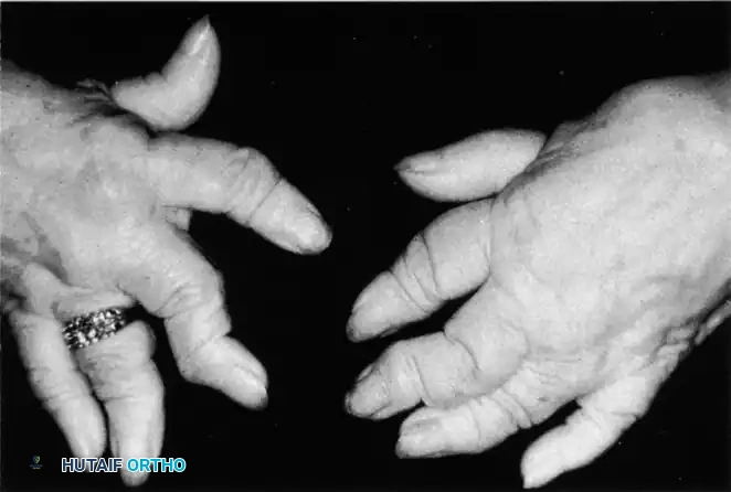
Rheumatoid Arthritis: Swan-Neck and Boutonnière Deformities
Involvement of the MCP and PIP joints frequently results in laxity of the capsuloligamentous structures, leading to predictable collapse patterns.
The Swan-Neck Deformity (seen above) is characterized by PIP joint hyperextension and DIP joint flexion. In RA, this is often initiated by terminal tendon rupture or volar plate laxity at the PIP joint, exacerbated by intrinsic muscle tightness. Surgical management depends on joint flexibility. For a flexible deformity, soft-tissue procedures such as flexor digitorum superficialis (FDS) tenodesis or lateral band mobilization are indicated to restrict PIP hyperextension. If the PIP joint is fixed and arthritic, arthrodesis or surface replacement arthroplasty is required.
Conversely, the Boutonnière Deformity involves PIP joint flexion and DIP joint hyperextension. This occurs when the central slip of the extensor tendon ruptures or attenuates, allowing the lateral bands to subluxate volar to the axis of rotation of the PIP joint.
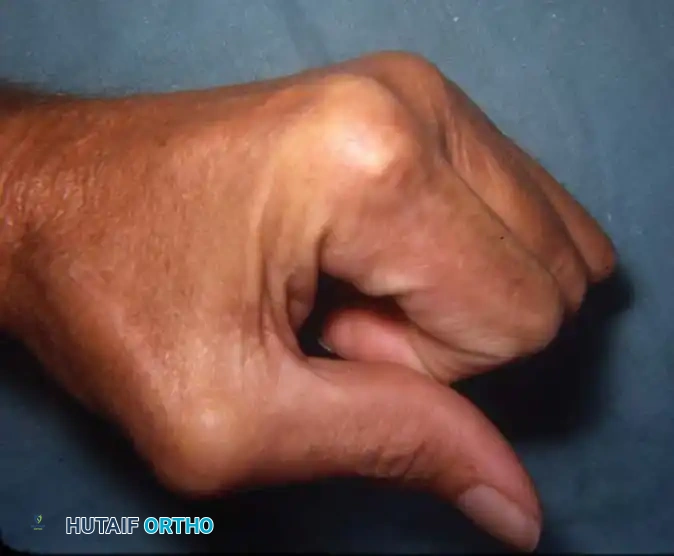
For a fixed rheumatoid boutonnière deformity of the thumb (Type I, as seen above), soft-tissue reconstruction alone is universally doomed to fail due to the massive forces across the joint and poor tissue quality. Arthrodesis of the MCP joint combined with IP joint release or pinning is the most reliable method to restore a stable pinch mechanism. The MCP joint is typically fused in 10 to 15 degrees of flexion using crossed Kirschner wires, a tension band construct, or a dedicated compression plate.
Advanced Erosive Disease: "Main en Lorgnette"
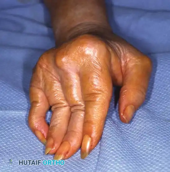
In severe, progressive, and neglected rheumatoid disease, profound osteolysis and articular destruction occur. The phalanges and metacarpals telescope into one another, creating redundant, folded skin. This end-stage presentation is known as the "main en lorgnette" or opera glass hand.
Surgical management at this stage is highly complex. The primary goal is to restore longitudinal stability to the digits. This often requires multiple joint arthrodeses using autologous bone grafting (frequently harvested from the iliac crest or distal radius) to bridge massive segmental bone defects. Extensive tenosynovectomies are performed concurrently to salvage any remaining functional tendons. Silicone arthroplasty (Swanson implants) may be utilized at the MCP joints to maintain a functional arc of motion, but only if sufficient diaphyseal bone stock remains to support the implant stems.
Systemic Lupus Erythematosus: Soft-Tissue Realignment and Sympathectomy
Because SLE primarily causes capsuloligamentous attenuation with preserved cartilage, joint-sacrificing procedures are delayed. For reducible MCP ulnar drift (Jaccoud's arthropathy), soft-tissue realignment is indicated. A transverse dorsal incision is made over the metacarpal heads. The ulnar sagittal bands are released, allowing centralization of the extensor tendons over the MCP joints. An ulnar intrinsic release is performed to remove the deforming force pulling the digits into ulnar deviation. Finally, the attenuated radial collateral ligaments and dorsal capsule are imbricated or reconstructed to restore stability.
For patients with SLE suffering from severe, medically refractory Raynaud phenomenon, a periarterial digital sympathectomy is indicated. Using an operating microscope, the common digital arteries are exposed in the palm via a zigzag (Bruner) incision. The adventitia of the artery, which houses the sympathetic nerve fibers, is meticulously stripped over a distance of 1 to 2 centimeters. Any constricting fascial bands or transverse retinacular ligaments compressing the neurovascular bundles are released. This delicate microsurgical procedure disrupts the vasospastic reflex arc, dramatically improving baseline perfusion and promoting ulcer healing.
Complications, Incidence Rates, and Salvage Management
Surgical intervention in the arthritic hand is fraught with potential complications, ranging from minor wound healing issues to catastrophic implant failure. The surgeon must be intimately familiar with these risks and possess the technical armamentarium to manage them effectively.
In TMC LRTI procedures, the most common radiographic complication is proximal subsidence of the first metacarpal. While mild subsidence is nearly universal and often asymptomatic, severe subsidence (>50% of the trapezial space) can lead to impingement of the metacarpal base against the scaphoid, causing recurrent pain. Salvage management for symptomatic subsidence involves revision suspensionplasty using an alternative tendon (such as the APL or ECRL) or, in recalcitrant cases, conversion to a TMC arthrodesis. Complex Regional Pain Syndrome (CRPS) is another devastating complication following hand surgery, characterized by disproportionate pain, swelling, stiffness, and sudomotor changes. Early recognition and aggressive multimodal intervention, including sympathetic blocks, gabapentinoids, and intensive hand therapy, are critical.
In rheumatoid reconstruction, particularly with silicone MCP arthroplasties, implant fracture and silicone particulate synovitis are significant concerns. Modern high-performance silicone elastomers have reduced the incidence of fracture, but it remains a risk in high-demand patients. Silicone synovitis presents as a painful, swollen joint with radiographic evidence of progressive osteolysis around the implant stems. Salvage requires complete implant removal, aggressive debridement of the reactive synovium and particulate debris, and either placement of a new implant (if bone stock permits) or conversion to an excision arthroplasty or arthrodesis.
Infection is a profound risk in patients with systemic arthropathies due to their underlying immunocompromised state and the concurrent use of biologic agents and corticosteroids. Superficial infections can often be managed with oral antibiotics and local wound care. Deep infections involving the joint space or implants require emergent surgical irrigation and debridement. If an implant is present, it must typically be removed, and an antibiotic-impregnated cement spacer is placed until the infection is eradicated, followed by a delayed secondary reconstruction.
Complications and Salvage Management Table
| Complication | Estimated Incidence | Associated Procedure | Salvage Management / Treatment Strategy |
|---|---|---|---|
| Metacarpal Subsidence | 15% - 30% (Radiographic) | TMC LRTI / Trapeziectomy | Often asymptomatic. If painful: revision suspensionplasty, interposition of allograft, or TMC arthrodesis. |
| Silicone Implant Fracture | 10% - 20% (at 10 years) | MCP/PIP Arthroplasty | Implant removal, debridement, and revision arthroplasty vs. arthrodesis depending on bone stock. |
| Silicone Synovitis | 5% - 10% | MCP/PIP Arthroplasty | Aggressive synovectomy, removal of particulate debris, implant extraction, and bone grafting of osteolytic cysts. |
| Recurrent Deformity | 20% - 40% (Long-term) | SLE Soft-Tissue Realignment | Progression of disease. May eventually require joint-sacrificing procedures (arthrodesis/arthroplasty) once cartilage degrades. |
| Deep Joint Infection | 2% - 5% (Higher in RA/SLE) | All arthroplasties/fusions | Emergent I&D, implant removal, placement of antibiotic spacer, 6 weeks IV antibiotics, delayed reconstruction. |
| CRPS (Type I) | 3% - 7% | All hand procedures | Early recognition is key. Stellate ganglion blocks, high-dose Vitamin C, aggressive desensitization therapy. |
Phased Post-Operative Rehabilitation Protocols
The ultimate success of any reconstructive hand procedure is inextricably linked to the quality of postoperative rehabilitation. The hand is exceptionally prone to stiffness, and a delicate balance must be struck between protecting the surgical repair and initiating early motion to prevent debilitating adhesions. Rehabilitation protocols must be tailored to the specific procedure performed and the underlying pathology of the patient.
Rehabilitation following TMC LRTI
- Phase I (Weeks 0-4): Protection and Healing. The patient is placed in a rigid, custom-molded thermoplastic thumb spica orthosis, immobilizing the wrist and thumb (MCP and TMC joints) while leaving the IP joint free. Strict elevation and edema control are emphasized. Active range of motion (AROM) of the uninvolved digits, elbow, and shoulder is initiated immediately to prevent proximal stiffness.
- Phase II (Weeks 4-8): Early Motion. The rigid orthosis is transitioned to a removable splint, worn at all times except during therapy. Under the guidance of a certified hand therapist (CHT), the patient begins gentle AROM of the thumb, focusing on palmar abduction, radial abduction, and opposition to the small finger. Forceful pinch and grasp are strictly prohibited to protect the ligament reconstruction.
- Phase III (Weeks 8-12): Strengthening. The splint is gradually weaned during the day but maintained at night. Progressive strengthening exercises are introduced, utilizing therapy putty and hand grippers. Isotonic and isometric exercises are advanced as tolerated.
- Phase IV (Months 3-6): Return to Function. Unrestricted activity is typically permitted after 12 weeks. Patients are advised that maximal grip and pinch strength, as well as complete resolution of surgical site ache, may take up to 6 to 12 months to achieve.
Rehabilitation following MCP Silicone Arthroplasty (RA)
Rehabilitation following MCP arthroplasty in the rheumatoid hand is highly specialized. The goal is to maintain the corrected alignment while allowing the formation of a stable, functional pseudocapsule around the silicone implant.
- Phase I (Days 3-5 to Week 4): Dynamic Splinting. The bulky postoperative dressing is removed early. A custom dynamic extension splint is fabricated. This orthosis supports the wrist in slight extension and provides dynamic outrigger traction to maintain the MCP joints in neutral alignment (preventing ulnar drift) while allowing active flexion. The patient performs active flexion against the dynamic traction, and the rubber bands passively extend the digits.
- Phase II (Weeks 4-6): Weaning and Active Motion. The dynamic splint is worn primarily at night and during periods of rest. Active and active-assisted range of motion exercises are advanced. The therapist focuses on intrinsic stretching and maintaining the centralization of the extensor tendons.
- Phase III (Weeks 6-12): Functional Integration. Gentle strengthening is initiated. However, due to the inherent vulnerability of the silicone implants and the underlying rheumatoid disease, heavy resistive exercises and forceful repetitive grasping are permanently discouraged to maximize the longevity of the arthroplasties.
Summary of Landmark Literature and Clinical Guidelines
The operative management of hand arthritis has evolved significantly over the past half-century, guided by several landmark anatomical, biomechanical, and clinical studies. A comprehensive understanding of this literature is essential for the academic orthopedic surgeon.
The foundational principles of trapeziometacarpal osteoarthritis management were established by Eaton and Littler (1973), who described the critical role of the anterior oblique ligament in preventing dorsal subluxation of the first metacarpal. They introduced the concept of ligament reconstruction using a slip of the FCR tendon, a technique that remains highly relevant today. Their staging system for TMC arthritis (Stages I-IV) remains the universally accepted standard for radiographic classification and surgical decision-making.
The modern LRTI technique was popularized by Burton and Pellegrini (1986). In their seminal paper published in the Journal of Hand Surgery, they detailed the technique of complete trapeziectomy, FCR ligament reconstruction through a metacarpal bone tunnel, and tendon interposition. Their long-term follow-up studies demonstrated excellent pain relief, restoration of pinch strength, and prevention of severe metacarpal subsidence, establishing the LRTI as the gold standard against which all other basal joint procedures are measured.
In the realm of rheumatoid hand reconstruction, the contributions of Alfred B. Swanson (1972) cannot be overstated. Swanson pioneered the concept of flexible implant arthroplasty using silicone elastomers. His extensive research demonstrated that the silicone implant acts not as a true joint replacement, but as a dynamic spacer that maintains alignment while a functional, fibrous pseudocapsule forms around it. Despite the advent of pyrolytic carbon and surface replacement arthroplasties, the Swanson silicone implant remains the most reliable and widely utilized device for rheumatoid MCP joint destruction.
For systemic lupus erythematosus, the literature emphasizes the critical distinction between erosive and non-erosive arthropathy. Bywaters (1950) provided the classic description of Jaccoud's arthropathy, detailing the profound capsuloligamentous laxity and reducible deformities that characterize the condition. Contemporary clinical guidelines from the American Society for Surgery of the Hand (ASSH) strongly advocate for joint-preserving soft-tissue realignments in SLE patients, reserving arthrodesis only for end-stage, fixed deformities where cartilage has secondarily degraded.
In conclusion, the operative management of hand arthritis requires a profound understanding of the underlying systemic or mechanical pathology. Whether performing a complex ligament reconstruction for basal
