Operative Management of Gout and Scleroderma in the Hand
Key Takeaway
Gout and scleroderma present complex reconstructive challenges in hand surgery. While acute gout mimics infection, chronic tophaceous gout causes severe tendon and joint destruction requiring meticulous debridement. Scleroderma leads to profound contractures and ischemia, necessitating joint arthrodesis, resection arthroplasty, and digital sympathectomy. This guide details the evidence-based surgical indications, operative techniques, and postoperative protocols for managing these debilitating systemic arthropathies.
Comprehensive Introduction and Patho-Epidemiology
The surgical management of systemic arthropathies and connective tissue diseases—specifically gout, calcium pyrophosphate deposition disease (pseudogout), and scleroderma—requires a profound understanding of both the underlying medical pathophysiology and the complex biomechanical alterations they inflict upon the hand and wrist. Unlike primary osteoarthritis, which is largely a mechanically driven degenerative process, these conditions are characterized by aggressive soft tissue infiltration, profound inflammatory cascades, and severe microvascular compromise. The orthopedic hand surgeon must navigate a delicate balance between aggressive debridement or reconstruction and the preservation of inherently tenuous vascular supplies. Operating on these patients demands not only technical precision but also an exhaustive comprehension of the systemic disease processes that dictate local tissue healing and long-term functional outcomes.
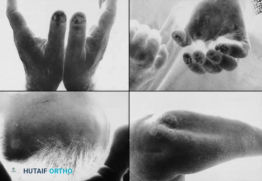
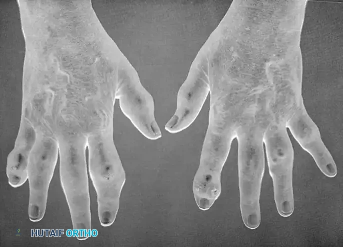
Gout is a crystal-induced arthropathy caused by the deposition of monosodium urate (MSU) crystals within joints, periarticular soft tissues, and tendon sheaths. The pathophysiological hallmark is hyperuricemia, leading to the precipitation of MSU crystals when local tissue saturation points are exceeded. These crystals are intensely pro-inflammatory, directly activating the NLRP3 inflammasome within local macrophages and triggering a massive release of interleukin-1β (IL-1β). While gout typically presents as an acute, exquisitely painful, erythematous monoarthropathy in men (classically podagra), its chronic tophaceous form frequently involves the hand and wrist, particularly in postmenopausal women. The sheer volume of these tophaceous deposits mechanically disrupts the delicate architecture of the hand, eroding bone, attenuating tendons, and compromising the overlying dermal capillary networks.
Scleroderma, or systemic sclerosis, presents an entirely different, yet equally devastating, pathophysiological challenge. It is a complex autoimmune connective tissue disease characterized by a triad of microvascular endothelial dysfunction, profound immune dysregulation, and excessive, uncontrolled collagen deposition (primarily Types I and III) in the skin and internal organs. Hand surgeons are most frequently consulted for patients with limited cutaneous scleroderma, historically termed CREST syndrome (Calcinosis, Raynaud phenomenon, Esophageal dysmotility, Sclerodactyly, and Telangiectasia). The age of onset is typically over 40 years, with a strong female predominance. Unlike rheumatoid arthritis, synovial thickening in scleroderma is minimal. The primary pathology lies in the fibrotic transformation of the skin, subcutaneous tissues, and tendon sheaths, leading to relentless ischemic contractures and digital auto-amputation.
When evaluating destructive arthropathies of the hand, the surgeon must definitively differentiate gout and scleroderma from other inflammatory conditions, notably psoriatic arthritis and pseudogout. Psoriatic arthritis frequently involves the distal interphalangeal (DIP) joints and is characterized by "pencil-in-cup" radiographic deformities, dactylitis (sausage digits), and characteristic dermatologic findings. Recognizing the cutaneous manifestations—such as erythematous plaques on the extensor surfaces and severe nail pitting or onycholysis—is critical for differentiating this autoimmune condition from crystal arthropathies. Conversely, pseudogout, caused by calcium pyrophosphate dihydrate (CPPD) crystal deposition, frequently involves the wrist and hand. It mimics acute gout or septic arthritis with intermittent, acute inflammatory attacks and can cause severe flexor tenosynovitis leading to acute median nerve compression.
Detailed Surgical Anatomy and Biomechanics
The surgical anatomy of the hand in the context of gouty arthropathy is defined by the predilection of monosodium urate crystals to precipitate in specific synovial and avascular environments. Tophi exhibit a strong affinity for the flexor and extensor tendon sheaths, the carpal tunnel, and the collateral ligaments of the interphalangeal joints. Within the extensor compartments, the third dorsal compartment is particularly vulnerable. The extensor pollicis longus (EPL) tendon, as it acutely angulates around Lister's tubercle, is subjected to immense mechanical friction. When infiltrated by MSU crystals, the already tenuous watershed vascularity of the EPL is further compromised, leading to spontaneous rupture. Similarly, within the rigid confines of the carpal tunnel, massive tophaceous infiltration of the flexor tenosynovium exponentially increases interstitial pressure, resulting in acute or chronic median nerve ischemia and profound carpal tunnel syndrome.
In scleroderma, the anatomic alterations are driven by severe microvascular obliteration and dermal fibrosis. The superficial and deep palmar arches, along with the common and proper digital arteries, undergo profound intimal hyperplasia and adventitial fibrosis. This chronic ischemia is exacerbated by Raynaud's phenomenon, a hyperactive sympathetic vasoconstrictive response. The sympathetic nerve fibers responsible for this vasospasm travel directly within the adventitial layer of the digital arteries. This anatomic relationship forms the basis for digital sympathectomy, wherein the adventitia is microsurgically stripped from the vessel wall to abolish sympathetic tone and restore distal perfusion. Furthermore, the relentless deposition of dense collagen in the dermis tethers the skin directly to the underlying extensor apparatus, obliterating the normal gliding planes and resulting in the characteristic "sclerodactyly."
Biomechanically, these diseases produce distinct patterns of structural collapse. In chronic tophaceous gout, the expanding tophi act as space-occupying lesions that mechanically block joint excursion and physically erode the collateral ligaments and volar plates. This leads to profound, multidirectional joint instability and subluxation. The chalky MSU deposits infiltrate the tendon substance itself, mechanically separating the collagen fascicles and drastically reducing the tensile strength of the tendon, predisposing it to catastrophic failure under minimal physiological loads.
The biomechanical collapse in scleroderma is characterized by rigid, unyielding contractures due to the global fibrosis of all soft tissue envelopes. The proximal interphalangeal (PIP) joints invariably develop severe, fixed flexion contractures. The metacarpophalangeal (MCP) joints, however, present a more complex biomechanical paradox. They may present with either a severe flexion deformity or a paradoxical hyperextension deformity. The hyperextension deformity at the MCP joint is often a secondary biomechanical consequence of the severe PIP joint flexion contractures, coupled with the fibrotic retraction of the intrinsic musculature and the dorsal skin envelope. Additionally, the first web space undergoes a profound adduction contracture due to the fibrotic shortening of the adductor pollicis aponeurosis and the overlying skin, completely abolishing the patient's ability to perform functional grasp or pinch maneuvers.
Exhaustive Indications and Contraindications
The decision to proceed with surgical intervention in patients with systemic arthropathies is highly nuanced. In gout, surgery is rarely indicated for acute flares; medical management (NSAIDs, colchicine, corticosteroids, and subsequent urate-lowering therapy) remains the absolute gold standard. However, surgical intervention becomes mandatory when chronic tophaceous deposits threaten the structural integrity of the hand or compromise critical neurovascular structures. In scleroderma, the severe contracture of all soft tissue structures limits the potential for functional movement, meaning surgical intervention must be carefully planned, prioritizing stable, functional positions over the unrealistic restoration of full mobility.
🚨 SURGICAL WARNING: The "Unsuspecting Surgeon" Pitfall
Acute gouty arthritis of the hand or wrist can perfectly mimic a closed space infection, abscess, or septic tenosynovitis. An unsuspecting surgeon may hastily perform an incision and drainage (I&D). Inappropriate surgical intervention during an acute gout flare can lead to delayed wound healing, secondary bacterial infection, and chronic sinus tract formation. Always rule out crystal arthropathy via aspiration before proceeding with I&D in atypical presentations.
The primary indications for surgical intervention in gout include the compression of critical neurovascular structures, such as acute or refractory carpal tunnel syndrome secondary to tophaceous flexor tenosynovitis. Impending or actual tendon rupture, most commonly of the EPL or digital extensors, necessitates operative reconstruction. Furthermore, severe mechanical blocks to joint motion caused by massive tophi, progressive skin necrosis or ulceration over an expanding tophus, and intractable pain in a completely destroyed joint are clear indications for surgical debulking, arthrodesis, or amputation.
For scleroderma patients, the indications are driven by the need to salvage function and prevent catastrophic ischemic tissue loss. Debilitating digital contractures that prevent basic activities of daily living, particularly severe first web space adduction contractures, warrant surgical release and reconstruction. Refractory, exquisitely painful fingertip ulcerations that fail exhaustive conservative management are indications for digital sympathectomy. Additionally, the eruption of painful, infected calcinosis cutis deposits through the compromised dermal layer requires surgical excision to achieve wound closure and eradicate chronic infection.
| Condition | Primary Surgical Indications | Absolute Contraindications | Relative Contraindications |
|---|---|---|---|
| Gouty Arthropathy | Acute/refractory nerve compression (e.g., CTS) Impending/actual tendon rupture Massive tophi causing mechanical block Skin necrosis/ulceration over tophus Intractable pain in destroyed joint |
Acute gouty flare (unless concurrent septic arthritis is proven) Medically unstable patient Uncontrolled hyperuricemia (for elective cases) |
Poor overlying skin envelope (high risk of necrosis) Patient non-compliance with urate-lowering therapy Severe peripheral vascular disease |
| Scleroderma (CREST) | Refractory ischemic fingertip ulcers Severe first web space adduction contracture Rigid, non-functional PIP/MCP contractures Painful, erupting calcinosis cutis Intractable Raynaud's pain |
Active smoking/nicotine use Acute systemic crisis (renal, pulmonary) Profound, irreversible digital ischemia (auto-amputation preferred) |
High-dose systemic corticosteroids (poor healing) Severe pulmonary hypertension (anesthesia risk) Unrealistic patient expectations regarding ROM |
> SURGICAL WARNING: Smoking Cessation
Absolute cessation of smoking is a non-negotiable prerequisite for any vascular or reconstructive surgery in the scleroderma patient. Nicotine-induced vasoconstriction will universally doom local flaps, skin grafts, and sympathectomy outcomes.
Pre-Operative Planning, Templating, and Patient Positioning
Pre-operative evaluation of the patient with gouty arthropathy must begin with definitive diagnostic confirmation. The diagnosis of gout is definitively established only by joint or tendon sheath aspiration. Polarized light microscopy will reveal negatively birefringent, needle-shaped monosodium urate crystals. Imaging plays a critical role in pre-operative templating. High-resolution plain radiographs will demonstrate the pathognomonic "punched-out" periarticular erosions with overhanging sclerotic margins (Martel's sign). In complex cases, Dual-Energy Computed Tomography (DECT) is invaluable for mapping the exact volumetric burden and anatomic distribution of urate crystals, allowing the surgeon to precisely plan the extent of the required tenosynovectomy or debulking procedure.
CLINICAL PEARL: Serum Uric Acid Reliability
The presence of hyperuricemia alone does not establish the diagnosis of gout, as many hyperuricemic patients never experience an attack. Conversely, during an acute gout flare, serum uric acid levels may be entirely normal due to the acute phase response increasing renal urate excretion. Rely on synovial fluid analysis, not serum labs, for definitive diagnosis.
In the scleroderma patient, pre-operative planning is overwhelmingly focused on assessing the profound microvascular compromise. A meticulous vascular examination, including a digital Allen test, is mandatory. Non-invasive vascular studies, such as digital brachial indices (DBI) and arterial Doppler ultrasound, are utilized to map the patency of the superficial palmar arch and digital vessels. In cases planned for digital sympathectomy, preoperative angiography or high-resolution magnetic resonance angiography (MRA) may be indicated to confirm the presence of targetable vessels. Furthermore, rigorous rheumatologic and pulmonologic clearance is required, as these patients frequently suffer from concurrent interstitial lung disease and pulmonary arterial hypertension, significantly elevating their perioperative anesthetic risk.
Patient positioning and anesthetic considerations are critical components of the surgical plan. The patient is typically positioned supine with the operative extremity extended on a radiolucent hand table. Regional anesthesia (axillary or supraclavicular brachial plexus block) is highly preferred over general anesthesia. In scleroderma patients, a regional block provides a profound, immediate sympathectomy effect, maximizing vasodilation and improving digital perfusion during the procedure. A well-padded pneumatic tourniquet is applied to the upper arm; however, in scleroderma patients with severe vascular disease, the tourniquet time must be strictly minimized, and the pressure should be set to the lowest effective level (typically 50-75 mmHg above systolic pressure) to prevent irreversible endothelial crush injury.
When planning surgical incisions, the surgeon must respect the compromised dermal vascularity inherent to both diseases. In gout, incisions should be planned to allow extensile exposure while preserving broad, full-thickness vascularity to the skin flaps. Tophi often adhere intimately to the dermis, and aggressive thinning of the skin flaps will inevitably lead to full-thickness necrosis. In scleroderma, incisions should be meticulously planned to avoid crossing flexion creases whenever possible, and the use of continuous, tension-free dermal closures is paramount. The surgeon must anticipate the need for local tissue rearrangements (Z-plasties) or full-thickness skin grafts, particularly when releasing severe first web space contractures.
Step-by-Step Surgical Approach and Fixation Technique
Surgical Management of Gouty Arthropathy
The operative approach to tophaceous gout focuses on meticulous debulking and the preservation of vital neurovascular structures. The chalky, toothpaste-like MSU deposits are carefully curetted and irrigated. The surgeon must recognize that complete, oncologic-level excision is often impossible without sacrificing vital structures; the goal is mechanical debulking to relieve pressure and restore gliding planes. If the flexor or extensor tendons are heavily infiltrated, a meticulous tenosynovectomy is performed under loupe magnification. The epitenon is preserved whenever possible to maintain tendon vascularity.
In cases of tendon rupture, primary repair is rarely possible due to massive substance loss and extreme tissue friability. Tendon transfers are the preferred reconstructive option. For the frequently ruptured EPL tendon, the Extensor Indicis Proprius (EIP) to EPL transfer is the workhorse procedure. The EIP is harvested proximal to the extensor hood, routed subcutaneously, and woven into the distal stump of the EPL using a Pulvertaft weave. Tensioning is set with the wrist in neutral and the thumb in full extension. In cases of extreme bony disruption and joint destruction caused by massive intra-articular tophi, arthrodesis is required. The articular surfaces are aggressively debrided of all crystalline material down to bleeding subchondral bone, and rigid fixation is achieved using crossed K-wires, tension band constructs, or low-profile dorsal plates, depending on the quality of the surrounding soft tissue envelope.
Surgical Management of Scleroderma
Because the severe contracture of all soft tissue structures limits the potential for functional movement, surgical intervention in scleroderma must prioritize stable, functional positions over the restoration of full mobility. At the Distal Interphalangeal (DIP) joint, severe skin ulceration, fixed flexion contractures, dry gangrene, and secondary osteomyelitis are common. While arthrodesis is an option, healing is highly unpredictable due to severe vascular compromise. Terminal amputation is often the most reliable method to rapidly eliminate pain, eradicate infection, and provide a durable soft tissue envelope.
Scleroderma invariably leads to rigid PIP joint flexion contractures. Soft tissue releases (volar plate release, checkrein ligament excision) are universally unsuccessful due to the diffuse fibrotic nature of the disease. Arthrodesis is the procedure of choice. The joint is approached dorsally, and the articular surfaces are prepared. The joint is fused in a functional position (typically 30 to 40 degrees of flexion, increasing from the index to the small finger). Tension-band wiring or buried K-wires are heavily preferred over bulky plates, which the atrophic, tightly bound skin simply cannot accommodate without catastrophic wound breakdown.
The Metacarpophalangeal (MCP) joint may present with a severe flexion deformity or a paradoxical hyperextension deformity. Resection arthroplasty has proven to be a highly effective method for preserving motion and correcting alignment at this joint, as championed by Nalebuff, Melone, McLoughlin, and Beldner. If the primary deformity is MCP flexion, the joint is approached through a standard dorsal longitudinal incision. The metacarpal head is resected at the metaphyseal flare to completely decompress the joint and allow extension. If the MCP joint is hyperextended (often accompanied by severe PIP flexion), Nalebuff recommends a palmar approach. A transverse or zigzag palmar incision is made, the flexor tendons are retracted, and the metacarpal head is resected from the volar aspect. This volar approach simultaneously releases the contracted volar skin and allows for concurrent dorsal fusion of the PIP joint to correct the complex multi-joint collapse pattern.
Management of Ischemia and Web Space Contractures
Adduction contracture of the first web space is a profoundly disabling complication. Release of the adductor pollicis aponeurosis is performed through a dorsal or palmar approach. If the contracture is severe and involves the carpometacarpal joint, Nalebuff advocates for concomitant trapezial excision to thoroughly decompress the first ray. Thumb MCP and IP joint fusions may be added to provide a stable post for pinch. Full-thickness skin grafting or local Z-plasties of the web space are frequently required to close the soft tissue defect.
For refractory ischemic ulcers and severe Raynaud's pain, digital sympathectomy is indicated. As described by Flatt, Wilgis, and Jones, this involves microsurgical stripping of the adventitia (which contains the sympathetic nerve fibers) from the common and proper digital arteries. Operating under operating microscope magnification, a 2 to 3 cm segment of the adventitia is meticulously excised. Intra-arterial injection of vasodilating drugs (e.g., papaverine, botulinum toxin A, or calcium channel blockers) during surgery helps dilate the spastic vessels. Subcutaneous calcium deposits (calcinosis cutis) frequently erupt through the eroded pulps of the fingers. These deposits are excised through a mid-lateral incision to avoid placing a scar on the tactile volar pulp.
Complications, Incidence Rates, and Salvage Management
Surgical intervention in the setting of systemic arthropathies and connective tissue diseases is fraught with a high incidence of postoperative complications. The underlying pathophysiology of both gout and scleroderma inherently compromises the normal phases of wound healing. In gout, the primary complications revolve around wound breakdown over massive tophaceous deposits and the recurrence of the disease. The skin overlying chronic tophi is often paper-thin, ischemic, and intimately adherent to the underlying crystal mass. Even with meticulous dissection, marginal skin necrosis is common, occurring in up to 15-20% of extensive debulking procedures.
In scleroderma, the complication profile is dominated by the profound microvascular ischemia. Flap necrosis, failure of full-thickness skin grafts, and recurrent digital ulceration are pervasive risks. The atrophic, fibrotic skin has a drastically reduced capacity for neovascularization. Furthermore, the rate of nonunion following arthrodesis in scleroderma patients is significantly elevated compared to the general population, primarily due to the avascular nature of the prepared bone ends. Infection, particularly secondary to erupting calcinosis cutis or ischemic dry gangrene, can rapidly progress to deep space infections or osteomyelitis if not aggressively managed.
Salvage management requires a multidisciplinary approach and a willingness to accept functional compromises. For recalcitrant wound breakdown in gout, prolonged local wound care, negative pressure wound therapy (NPWT), and secondary intention healing are often required, as local tissue transfers are frequently unviable. In cases of infected nonunion or progressive ischemic necrosis in scleroderma, ray amputation or terminal digital amputation is often the definitive salvage procedure. Free tissue transfer is theoretically possible but is considered exceptionally high-risk and generally contraindicated due to the severe systemic microvascular disease affecting potential recipient vessels.
| Complication | Estimated Incidence | Pathophysiologic Driver | Salvage Management Strategy |
|---|---|---|---|
| Wound Dehiscence / Necrosis | 15-25% (Scleroderma) 10-20% (Gout) |
Microvascular ischemia; dermal adherence to tophi; poor collagen synthesis | Prolonged local wound care; NPWT; secondary intention; split-thickness skin grafting (if bed is vascularized) |
| Arthrodesis Nonunion | 10-15% (Scleroderma) | Avascular subchondral bone; inability to use rigid compression plating | Prolonged immobilization; revision with autologous bone grafting; conversion to terminal amputation |
| Recurrent Ischemic Ulceration | 30-40% (post-sympathectomy at 5 yrs) | Progressive intimal hyperplasia; relentless autoimmune vasculopathy | Optimization of systemic vasodilators (prostacyclins, PDE5 inhibitors); repeat botulinum toxin injections; amputation |
| Post-Operative Acute Gout Flare | 20-30% | Surgical stress response; fluid shifts altering serum urate concentration | Immediate initiation of high-dose NSAIDs, oral corticosteroids, or intra-articular steroid injection |
| Secondary Infection / Osteomyelitis | 5-10% | Erupting calcinosis cutis acting as a nidus; chronic open ischemic ulcers | Aggressive surgical debridement; targeted intravenous antibiotic therapy; ray amputation for definitive source control |
Phased Post-Operative Rehabilitation Protocols
The postoperative management of systemic arthropathies requires a meticulously coordinated, multidisciplinary approach involving the orthopedic surgeon, the rheumatologist, and a specialized certified hand therapist (CHT). The rehabilitation protocols must be carefully tailored to respect the compromised biology of the soft tissues while preventing the rapid recurrence of debilitating contractures. Postoperative flares of gout are exceedingly common due to the physiological stress of surgery and massive fluid shifts. Consequently, prophylactic medical management (e.g., colchicine, NSAIDs, or low-dose corticosteroids) must be optimized perioperatively in conjunction with the rheumatology service.
Phase 1: Immediate Post-Operative Period (0-2 Weeks)
Following arthrodesis, resection arthroplasty, or extensive tenosynovectomy, the hand is placed in rigid immobilization utilizing a bulky, non-compressive dressing to allow for initial soft tissue healing and to manage profound postoperative edema. Strict elevation is mandatory. In scleroderma patients, the surgeon must be hyper-vigilant regarding dressing tension; even mild compression can precipitate catastrophic digital ischemia. Wound care is paramount. In scleroderma patients, sutures must be left in place significantly longer than usual—often 3 to 4 weeks—due to delayed collagen synthesis and poor vascularity. Early removal will almost certainly result in wound dehiscence.
Phase 2: Early Mobilization and Splinting (2-6 Weeks)
For gout patients who have undergone tenosynovectomy or tendon transfer (e.g., EIP to EPL), early protected active range of motion is initiated under the strict guidance of a hand therapist to prevent dense peritendinous adhesions. Dynamic or static progressive splinting is utilized to protect tendon repairs while allowing controlled excursion. For scleroderma patients, therapy focuses intensely on maintaining the functional gains achieved in the operating room. Custom thermoplastic orthoses are fabricated to hold the digits in the newly achieved, corrected positions (e.g., maintaining MCP extension following volar resection arthroplasty). Active ROM is encouraged within the limits of pain and wound healing, but aggressive passive stretching is avoided to prevent micro-tearing of the fragile skin.
Phase 3: Long-Term Maintenance and Systemic Optimization (6+ Weeks)
As the soft tissues stabilize, the focus shifts to maximizing functional grasp and pinch. Strengthening exercises are gradually introduced, though patients with advanced systemic disease will likely never return to baseline grip strength. For scleroderma patients, long-term nighttime splinting is often necessary indefinitely to prevent the insidious recurrence of flexion contractures. Thermal management becomes critical; patients must be educated on the strict avoidance of cold exposure and the continuous use of thermal gloves to mitigate Raynaud's attacks. Systemic vasodilators (calcium channel blockers, phosphodiesterase inhibitors) must be rigorously maintained. Regular follow-up is required to monitor for the recurrence of tophi in gout patients or the development of new ischemic ulcers in scleroderma patients, allowing for prompt, conservative intervention before surgical salvage becomes necessary.
Summary of Landmark Literature and Clinical Guidelines
The surgical management of systemic arthropathies in the hand is guided by several landmark contributions that have defined the modern operative approach. In the realm of scleroderma, the fundamental principles of joint reconstruction were established by Nalebuff, whose seminal work described the paradoxical biomechanics of the MCP joint. Nalebuff's advocacy for the palmar approach to MCP resection arthroplasty in the setting of severe hyperextension deformities remains a cornerstone technique, allowing for simultaneous volar skin release and joint decompression. This work was further expanded upon by Melone, McLoughlin, and Beldner, who validated the long-term functional benefits of aggressive metacarpal head resection in restoring functional grasp.
The management of profound digital ischemia in scleroderma was revolutionized by the anatomical and clinical studies of Flatt, Wilgis, and Jones. Their pioneering descriptions of the digital sympathetic anatomy—specifically the localization of sympathetic nerve fibers within the adventitia of the proper digital arteries—provided the anatomical rationale for peripheral digital sympathectomy. Their long-term outcome studies demonstrated that while adventitial stripping may not permanently halt the progression of the underlying autoimmune vasculopathy, it provides critical, immediate vasodilation that is highly effective for healing recalcitrant ischemic ulcers and providing sustained pain relief.
In the management of gouty arthropathy, current clinical guidelines heavily emphasize the primacy of medical management. The American College of Rheumatology (ACR) guidelines strictly dictate that surgical intervention should be reserved for cases of structural failure, impending tendon rupture, or severe neurovascular compromise. The literature consistently demonstrates that aggressive surgical debulking of tophi in the absence of mechanical compromise carries an unacceptably high rate of wound complications. Furthermore, contemporary rheumatologic guidelines mandate the perioperative use of prophylactic agents (colchicine or systemic steroids) to mitigate the well-documented phenomenon of surgically induced acute gout flares, ensuring that the orthopedic intervention does not inadvertently precipitate a systemic inflammatory crisis.
