Glenohumeral Rheumatoid Arthritis & Septic Arthritis: Diagnosis, Anatomy & Management
Key Takeaway
Diagnosing septic arthritis in glenohumeral rheumatoid arthritis (RA) is challenging due to overlapping symptoms and high patient susceptibility. Early recognition is paramount. It requires careful assessment of RA-specific risk factors (e.g., immunosuppression), considering distinct surgical anatomy and altered biomechanics. Joint aspiration is often crucial for timely management and preserving function.
A 58-year-old female with long-standing rheumatoid arthritis (RA) presents with acute worsening of right shoulder pain, malaise, and a temperature of 38.1°C. She is currently on methotrexate and a TNF-alpha inhibitor. You suspect septic arthritis of the glenohumeral joint. How do you approach the initial diagnostic workup?
Candidate: I would perform a thorough history and physical exam, check inflammatory markers (ESR/CRP), get blood cultures, and perform an urgent joint aspiration under ultrasound guidance to send for cell count, Gram stain, and culture. I would also consider withholding her DMARDs/biologics.
Failing to emphasize that RA patients have a high baseline level of inflammation and joint pain, which masks infection. Borderline candidates often treat the synovial fluid analysis like that of an osteoarthritis patient; in RA, the threshold for suspicion of septic arthritis must be lower, and the differential must include crystalline arthropathy (e.g., gout/pseudogout).
Start with a structured, multi-disciplinary approach: (1) Clinical: Distinguish systemic vs. local symptoms; (2) Labs: Serial ESR/CRP, WBC, and blood cultures; (3) The Gold Standard: US-guided aspiration. Crucially, mention that cell counts >50,000/µL are diagnostic, but one must interpret this in the context of her baseline RA synovitis. Add the necessity of polarizing light microscopy to rule out crystal-induced arthropathy, which is a key mimic in this population.
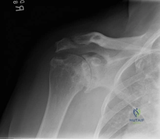
You have diagnosed septic arthritis of the shoulder in this patient. Radiographs reveal advanced destruction of the humeral head and glenoid, with superior migration of the humeral head. What are your surgical management considerations?
Candidate: I would take her to the operating room for an urgent irrigation and debridement (I&D) and synovectomy. I would avoid placing an implant at this stage because of the active infection, and I would start her on culture-directed intravenous antibiotics.
Suggesting a single-stage procedure or rushing to discuss arthroplasty too early. Failing to address the "cuff tear arthropathy" pattern (superior migration) seen on the images, which implies that if she eventually requires reconstruction, a standard TSA is inappropriate; it would have to be an rTSA.
Emphasize "Source Control." Mandatory urgent open or arthroscopic I&D and synovectomy. Acknowledge that the superior migration indicates irreparable rotator cuff deficiency, mandating a future reverse total shoulder arthroplasty (rTSA) once infection is eradicated. State the strict contraindication for primary arthroplasty during active infection. Define the "cool down" period: 6–12 months of monitoring before considering definitive reconstruction.
The patient has been cleared of infection 9 months post-debridement. She has persistent pain and functional limitation. You are planning a reverse total shoulder arthroplasty. What are the key technical challenges in an RA patient compared to a primary OA patient?
Candidate: In RA patients, the bone quality is usually much poorer due to osteopenia. The glenoid anatomy is also frequently distorted with eccentric bone loss, making baseplate fixation difficult. I would also be concerned about the quality of the deltoid muscle and potential prior capsular scarring.
Missing the importance of the subscapularis. Candidates often forget to mention how the subscapularis status in RA is often poor, and while rTSA doesn't strictly require it, surgeons must account for the impact on anterior stability.
Systematically address: (1) Bone Stock: Management of severe glenoid bone loss (augmented baseplates or bone grafting); (2) Soft Tissues: The need for thorough capsular releases to restore tension, despite chronic contractures; (3) Implant Choice: The reliance on the deltoid as the primary motor, noting that RA often involves generalized systemic atrophy; (4) Infection Risk: Acknowledge the heightened risk of PJI in RA and the need for meticulous soft tissue handling and post-operative vigilance.
