Dysplasia Epiphysealis Hemimelica (DEH) MCQs - Arab Board
Dysplasia Epiphysealis Hemimelica (DEH) MCQs - Arab Board
Comprehensive 100-Question Exam
00:00
Start Quiz
Question 1
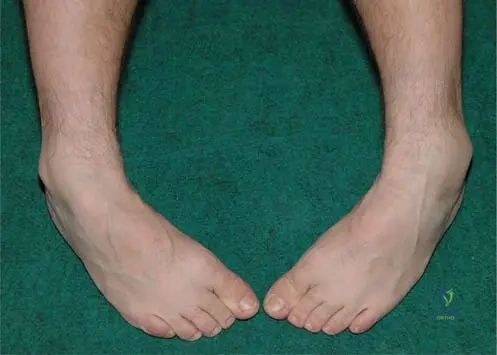
A 6-year-old boy presents with a painless, hard swelling on the lateral aspect of his left ankle, as shown in the clinical photograph below. He has a limited range of motion in the affected joint. Given the most likely diagnosis, which of the following is true regarding the typical epidemiological profile of this condition?
Explanation
Correct Answer: C
The clinical image demonstrates a bone-hard swelling typical of Dysplasia Epiphysealis Hemimelica (DEH), also known as Trevor's disease. DEH is a rare skeletal developmental disorder that typically presents in young children between the ages of 2 and 14 years. It has an incidence of 1 in 1,000,000, affects males twice as frequently as females, and predominantly involves the lower limbs (distal femur, distal tibia, and talus).
Question 2
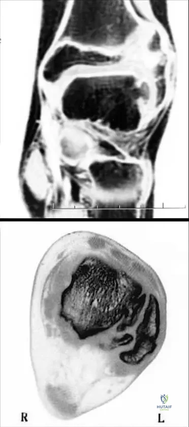
A 9-year-old boy presents with chronic ankle pain and stiffness. The MRI and CT scans of his hindfoot are provided below. Based on the imaging findings demonstrating a lesion arising from the epiphysis, what is the most likely histological diagnosis of this mass?
Explanation
Correct Answer: C
The imaging shows Dysplasia Epiphysealis Hemimelica (DEH) localized on the lateral side of the talus. Histologically, DEH is identical to an osteochondroma. The key distinguishing feature is its location: DEH arises from the epiphysis, whereas a classic osteochondroma arises from the metaphysis or diaphysis.
Question 3
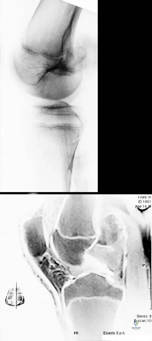
A 12-year-old male presents with a palpable mass behind his knee and restricted knee flexion. Imaging is shown below. The mass is seen protruding from the distal femoral epiphysis. Which of the following best describes the characteristic growth pattern of this pathology?
Explanation
Correct Answer: B
The images demonstrate DEH of the distal femur. Characteristically, the involvement in DEH is 'hemimelic', meaning it affects only one half (either the medial or the lateral side) of the epiphysis. It does not typically involve the entire epiphysis symmetrically, nor does it originate in the metaphysis or diaphysis.
Question 4
Dysplasia epiphysealis hemimelica (DEH) shares identical histological features with which of the following benign bone tumors, and how is it primarily distinguished from it clinically and radiographically?
Explanation
Correct Answer: B
Histologically, the lesion in Dysplasia Epiphysealis Hemimelica (DEH) is similar to an osteochondroma (composed of bone covered by a cartilage cap). However, they are distinguished by their site of origin: osteochondromas arise from the metaphysis or diaphysis, whereas DEH uniquely arises from the epiphysis.
Question 5
Which of the following statements accurately reflects the epidemiology and etiology of Dysplasia Epiphysealis Hemimelica (DEH)?
Explanation
Correct Answer: C
DEH is a very rare disorder with an incidence of 1 in 1,000,000. It affects males twice as frequently as females. The etiology remains unknown (unlike multiple hereditary exostoses which are linked to EXT genes). The age of onset is usually between 2 and 14 years, and it predominantly affects the lower extremities.
Question 6
A 5-year-old boy is being evaluated for a hard, asymmetrical mass around his knee. Radiographs are obtained. Which of the following radiographic findings is most characteristic of Dysplasia Epiphysealis Hemimelica?
Explanation
Correct Answer: B
On radiographs, DEH characteristically shows asymmetric epiphyseal enlargement with multiple ossification centers. This reflects the abnormal, hemimelic overgrowth of the epiphyseal cartilage and its subsequent irregular ossification.
Question 7
Dysplasia epiphysealis hemimelica (DEH) most frequently affects the lower extremities. Which of the following groups of bones represents the most common sites of involvement?
Explanation
Correct Answer: B
DEH usually occurs in the lower limb. The most commonly affected sites are the distal femur, distal tibia, and the talus. Upper limb involvement is extremely rare.
Question 8
A 7-year-old male is brought to the orthopedic clinic by his parents. Which of the following constellations of symptoms is most consistent with a presentation of Dysplasia Epiphysealis Hemimelica (DEH)?
Explanation
Correct Answer: C
The most common presenting symptoms of DEH include the presence of a mass with the consistency of bone, joint deformity, aching pains, and a limited range of motion due to the mechanical block caused by the epiphyseal overgrowth.
Question 9
Review the provided MRI and CT images of a pediatric patient presenting with hindfoot pain and restricted motion. The pathology is localized to the lateral side of the affected bone. Based on the typical distribution of this disease, which bone is primarily affected in this image?
Explanation
Correct Answer: C
The images (MRI frontal plane and CT slides) demonstrate DEH localized on the lateral side of the talus. The talus is one of the three most common locations for DEH, alongside the distal femur and distal tibia.
Question 10
The images below show a lateral radiograph and a sagittal MRI of a knee in a young patient with aching pain and limited range of motion. The mass originates from the distal femoral epiphysis. Into which anatomical space is the bone mass predominantly protruding?
Explanation
Correct Answer: B
The sagittal MRI slide clearly shows a protruding bone mass extending posteriorly from the distal femoral epiphysis directly into the popliteal fossa. This posterior extension can cause significant mechanical block to knee flexion and aching pain.
Question 11
A 7-year-old boy presents with the clinical finding shown below. Radiographs reveal an underlying epiphyseal lesion. Histological examination of this mass would most likely reveal features identical to which of the following conditions?
Explanation
Correct Answer: Osteochondroma
The image shows a moderate, bone-hard swelling typical of Dysplasia Epiphysealis Hemimelica (DEH). Histologically, DEH is indistinguishable from an osteochondroma, featuring a cartilage cap with underlying trabecular bone. The key difference is anatomical location: DEH arises from the epiphysis, whereas osteochondromas arise from the metaphysis or diaphysis.
Question 12
Review the provided MRI and CT images of a pediatric patient's ankle. What is the primary anatomical feature that distinguishes this specific pathology from a solitary osteochondroma?
Explanation
Correct Answer: It arises from the epiphysis rather than the metaphysis
The imaging demonstrates Dysplasia Epiphysealis Hemimelica (DEH) localized to the lateral side of the talus. While histologically similar to an osteochondroma, DEH is uniquely characterized by its origin from the epiphysis. It is typically unilateral, asymmetric (hemimelic), and more common in males.
Question 13
The imaging below demonstrates a protruding bone mass in the posterior aspect of the knee in a 10-year-old child. Based on the typical behavior of this disease, which of the following statements regarding its anatomical distribution is most accurate?
Explanation
Correct Answer: It characteristically involves either the medial or lateral half of the epiphysis
The images show DEH of the distal femur. The term 'hemimelica' refers to the characteristic involvement of only one half (either medial or lateral) of the affected epiphysis. It rarely affects the upper extremities and does not typically involve the entire epiphysis symmetrically.
Question 14
Dysplasia epiphysealis hemimelica (DEH) is a rare developmental disorder. Which of the following best describes the epidemiological profile of this condition?
Explanation
Correct Answer: Incidence of 1 in 1,000,000 with male predominance
DEH is an extremely rare skeletal developmental disorder with an estimated incidence of 1 in 1,000,000. Males are affected twice as frequently as females.
Question 15
A 9-year-old male presents with aching pain, deformity, and limited range of motion in his right knee. Radiographs show asymmetric epiphyseal enlargement of the distal femur with multiple ossification centers. What is the most likely diagnosis?
Explanation
Correct Answer: Dysplasia Epiphysealis Hemimelica
The clinical presentation of aching pain, deformity, and limited range of motion combined with the hallmark radiographic finding of asymmetric epiphyseal enlargement with multiple ossification centers is classic for Dysplasia Epiphysealis Hemimelica (DEH).
Question 16
Regarding the age of onset for Dysplasia Epiphysealis Hemimelica (DEH), during which of the following age ranges do patients most commonly present with initial symptoms?
Explanation
Correct Answer: 2 to 14 years
DEH is a developmental disorder affecting the epiphyses in young children. The age of onset is usually between 2 and 14 years, corresponding to the period of active epiphyseal growth and ossification.
Question 17
A pediatric orthopedic surgeon is evaluating a child with suspected Dysplasia Epiphysealis Hemimelica (DEH). Which of the following anatomical locations is the LEAST likely to be the primary site of involvement?
Explanation
Correct Answer: Proximal humerus
DEH usually occurs in the lower limb, with the distal femur, distal tibia, and talus being the most commonly affected sites. Upper limb involvement, such as the proximal humerus, is extremely rare.
Question 18
Which of the following radiographic findings is considered the hallmark of Dysplasia Epiphysealis Hemimelica in a growing child?
Explanation
Correct Answer: Asymmetric epiphyseal enlargement with multiple ossification centers
The characteristic radiographic appearance of DEH is asymmetric epiphyseal enlargement (hemimelic involvement) accompanied by multiple ossification centers within the affected area.
Question 19
The advanced imaging provided shows a lesion characteristic of Dysplasia Epiphysealis Hemimelica (DEH). Given the intra-articular nature of this pathology, what is the most common clinical presentation associated with this specific location?
Explanation
Correct Answer: Presence of a bone-hard mass with aching pains and limited range of motion
The most common presenting symptoms of DEH include the presence of a mass with the consistency of bone, deformity, aching pains, and limited range of motion due to the intra-articular and epiphyseal nature of the overgrowth.
Question 20
The sagittal MRI provided demonstrates a mass protruding from the distal femoral epiphysis into the popliteal fossa. In the context of Dysplasia Epiphysealis Hemimelica, what is the fundamental etiology of this condition?
Explanation
Correct Answer: Unknown etiology
Despite its histological similarity to osteochondromas (which can be linked to EXT1/EXT2 mutations in multiple hereditary exostoses), the etiology of Dysplasia Epiphysealis Hemimelica (DEH) remains unknown.
Question 21
A 7-year-old boy presents with the clinical finding shown below. Palpation reveals a painless, bone-hard swelling. Based on the most likely diagnosis, what is the histological appearance of this lesion most similar to?
Explanation
Correct Answer: Osteochondroma
The clinical image demonstrates a bone-hard swelling on the lateral side of the ankle, characteristic of Dysplasia Epiphysealis Hemimelica (DEH). Histologically, DEH is identical to an osteochondroma. The key differentiating factor is its anatomical origin: DEH arises from the epiphysis, whereas a classic osteochondroma arises from the metaphysis or diaphysis.
Question 22
Review the coronal MRI and CT images of a pediatric patient's ankle below. The lesion is localized to the lateral aspect of the talus. Which of the following terms best describes this characteristic pattern of involvement?
Explanation
Correct Answer: Hemimelic
The images show Dysplasia Epiphysealis Hemimelica (DEH) localized to the lateral side of the talus. Characteristically, the involvement in this condition is 'hemimelic', meaning it affects only one half (either the medial or lateral side) of the epiphysis.
Question 23
A 10-year-old male presents with aching pain and limited knee range of motion. The lateral radiograph and sagittal MRI are shown below. The mass protrudes into the popliteal fossa. From which specific anatomical region does this pathology primarily arise?
Explanation
Correct Answer: Epiphysis
The images demonstrate Dysplasia Epiphysealis Hemimelica (DEH) of the knee. Unlike osteochondromas which arise from the metaphysis, DEH is a developmental disorder that specifically arises from the epiphysis, leading to asymmetric epiphyseal overgrowth and joint deformity.
Question 24
Dysplasia epiphysealis hemimelica (DEH) is a rare skeletal developmental disorder. Which of the following demographic profiles is most consistent with the typical presentation of DEH?
Explanation
Correct Answer: A 4-year-old male
The age of onset for DEH is usually between 2 and 14 years. Furthermore, males are affected twice as frequently as females, making a young male child the most typical demographic presentation.
Question 25
When evaluating a child with suspected Dysplasia Epiphysealis Hemimelica (DEH), what is the classic radiographic hallmark observed in the affected joint?
Explanation
Correct Answer: Asymmetric epiphyseal enlargement with multiple ossification centers
On radiographs, DEH characteristically presents as asymmetric epiphyseal enlargement (due to its hemimelic nature) accompanied by multiple ossification centers within the affected epiphyseal region.
Question 26
A biopsy is obtained from a bony mass in the distal tibia of an 8-year-old boy. The pathologist reports that the histology is identical to an osteochondroma. Which of the following clinical or radiographic findings would definitively point to a diagnosis of Dysplasia Epiphysealis Hemimelica (DEH) rather than a standard osteochondroma?
Explanation
Correct Answer: The lesion arises from the epiphysis
While DEH and osteochondroma share identical histological features (including a cartilage cap), their anatomical origins differ. Osteochondromas arise from the metaphysis or diaphysis, whereas DEH arises exclusively from the epiphysis.
Question 27
Dysplasia epiphysealis hemimelica (DEH) predominantly affects the lower extremities. Which of the following groups of bones represents the most common sites of involvement?
Explanation
Correct Answer: Distal femur, distal tibia, and talus
DEH usually occurs in the lower limb. The most commonly affected anatomical sites are the distal femur, distal tibia, and the talus. Upper limb involvement is considered extremely rare.
Question 28
Dysplasia epiphysealis hemimelica (DEH) is an extremely rare condition. According to epidemiological data, what is the estimated incidence of this disorder?
Explanation
Correct Answer: 1 in 1,000,000
DEH is a very rare skeletal developmental disorder with an estimated incidence of 1 in 1,000,000 in the general population.
Question 29
A 9-year-old boy is evaluated for a mass around his ankle. Which of the following is NOT a typical presenting symptom or sign of Dysplasia Epiphysealis Hemimelica (DEH)?
Explanation
Correct Answer: Rapidly progressive overlying skin erythema and warmth
DEH typically presents with a bone-hard mass, deformity, aching pains, and limited range of motion. Rapidly progressive erythema and warmth are signs of acute inflammation or infection (e.g., osteomyelitis or septic arthritis) and are not characteristic of DEH.
Question 30
A resident is reviewing cases of Dysplasia Epiphysealis Hemimelica (DEH) for a departmental presentation. They note a specific pattern regarding the anatomical distribution of the lesions. Which of the following statements is true regarding the anatomical distribution of DEH?
Explanation
Correct Answer: Upper limb involvement is extremely rare.
DEH is predominantly a condition of the lower extremities (distal femur, distal tibia, talus). Upper limb involvement is documented in the literature but is considered extremely rare.
Question 31
A 6-year-old boy presents with the clinical finding shown below. Based on the most likely diagnosis, what is the typical age of onset for this condition?
Explanation
Correct Answer: 2 to 14 years
The image demonstrates a moderate, painless, bone-hard swelling at the lateral side of the ankle, characteristic of Dysplasia Epiphysealis Hemimelica (DEH). According to the text, the age of onset for DEH is usually between 2 and 14 years.
Question 32
The imaging below shows a lesion localized on the lateral side of the talus. Histologically, this lesion is identical to an osteochondroma. What is the primary anatomical difference between this condition and a classic osteochondroma?
Explanation
Correct Answer: This lesion arises from the epiphysis
The imaging shows Dysplasia Epiphysealis Hemimelica (DEH) of the talus. Histologically, DEH is similar to an osteochondroma. However, the key distinguishing feature is that an osteochondroma arises from the metaphysis or diaphysis, whereas DEH arises directly from the epiphysis.
Question 33
A 9-year-old male presents with limited range of motion in the knee. Imaging is shown below. Which of the following statements is true regarding the epidemiology of this condition?
Explanation
Correct Answer: Males are affected twice as frequently as females
The images display DEH of the distal femoral epiphysis protruding into the popliteal fossa. DEH is a rare skeletal developmental disorder with an incidence of 1 in 1,000,000. Epidemiologically, males are affected twice as frequently as females.
Question 34
Dysplasia epiphysealis hemimelica (DEH) is characterized by asymmetric overgrowth. Which of the following best describes the typical radiographic appearance of DEH?
Explanation
Correct Answer: Asymmetric epiphyseal enlargement with multiple ossification centers
Characteristically, DEH lesions show on radiographs as asymmetric epiphyseal enlargement with multiple ossification centers. This reflects the irregular, hemimelic overgrowth of the affected epiphysis.
Question 35
A 10-year-old boy is diagnosed with Dysplasia Epiphysealis Hemimelica (DEH). Which of the following anatomical locations is MOST commonly affected by this disorder?
Explanation
Correct Answer: Distal femur
DEH usually occurs in the lower limb. The distal femur, distal tibia, and talus are the most commonly affected sites. Upper limb involvement is considered extremely rare.
Question 36
The term 'hemimelica' in Dysplasia Epiphysealis Hemimelica refers to which of the following characteristic patterns of involvement?
Explanation
Correct Answer: Involvement of either the medial or lateral side of the epiphysis
The text explicitly states that characteristically the involvement is hemimelic, meaning that either the medial or the lateral epiphyseal side is involved, rather than the entire epiphysis symmetrically.
Question 37
A 7-year-old child presents with a painless, bone-hard swelling at the lateral side of the left ankle, as shown below. What is the estimated incidence of the suspected condition?
Explanation
Correct Answer: 1 in 1,000,000
The clinical image shows a typical presentation of Dysplasia Epiphysealis Hemimelica (DEH). DEH is a very rare skeletal developmental disorder with an estimated incidence of 1 in 1,000,000.
Question 38
Regarding the clinical presentation of Dysplasia Epiphysealis Hemimelica (DEH), which of the following is considered extremely rare?
Explanation
Correct Answer: Involvement of the upper limb
DEH predominantly affects the lower extremities (distal femur, distal tibia, and talus). The text notes that upper limb involvement is extremely rare. Presentation with a bone-hard mass and aching pains are common symptoms.
Question 39
A patient presents with a protruding bone mass from the distal femoral epiphysis to the popliteal fossa, as seen in the sagittal MRI below. If a biopsy were performed on this mass, the histological findings would most closely resemble which of the following?
Explanation
Correct Answer: Osteochondroma
The MRI shows DEH of the distal femur. Histologically, DEH is similar to an osteochondroma (featuring a cartilage cap over bony trabeculae). The main difference is anatomical origin: DEH arises from the epiphysis, whereas osteochondromas arise from the metaphysis or diaphysis.
Question 40
A 12-year-old boy presents with a deformity and limited range of motion in his right ankle. Imaging reveals an asymmetric epiphyseal enlargement with multiple ossification centers on the medial side of the distal tibia. Which of the following is the most likely diagnosis?
Explanation
Correct Answer: Dysplasia Epiphysealis Hemimelica
The clinical presentation of a young male with limited ROM, deformity, and radiographic findings of asymmetric epiphyseal enlargement with multiple ossification centers on one side (hemimelic) of the distal tibia is the classic description of Dysplasia Epiphysealis Hemimelica (DEH).
Question 41
A 6-year-old boy presents with the clinical finding shown below. Palpation reveals a bone-hard, painless mass. Given the most likely diagnosis of Dysplasia Epiphysealis Hemimelica (DEH), which of the following statements regarding the epidemiology of this condition is most accurate?
Explanation
Correct Answer: C
Dysplasia Epiphysealis Hemimelica (DEH), also known as Trevor's disease, typically presents in young children with an age of onset usually between 2 and 14 years. It is extremely rare (incidence of 1 in 1,000,000), affects males twice as frequently as females, and predominantly involves the lower limbs (distal femur, distal tibia, and talus). It presents as a mass with the consistency of bone, not a soft fluctuant mass.
Question 42
A 9-year-old male presents with restricted ankle motion and a palpable hard mass. The imaging studies below are obtained. Based on the typical characteristics of the demonstrated pathology, which of the following best describes the expected histological findings?
Explanation
Correct Answer: B
The imaging shows Dysplasia Epiphysealis Hemimelica (DEH) localized on the lateral side of the talus. Histologically, DEH is indistinguishable from an osteochondroma. The critical differentiating factor is anatomical location: osteochondromas arise from the metaphysis or diaphysis, whereas DEH arises from the epiphysis.
Question 43
A 12-year-old boy presents with aching pain and limited range of motion in his knee. The imaging below reveals a protruding bone mass extending into the popliteal fossa. Which of the following is the most likely origin of this lesion?
Explanation
Correct Answer: C
The images demonstrate Dysplasia Epiphysealis Hemimelica (DEH) of the knee. By definition, DEH is a developmental disorder affecting the epiphyses. In this specific case, the protruding bone mass originates from the distal femoral epiphysis and extends into the popliteal fossa.
Question 44
When evaluating a pediatric patient suspected of having Dysplasia Epiphysealis Hemimelica (DEH), what is the hallmark radiographic feature typically observed?
Explanation
Correct Answer: B
Characteristically, DEH lesions show on radiographs as asymmetric epiphyseal enlargement with multiple ossification centers. The involvement is typically hemimelic, meaning it affects either the medial or lateral side of the epiphysis, leading to this asymmetric appearance.
Question 45
Which of the following anatomical locations is considered extremely rare for the development of Dysplasia Epiphysealis Hemimelica (DEH)?
Explanation
Correct Answer: D
DEH usually occurs in the lower limb, with the distal femur, distal tibia, and talus being the most commonly affected sites. Upper limb involvement (such as the distal radius) is considered extremely rare.
Question 46
The term 'hemimelica' in Dysplasia Epiphysealis Hemimelica refers to which of the following characteristic patterns of involvement?
Explanation
Correct Answer: C
The term 'hemimelica' denotes that the involvement is characteristically limited to one half of the epiphysis—either the medial or the lateral epiphyseal side is involved, leading to asymmetric growth and deformity.
Question 47
A pediatric orthopedic surgeon is counseling the parents of a child recently diagnosed with Dysplasia Epiphysealis Hemimelica (DEH). When discussing the rarity of the condition, what is the accepted estimated incidence?
Explanation
Correct Answer: E
Dysplasia Epiphysealis Hemimelica (DEH) is an exceptionally rare skeletal developmental disorder. The reported incidence in the literature is approximately 1 in 1,000,000.
Question 48
A 7-year-old boy presents with a bony mass around the knee. Radiographs show an exostosis. The surgeon is differentiating between an osteochondroma and Dysplasia Epiphysealis Hemimelica (DEH). Which of the following features definitively distinguishes DEH from a classic osteochondroma?
Explanation
Correct Answer: C
Histologically, DEH is similar or identical to an osteochondroma. The definitive distinguishing feature is the anatomical origin: an osteochondroma arises from the metaphysis or diaphysis, whereas DEH arises specifically from the epiphysis.
Question 49
Review the advanced imaging of the ankle provided below. The pathology is localized to the lateral side of the talus. In a patient with this specific presentation of Dysplasia Epiphysealis Hemimelica (DEH), which of the following clinical symptoms is most likely to be the primary complaint alongside the palpable mass?
Explanation
Correct Answer: B
The most common presenting symptoms of DEH include the presence of a mass with the consistency of bone, deformity, aching pains, and limited range of motion. Neurological deficits, nocturnal pain relieved by NSAIDs (typical of osteoid osteoma), and pathological fractures are not classic primary presentations of DEH.
Question 50
A resident is reviewing a case series of patients with Dysplasia Epiphysealis Hemimelica (DEH). Based on the established epidemiological profile of this disorder, which of the following demographic presentations is most typical?
Explanation
Correct Answer: B
DEH typically affects young children, with the age of onset usually between 2 and 14 years. Furthermore, males are affected twice as frequently as females. Therefore, an 8-year-old male fits the classic demographic profile perfectly.
Question 51
A 4-year-old boy is brought to the orthopedic clinic with a painless, bony prominence on the medial aspect of his right knee. Radiographs reveal an irregular, multi-centric ossification center arising from the medial distal femoral epiphysis. Which of the following is true regarding the genetic etiology of this condition?
Explanation
Question 52
A 6-year-old child is diagnosed with Dysplasia Epiphysealis Hemimelica of the ankle.
The parents inquire about the potential for malignant transformation of this lesion over the child's lifetime. What is the most accurate information to provide?
Explanation
Question 53
A 7-year-old boy presents with an enlarging mass on the medial aspect of his right foot and ankle.
He complains of a mechanical block to ankle dorsiflexion. Based on the pathogenesis of Dysplasia Epiphysealis Hemimelica, this lesion is histologically indistinguishable from which of the following bone tumors?
Explanation
Question 54
Which of the following bones is statistically the most frequently affected site in Dysplasia Epiphysealis Hemimelica?
Explanation
Question 55
A 5-year-old boy is incidentally found to have an irregular calcified mass arising from the lateral aspect of the proximal tibial epiphysis. He has full range of motion, no pain, and no angular deformity. What is the most appropriate initial management?
Explanation
Question 56
In patients diagnosed with Dysplasia Epiphysealis Hemimelica, the term 'hemimelica' specifically refers to which characteristic clinical feature?
Explanation
Question 57
A 10-year-old male with a known diagnosis of Trevor's disease presents with increasing varus deformity of the knee. The lesion originates from the medial femoral epiphysis. When does this mass typically cease its active growth?
Explanation
Question 58
Which of the following epidemiological profiles best fits the typical presentation of Dysplasia Epiphysealis Hemimelica?
Explanation
Question 59
According to the Azouz classification for Dysplasia Epiphysealis Hemimelica, a patient presenting with involvement of the distal femur, proximal tibia, and talus in the same lower extremity is classified as which of the following?
Explanation
Question 60
A 4-year-old boy is scheduled for excision of a symptomatic epiphyseal lesion of the ankle.
The surgeon carefully plans to resect the overgrowth while protecting the normal epiphysis. What is the most common reason for recurrence following surgical excision of DEH?
Explanation
Question 61
A 3-year-old is being evaluated for a hard, asymmetrical swelling of the ankle. Magnetic Resonance Imaging (MRI) is ordered. What is the primary advantage of MRI over plain radiography in early stages of Dysplasia Epiphysealis Hemimelica?
Explanation
Question 62
Which of the following clinical findings best distinguishes Dysplasia Epiphysealis Multiplex (Multiple Epiphyseal Dysplasia) from Dysplasia Epiphysealis Hemimelica?
Explanation
Question 63
A 9-year-old male with untreated DEH of the medial talus presents with worsening pain during ambulation. What is the most likely long-term sequela if the mechanical incongruity of the joint is left unaddressed?
Explanation
Question 64
Trevor's disease was originally described in 1950 under a different nomenclature. What historical term did Trevor use to describe this pathology?
Explanation
Question 65
When planning an excision for a symptomatic DEH lesion of the distal femur, which of the following describes the most significant challenge specific to this condition compared to a traditional metaphyseal osteochondroma?
Explanation
Question 66
A 5-year-old boy presents with a hard mass on the lateral aspect of his ankle. Plain radiographs demonstrate an eccentric ossific mass adjacent to the lateral talar body. Which of the following conditions presents as an intra-epiphyseal lytic lesion rather than an exostotic mass, distinguishing it from DEH?
Explanation
Question 67
Which zone of the epiphyseal cartilage is theorized to be the origin of the abnormal proliferation in Dysplasia Epiphysealis Hemimelica?
Explanation
Question 68
A 12-year-old boy undergoes surgical excision of a DEH mass from his medial proximal tibia. Postoperatively, what secondary deformity must the surgeon closely monitor for during his remaining growing years?
Explanation
Question 69
In evaluating a new patient with suspected DEH, which of the following systemic syndromic associations should the clinician screen for?
Explanation
Question 70
A pediatric orthopedic surgeon reviews a plain radiograph of a 6-year-old ankle showing a multi-lobulated, stippled ossification center distinct from, but adjacent to, the main talar ossification center. Over time, what is the expected natural radiographic progression of this lesion?
Explanation
Question 71
A 4-year-old boy presents with a painless, hard swelling on the medial aspect of his ankle. Radiographs reveal irregular, multicentric opacities adjacent to the medial aspect of the talar epiphysis. Histological examination of a biopsy from the lesion would most likely reveal which of the following?
Explanation
Question 72
Dysplasia Epiphysealis Hemimelica (DEH) is characterized by asymmetric cartilaginous overgrowth. Which of the following best describes the most common anatomical distribution of this disease?
Explanation
Question 73
A 7-year-old girl is diagnosed with Trevor's disease affecting her distal femur. Which of the following statements regarding the genetic profile of her condition is most accurate?
Explanation
Question 74
What is the fundamental pathophysiological mechanism underlying the development of Dysplasia Epiphysealis Hemimelica?
Explanation
Question 75
Which of the following classification systems is utilized to describe the anatomical extent and multiplicity of lesions in Dysplasia Epiphysealis Hemimelica?
Explanation
Question 76
A 5-year-old child presents with a mass around the knee. Imaging reveals an epiphyseal bony outgrowth consistent with DEH. Which of the following is the most appropriate primary indication for surgical excision?
Explanation
Question 77
When surgical intervention is performed for a symptomatic DEH lesion of the distal femur, what is the most significant potential iatrogenic complication?
Explanation
Question 78
Which of the following imaging modalities is considered the most accurate for delineating the unossified cartilage cap and determining the extent of articular surface involvement in DEH?
Explanation
Question 79
A 6-year-old boy has an irregular, multi-centric ossification mass adjacent to the medial talar epiphysis.
According to the natural history of this condition, when is the progressive growth of the lesion expected to cease?
Explanation
Question 80
In differentiating Dysplasia Epiphysealis Hemimelica (DEH) from Multiple Hereditary Exostoses (MHE), which of the following is a key distinguishing feature of DEH?
Explanation
Question 81
A 4-year-old boy presents with painless swelling of the medial ankle. Imaging shows irregular ossification centers adjacent to the medial malleolus. What is the classic eponymous name for this exact condition?
Explanation
Question 82
Dysplasia Epiphysealis Hemimelica (DEH) typically presents in a specific epidemiological group. Which demographic is overwhelmingly most commonly affected?
Explanation
Question 83
An 8-year-old male child has established Trevor disease involving the medial aspect of the distal femur. If left untreated, what resulting limb deformity is most likely to develop as the child grows?
Explanation
Question 84
A 10-year-old boy with a history of DEH of the ankle develops worsening pain. Serial radiographs are obtained. Which of the following is true regarding the risk of malignant transformation in DEH?
Explanation
Question 85
An MRI of a knee in a 5-year-old with suspected DEH is evaluated.
Which of the following findings on MRI definitively confirms DEH and differentiates it from synovial chondromatosis?
Explanation
Question 86
The Azouz classification system for DEH describes three distinct anatomical forms. A patient presenting with involvement of both the talus and the distal tibia on the ipsilateral side would be classified as which of the following?
Explanation
Question 87
During the surgical excision of a DEH lesion causing a mechanical block in the knee, the surgeon must exercise extreme caution.
What specific anatomical structure is at the highest risk of iatrogenic damage that could lead to severe long-term joint dysfunction?
Explanation
Question 88
While Dysplasia Epiphysealis Hemimelica is most commonly found in the lower extremities, it can rarely involve the upper limb. When the upper limb is involved, which of the following areas is most frequently affected?
Explanation
Question 89
A 5-year-old boy presents with progressive right knee and ankle asymmetry. Radiographs reveal cartilaginous overgrowth involving the medial distal femoral epiphysis, the proximal tibial epiphysis, and the medial aspect of the talus, all confined to the right lower extremity. According to the Azouz classification for Dysplasia Epiphysealis Hemimelica (DEH), which category best describes this patient's presentation?
Explanation
Question 90
The parents of a 4-year-old boy recently diagnosed with Dysplasia Epiphysealis Hemimelica (Trevor's disease) of the left ankle ask about the risk of their future children inheriting the condition. Which of the following is the most accurate genetic counseling information to provide?
Explanation
Question 91
A 6-year-old child presents with a painless, restricted range of motion in the right ankle. An MRI is ordered to evaluate a suspected Dysplasia Epiphysealis Hemimelica (DEH) lesion of the talus. Which of the following MRI findings is most characteristic of this pathology?
Explanation
Question 92
A 7-year-old boy presents with an enlarging mass on the medial side of his knee and a progressive varus deformity. You suspect Dysplasia Epiphysealis Hemimelica (DEH). Epidemiologically and anatomically, which of the following best describes the most common presentation of this condition?
Explanation
Question 93
A 9-year-old male with known Dysplasia Epiphysealis Hemimelica (DEH) of the distal femur presents for a routine follow-up. The parents inquire about the long-term growth behavior of the lesion. Which of the following best describes the natural history of the mass in DEH?
Explanation
Question 94
A 10-year-old child with an untreated, large Dysplasia Epiphysealis Hemimelica (DEH) lesion of the talus presents with worsening foot pain. Radiographs demonstrate significant joint space narrowing and subchondral sclerosis in the tibiotalar joint. What is the most common long-term complication of unmanaged symptomatic DEH?
Explanation
Question 95
A 5-year-old boy undergoes an excisional biopsy of an irregular, hard mass protruding from the medial distal tibial epiphysis. The histopathology report indicates mature trabecular bone covered by a hyaline cartilage cap undergoing endochondral ossification. Based on these findings, how does the histology of Dysplasia Epiphysealis Hemimelica (DEH) compare to an osteochondroma?
Explanation
Question 96
A 6-year-old male child presents with a progressive valgus deformity of his right knee and a palpable mass. Reviewing the classic imaging of Dysplasia Epiphysealis Hemimelica
, what is the most appropriate primary surgical principle if a mechanical block and significant deformity are present?
Explanation
Question 97
During a resident teaching round, the attending asks for the defining anatomical distinction between Multiple Hereditary Exostoses (MHE) and Dysplasia Epiphysealis Hemimelica (DEH). While both present with osteochondroma-like lesions, DEH is uniquely characterized by its origin from which specific region of the developing bone?
Explanation
Question 98
An 8-year-old boy presents with progressive restricted subtalar motion and a firm medial hindfoot mass. Radiographs show irregular, multicentric ossification centers adjacent to the talar body that appear to be coalescing. In planning surgical excision for this suspected Dysplasia Epiphysealis Hemimelica, the surgeon must be aware that the mass often blends indistinguishably with which structure?
Explanation
None
