Full Question & Answer Text (for Search Engines)
Question 1:
During a posterior-stabilized total knee arthroplasty, the trial components are placed and joint kinematics are assessed. The knee is perfectly balanced in extension, but the flexion gap is excessively tight. Which of the following adjustments is most appropriate to correct this mismatch?
Options:
- Thicken the tibial polyethylene insert
- Resect more distal femur
- Increase the posterior slope of the tibial cut
- Upsize the femoral component
- Translate the femoral component posteriorly
Correct Answer: Increase the posterior slope of the tibial cut
Explanation:
A knee that is balanced in extension but tight in flexion requires an intervention that specifically addresses the flexion gap without altering the extension gap. Increasing the posterior slope of the tibial cut removes more posterior tibial bone, effectively opening the flexion gap while having a negligible effect on the extension gap. Upsizing the femoral component or translating it posteriorly would further tighten the flexion gap. Resecting more distal femur would loosen the extension gap.
Question 2:
A 35-year-old male sustains a 'terrible triad' injury of the elbow (elbow dislocation, radial head fracture, and coronoid fracture). According to standard surgical principles for restoring elbow stability, which of the following is the recommended sequence of surgical reconstruction?
Options:
- Radial head fixation/replacement, coronoid fixation, LCL repair, MCL repair
- Coronoid fixation, radial head fixation/replacement, LCL repair, MCL repair (if needed)
- LCL repair, radial head fixation/replacement, coronoid fixation, MCL repair
- MCL repair, coronoid fixation, radial head fixation/replacement, LCL repair
- Coronoid fixation, LCL repair, radial head fixation/replacement, MCL repair
Correct Answer: Coronoid fixation, radial head fixation/replacement, LCL repair, MCL repair (if needed)
Explanation:
The standard surgical protocol for a terrible triad injury, established by Pugh et al., involves a deep-to-superficial repair from the inside out: (1) Fixation of the coronoid process, (2) Fixation or replacement of the radial head, (3) Repair of the lateral collateral ligament (LCL) complex to the lateral epicondyle. Repair of the medial collateral ligament (MCL) or application of an external fixator is reserved for cases where the elbow remains unstable after the first three steps.
Question 3:
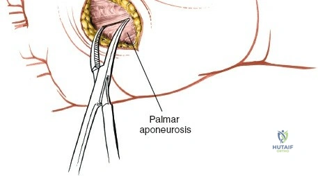
A patient presents with neglected pyogenic flexor tenosynovitis of the thumb. The infection is noted to have spread proximally and now directly involves the flexor sheath of the small finger, creating a 'horseshoe abscess.' This proximal communication occurs via which of the following anatomic structures?
Options:
- Space of Parona
- Midpalmar space
- Thenar space
- Guyon's canal
- Carpal tunnel
Correct Answer: Space of Parona
Explanation:
The flexor tendon sheath of the thumb (radial bursa) and the flexor tendon sheath of the small finger (ulnar bursa) communicate proximally in the distal forearm through the Space of Parona. This potential space is located between the pronator quadratus fascia and the deep flexor tendons, allowing an infection to spread from the thumb to the small finger, resulting in a classic horseshoe abscess.
Question 4:
A structural orthopedic implant is subjected to a constant load below its yield strength over a prolonged period. Over time, the material exhibits a gradual and continuous increase in deformation. This specific viscoelastic property is best described as:
Options:
- Stress relaxation
- Hysteresis
- Creep
- Fatigue
- Isotropy
Correct Answer: Creep
Explanation:
Creep is a viscoelastic property defined as the progressive deformation of a material over time when subjected to a constant load or stress. Stress relaxation is the decrease in stress over time when a material is held at a constant strain (deformation). Hysteresis refers to energy lost as heat during the loading and unloading cycles of a viscoelastic material.
Question 5:
A 45-year-old patient involved in a motor vehicle accident sustains a traumatic spondylolisthesis of the axis (Hangman's fracture). Radiographs reveal severe angulation of C2 on C3 with minimal translation (Levine-Edwards Type IIA). What is the mechanism of this specific injury pattern, and what is the appropriate initial management?
Options:
- Hyperextension and axial loading; application of rigid cervical collar
- Flexion-distraction; application of heavy skeletal traction
- Flexion-distraction; halo vest applied in slight compression and extension
- Hyperextension and axial loading; halo vest applied with heavy traction
- Axial loading; immediate occipitocervical fusion
Correct Answer: Flexion-distraction; halo vest applied in slight compression and extension
Explanation:
Levine and Edwards Type IIA Hangman's fractures result from a flexion-distraction mechanism. Radiographically, they are characterized by severe angulation with minimal translation. Traction is strictly contraindicated in Type IIA fractures because it worsens the distraction and deformity. The correct treatment is closed reduction with gentle compression and slight extension, typically followed by immobilization in a halo vest.
Question 6:
An infant is being treated with a Pavlik harness for developmental dysplasia of the hip (DDH). At the 2-week follow-up, the mother notes that the infant is no longer actively extending the knee on the treated side. What is the most likely cause of this complication?
Options:
- Excessive hip flexion causing femoral nerve palsy
- Excessive hip abduction causing obturator nerve palsy
- Excessive hip adduction causing sciatic nerve palsy
- Excessive hip flexion causing sciatic nerve palsy
- Excessive hip abduction causing avascular necrosis
Correct Answer: Excessive hip flexion causing femoral nerve palsy
Explanation:
Femoral nerve palsy is a known complication of Pavlik harness treatment, most commonly caused by excessive hip flexion, which stretches or compresses the femoral nerve. It presents as an inability to actively extend the knee. The treatment is to temporarily loosen the anterior straps to decrease hip flexion. Excessive hip abduction, on the other hand, puts the hip at increased risk for avascular necrosis (AVN) of the femoral head.
Question 7:
A 14-year-old boy presents with an aggressive, permeative diaphyseal lesion of the right femur with an 'onion-skin' periosteal reaction. A core needle biopsy confirms Ewing sarcoma. Which of the following specific chromosomal translocations is most frequently associated with this diagnosis?
Options:
- t(9;22)
- t(11;22)
- t(X;18)
- t(12;16)
- t(2;13)
Correct Answer: t(11;22)
Explanation:
Ewing sarcoma is classically associated with the chromosomal translocation t(11;22)(q24;q12), which results in the EWS-FLI1 fusion protein. This translocation is found in approximately 85% of cases. t(9;22) is the Philadelphia chromosome seen in CML. t(X;18) is associated with Synovial sarcoma. t(12;16) is seen in Myxoid liposarcoma. t(2;13) is characteristic of Alveolar rhabdomyosarcoma.
Question 8:
The Lisfranc ligament is a critical intra-articular stabilizer of the tarsometatarsal joint complex. What are the exact anatomic attachments of this ligament?
Options:
- Lateral aspect of the medial cuneiform to the medial base of the second metatarsal
- Medial aspect of the intermediate cuneiform to the medial base of the second metatarsal
- Lateral aspect of the medial cuneiform to the lateral base of the second metatarsal
- Plantar aspect of the medial cuneiform to the plantar base of the third metatarsal
- Dorsal aspect of the medial cuneiform to the dorsal base of the second metatarsal
Correct Answer: Lateral aspect of the medial cuneiform to the medial base of the second metatarsal
Explanation:
The Lisfranc ligament is a strong interosseous ligament that runs obliquely from the lateral surface of the medial cuneiform to the medial aspect of the base of the second metatarsal. It is critical for the stability of the midfoot because there is no direct transverse ligamentous connection between the bases of the first and second metatarsals.
Question 9:
A 24-year-old rugby player sustains a twisting knee injury. On physical examination, the 'dial test' reveals 15 degrees of increased external rotation of the tibia at 30 degrees of knee flexion compared to the uninjured side. However, at 90 degrees of knee flexion, the external rotation is symmetric bilaterally. What is the most likely diagnosis?
Options:
- Isolated posterior cruciate ligament (PCL) tear
- Combined anterior cruciate ligament (ACL) and PCL tear
- Combined PCL and posterolateral corner (PLC) tear
- Isolated posterolateral corner (PLC) tear
- Isolated medial patellofemoral ligament tear
Correct Answer: Isolated posterolateral corner (PLC) tear
Explanation:
The dial test assesses external rotation of the tibia on the femur. Increased external rotation (>10 degrees compared to the normal side) at 30 degrees of flexion but not at 90 degrees indicates an isolated injury to the posterolateral corner (PLC). If increased external rotation is present at both 30 degrees and 90 degrees of flexion, it indicates a combined injury to both the PLC and the posterior cruciate ligament (PCL).
Question 10:
A 30-year-old male sustains a severely comminuted fracture of the tibia. In the emergency department, he complains of severe leg pain out of proportion to the injury, exacerbated by passive stretch of the toes. Which of the following parameters is the most reliable objective threshold for diagnosing acute compartment syndrome and indicating urgent fasciotomy?
Options:
- Absolute compartment pressure > 20 mmHg
- Absolute compartment pressure > 30 mmHg
- Diastolic blood pressure minus compartment pressure <= 30 mmHg
- Mean arterial pressure minus compartment pressure <= 40 mmHg
- Systolic blood pressure minus compartment pressure <= 30 mmHg
Correct Answer: Diastolic blood pressure minus compartment pressure <= 30 mmHg
Explanation:
The most widely accepted and reliable objective criterion for diagnosing acute compartment syndrome is the differential pressure (Delta P). A Delta P (Diastolic Blood Pressure minus intracompartmental pressure) of 30 mmHg or less is a strong indicator for fasciotomy. Absolute compartment pressures are less reliable, especially in hypotensive patients.
Question 11:
A 60-year-old woman presents with persistent groin pain 5 years following a primary total hip arthroplasty. Her implant is a metal-on-polyethylene bearing with a 36-mm cobalt-chromium head on a titanium stem. Radiographs show no loosening, but an MRI with MARS sequencing reveals a large cystic pseudotumor. Blood tests reveal significantly elevated serum cobalt levels with normal serum chromium levels. Which of the following is the most likely etiology?
Options:
- Polyethylene wear debris
- Acetabular component loosening
- Trunnionosis (mechanically assisted crevice corrosion)
- Deep prosthetic joint infection
- Metal hypersensitivity (Type IV)
Correct Answer: Trunnionosis (mechanically assisted crevice corrosion)
Explanation:
The patient has Adverse Local Tissue Reaction (ALTR) secondary to trunnionosis (corrosion at the head-neck taper junction). This is classic in metal-on-polyethylene THAs (especially with large modular heads) where mechanically assisted crevice corrosion occurs at the trunnion. The hallmark laboratory finding is a disproportionately elevated serum cobalt level compared to chromium.
Question 12:
A 6-year-old child arrives at the trauma bay with a severely displaced Gartland type III supracondylar humerus fracture. On initial assessment, the hand is pulseless and pale. Following prompt closed reduction and percutaneous pinning in the operating room, the radial pulse remains unpalpable, but the hand becomes warm, pink, and has a capillary refill of 2 seconds. What is the most appropriate next step in management?
Options:
- Immediate exploration of the brachial artery
- CT angiography of the upper extremity
- Removal of pins and open reduction
- Observation and hospital admission for serial neurovascular checks
- Administration of intra-arterial vasodilators
Correct Answer: Observation and hospital admission for serial neurovascular checks
Explanation:
In the setting of a supracondylar humerus fracture, if the hand is 'pulseless but pink' (perfused via collateral circulation) following an adequate reduction and stabilization, the standard of care is close observation and hospital admission. Immediate vascular exploration is indicated if the hand remains 'pulseless and pale' (ischemic) despite reduction.
Question 13:
In elderly patients (age > 65) with a Type II odontoid fracture, which of the following radiographic findings is considered the strongest predictor for nonunion when managed nonoperatively with a hard cervical collar?
Options:
- Anterior displacement of 3 mm
- Initial displacement greater than 5 mm
- Concomitant C1 posterior arch fracture
- Presence of osteoporosis on DEXA scan
- A fracture gap of 1 mm
Correct Answer: Initial displacement greater than 5 mm
Explanation:
In Type II odontoid fractures, risk factors for nonunion with nonoperative management include initial displacement greater than 5 mm, posterior displacement, angulation > 10 degrees, age > 50 years, and delayed treatment. A displacement of > 5 mm is a highly significant predictor of failure with conservative management.
Question 14:
A 25-year-old male sustains a nondisplaced fracture of the scaphoid waist. He is warned about the risk of avascular necrosis (AVN) of the proximal pole due to its precarious retrograde blood supply. The primary blood supply to the proximal pole of the scaphoid is derived from which of the following vessels?
Options:
- Superficial palmar arch
- Dorsal carpal branch of the radial artery
- Palmar carpal branch of the radial artery
- Anterior interosseous artery
- Ulnar artery
Correct Answer: Dorsal carpal branch of the radial artery
Explanation:
The primary blood supply to the scaphoid (supplying 70-80% of the bone, including the proximal pole) comes from the dorsal carpal branch of the radial artery. These vessels enter the scaphoid via a dorsal ridge at or distal to the waist and provide retrograde flow to the proximal pole, explaining the high rate of AVN in proximal pole and waist fractures.
Question 15:
During an open repair of an acute Achilles tendon rupture using a posteromedial approach, the surgeon must be cautious to avoid injury to the sural nerve. At approximately what distance proximal to the calcaneal insertion does the sural nerve typically cross the lateral border of the Achilles tendon?
Options:
- 2-4 cm
- 5-7 cm
- 9-12 cm
- 15-18 cm
- 20-22 cm
Correct Answer: 9-12 cm
Explanation:
The sural nerve is at significant risk during Achilles tendon surgery, especially during percutaneous repairs or lateral approaches. Anatomic studies show that the sural nerve crosses the lateral border of the Achilles tendon at an average of 9 to 12 cm (roughly 10 cm) proximal to its insertion on the calcaneus.
Question 16:
Denosumab has emerged as an effective targeted medical therapy for locally advanced or recurrent giant cell tumors of bone (GCTB). What is the specific molecular mechanism of action of this medication?
Options:
- Inhibits vascular endothelial growth factor (VEGF)
- Binds to the RANK receptor on the surface of osteoclasts
- Binds to RANK ligand (RANKL), preventing its interaction with the RANK receptor
- Stimulates osteoprotegerin (OPG) production by osteoblasts
- Inhibits matrix metalloproteinases (MMPs)
Correct Answer: Binds to RANK ligand (RANKL), preventing its interaction with the RANK receptor
Explanation:
Denosumab is a fully human monoclonal antibody that directly binds to Receptor Activator of Nuclear factor Kappa-B Ligand (RANKL). By binding to RANKL, it prevents RANKL from binding to the RANK receptor on the surface of osteoclasts and their precursors, thereby inhibiting osteoclast formation, function, and survival. GCTB stromal cells express high levels of RANKL, which recruits the multinucleated giant cells.
Question 17:
Which of the following clinical and biomechanical scenarios best describes the prerequisites and process of primary bone healing?
Options:
- Endochondral ossification leading to a robust fracture callus
- Intramembranous ossification mediated by pluripotential stem cells from the periosteum
- Direct Haversian remodeling across a fracture site under conditions of absolute stability
- Cartilage template formation followed by vascular invasion and osteoblast differentiation
- Healing of a highly comminuted midshaft femur fracture treated with an intramedullary nail
Correct Answer: Direct Haversian remodeling across a fracture site under conditions of absolute stability
Explanation:
Primary (direct) bone healing occurs only under conditions of absolute stability and anatomic reduction (e.g., rigid plate fixation with compression). It proceeds via direct Haversian remodeling (cutting cones crossing the fracture site) without the formation of a cartilaginous intermediate or visible fracture callus. Options involving callus or endochondral ossification describe secondary bone healing.
Question 18:
When comparing the independent anteromedial (AM) portal technique to the traditional transtibial technique for femoral tunnel drilling in anterior cruciate ligament (ACL) reconstruction, the AM portal technique typically results in a femoral tunnel that is:
Options:
- More vertical and higher in the femoral notch
- More horizontal and closer to the anatomic footprint
- Identical in position but with a shorter tunnel length
- Anterior to the resident's ridge
- Associated with a higher risk of PCL impingement
Correct Answer: More horizontal and closer to the anatomic footprint
Explanation:
The independent anteromedial (AM) portal technique allows the surgeon to place the femoral tunnel independent of the tibial tunnel's trajectory. This typically results in a more horizontal femoral tunnel that is positioned lower in the notch, which better replicates the anatomic footprint of the native ACL. The transtibial technique often forces a more vertical, non-anatomic 'high noon' placement.
Question 19:
In the Young-Burgess classification of pelvic ring injuries, which of the following accurately describes the ligamentous disruption in an Anteroposterior Compression Type II (APC II) injury?
Options:
- Symphyseal diastasis less than 2.5 cm with intact posterior ligaments
- Symphyseal diastasis greater than 2.5 cm with disruption of both anterior and posterior sacroiliac ligaments
- Symphyseal diastasis with disruption of the anterior sacroiliac ligaments and intact posterior sacroiliac ligaments
- Vertical displacement of the hemipelvis with complete ligamentous disruption
- Internal rotation of the hemipelvis with a crush injury to the anterior sacrum
Correct Answer: Symphyseal diastasis with disruption of the anterior sacroiliac ligaments and intact posterior sacroiliac ligaments
Explanation:
An APC II pelvic ring injury is an 'open book' injury caused by an anteroposterior force. It involves symphyseal diastasis (usually > 2.5 cm), disruption of the sacrotuberous and sacrospinous ligaments, and tearing of the anterior sacroiliac (SI) ligaments. Crucially, the strong posterior SI ligaments remain intact, providing rotational instability but vertical stability. Complete posterior disruption defines an APC III injury.
Question 20:
According to the Loder classification system, which of the following is the defining clinical characteristic of an 'unstable' slipped capital femoral epiphysis (SCFE)?
Options:
- Slip angle greater than 50 degrees on the lateral radiograph
- Presence of a significant hip joint effusion on ultrasound
- Inability of the patient to ambulate, even with the use of crutches
- Duration of prodromal hip or knee symptoms less than 3 weeks
- Radiographic evidence of severe physeal widening and metaphyseal blanching
Correct Answer: Inability of the patient to ambulate, even with the use of crutches
Explanation:
The Loder classification divides SCFE into stable and unstable based entirely on clinical presentation. An 'unstable' SCFE is defined by the patient's inability to ambulate, either with or without crutches. This distinction is highly prognostic; unstable SCFE carries a much higher risk of avascular necrosis (AVN) of the femoral head (up to nearly 50%) compared to stable SCFE (near 0%).
Question 21:
Which of the following viscoelastic principles describes the phenomenon where articular cartilage experiences a progressive decrease in internal stress over time when subjected to a constant, maintained displacement?
Options:
- Creep
- Stress relaxation
- Hysteresis
- Anisotropy
- Fatigue failure
Correct Answer: Stress relaxation
Explanation:
Stress relaxation occurs when a viscoelastic material is subjected to a constant deformation (strain or displacement), resulting in a gradual decrease in internal stress over time. Conversely, 'creep' refers to the progressive deformation (strain) of a material over time when subjected to a constant load (stress). Hysteresis represents energy lost as heat during the loading and unloading cycles. Articular cartilage exhibits both creep and stress relaxation due to fluid exudation and macromolecular rearrangement.
Question 22:
A 28-year-old female presents with a palpable, painless mass behind her right knee. Radiographs demonstrate a heavily ossified, dense mass arising from the posterior cortex of the distal femur. A distinct radiolucent cleft is seen between portions of the mass and the underlying cortex ('string sign'). Histological evaluation reveals low-grade fibroblastic spindle cells with well-formed woven bone trabeculae. What is the most common genetic molecular abnormality associated with this tumor?
Options:
- EXT1 mutation
- t(11;22) translocation
- Amplification of MDM2 and CDK4
- t(X;18) translocation
- GNAS1 mutation
Correct Answer: Amplification of MDM2 and CDK4
Explanation:
The clinical and radiographic presentation is classic for a parosteal osteosarcoma, typically presenting as a surface lesion on the posterior distal femur with a radiolucent 'string sign' representing unmineralized periosteum. It is a low-grade surface osteosarcoma. Molecularly, it is characterized by supernumerary ring chromosomes and amplification of the 12q13-15 region, which contains the MDM2 and CDK4 genes. t(11;22) is seen in Ewing sarcoma, t(X;18) in synovial sarcoma, and GNAS1 mutations in fibrous dysplasia.
Question 23:
A 25-year-old motorcyclist is thrown from his bike, sustaining a massive traction injury to his right upper extremity. Radiographs reveal marked lateral displacement of the scapula with an intact glenohumeral joint and a displaced clavicle fracture. Examination shows a pulseless, flail right arm. Which of the following vascular injuries is most commonly associated with this specific skeletal injury pattern?
Options:
- Subclavian artery
- Axillary artery
- Brachial artery
- Suprascapular artery
- Posterior circumflex humeral artery
Correct Answer: Subclavian artery
Explanation:
Scapulothoracic dissociation is a devastating, high-energy closed traction injury involving complete disruption of the scapulothoracic articulation. It is essentially a closed forequarter amputation. The classic triad includes massive swelling/hematoma, lateral displacement of the scapula on chest X-ray, and an acromioclavicular/sternoclavicular/clavicle injury. It is highly associated with severe neurovascular injuries. The subclavian artery or axillary artery may be injured, but the subclavian artery is anatomically tethered as it crosses the first rib and is most commonly ruptured or avulsed in this specific high-energy distraction pattern, along with brachial plexus root avulsions.
Question 24:
In the natural progression of Scaphoid Nonunion Advanced Collapse (SNAC), which of the following carpal articulations is typically spared from degenerative changes due to its congruent spherical geometry and preserved kinematics?
Options:
- Radioscaphoid joint
- Capitolunate joint
- Scaphocapitate joint
- Radiolunate joint
- Scaphotrapezial joint
Correct Answer: Radiolunate joint
Explanation:
The radiolunate joint is characteristically spared in both SLAC (Scapholunate Advanced Collapse) and SNAC (Scaphoid Nonunion Advanced Collapse) wrists. The spherical geometry of the lunate and its congruent lunate fossa on the distal radius preserve contact area and mechanics, preventing early cartilage wear despite carpal collapse. SNAC staging typically progresses from the radial styloid-scaphoid joint (Stage I), to the scaphocapitate joint (Stage II), and finally the capitolunate joint (Stage III).
Question 25:
According to the Grauer modification of the Anderson and D'Alonzo classification for odontoid fractures, which specific fracture pattern is considered the best candidate for an anterior odontoid screw osteosynthesis, assuming the patient's anatomy and bone quality are favorable?
Options:
- Type I fracture
- Type IIA fracture
- Type IIB fracture
- Type IIC fracture
- Type III fracture
Correct Answer: Type IIB fracture
Explanation:
The Grauer modification breaks Type II odontoid fractures into three subtypes to guide treatment. Type IIA is a transverse fracture with minimal displacement, often treated with a halo. Type IIB is an oblique fracture extending anterosuperiorly to posteroinferiorly; this trajectory is perpendicular to an anteriorly placed lag screw, making it the ideal candidate for anterior odontoid screw fixation. Type IIC is comminuted or fractures from posterosuperior to anteroinferior, mechanically unfavorable for an anterior screw and better treated with posterior C1-C2 fusion.
Question 26:
A 12-year-old male presents with an acute-on-chronic slipped capital femoral epiphysis (SCFE) of the left hip. Radiographs of the right hip are completely normal. Which of the following conditions represents an absolute indication for prophylactic in situ pinning of the contralateral, asymptomatic right hip?
Options:
- Obesity with BMI > 95th percentile
- Hypothyroidism
- Male gender
- African American ethnicity
- Age greater than 14 years
Correct Answer: Hypothyroidism
Explanation:
Prophylactic pinning of the contralateral hip in SCFE is universally recommended for patients with endocrine disorders (such as hypothyroidism, growth hormone deficiency, or panhypopituitarism) and patients undergoing radiation therapy, due to the extremely high risk of bilateral involvement in these systemic conditions. While obesity and young age are risk factors for bilaterality, they remain relative indications depending on the surgeon's and family's shared decision-making, whereas endocrinopathies are considered strong/absolute indications.
Question 27:
The oxidation of ultra-high molecular weight polyethylene (UHMWPE) components in total joint arthroplasty significantly increases wear rates and leads to catastrophic failure. Which of the following sterilization methods carries the highest risk for in vivo oxidation?
Options:
- Gamma irradiation in an inert argon gas environment
- Gamma irradiation in air
- Ethylene oxide sterilization
- Gas plasma sterilization
- Gamma irradiation followed by remelting
Correct Answer: Gamma irradiation in air
Explanation:
Gamma irradiation of UHMWPE in air generates free radicals that react with oxygen to form hydroperoxides, leading to chain scission, decreased molecular weight, and severe oxidative degradation (shelf oxidation and in vivo oxidation). This dramatically reduces the mechanical properties and wear resistance of the plastic. To combat this, modern PE is sterilized in inert environments (like argon or vacuum), treated with ethylene oxide/gas plasma (which do not create free radicals), or irradiated and then thermally treated (remelted or annealed) to quench free radicals.
Question 28:
The medial patellofemoral ligament (MPFL) provides the primary soft tissue restraint against lateral patellar translation. At what degree of knee flexion does the MPFL contribute the greatest percentage of restraining force?
Options:
- 0 to 30 degrees
- 30 to 60 degrees
- 60 to 90 degrees
- 90 to 120 degrees
- Greater than 120 degrees
Correct Answer: 0 to 30 degrees
Explanation:
The MPFL provides approximately 50-60% of the restraint to lateral patellar displacement in the first 0 to 30 degrees of knee flexion. Beyond 30 degrees of flexion, the patella typically engages the trochlear groove, and bony architecture (the lateral trochlear facet) becomes the primary stabilizer against lateral translation. Therefore, MPFL insufficiency is most clinically evident in early flexion/extension.
Question 29:
A 30-year-old construction worker sustains a crush injury to his left foot. Radiographs and CT demonstrate lateral subluxation of the second, third, fourth, and fifth metatarsals relative to the cuneiforms and cuboid, while the first metatarsal remains anatomically aligned with the medial cuneiform. According to the Myerson classification of Lisfranc injuries, what type of injury pattern is this?
Options:
- Type A
- Type B1
- Type B2
- Type C1
- Type C2
Correct Answer: Type B2
Explanation:
In the Myerson classification of Lisfranc injuries: Type A is total incongruity (homolateral displacement of all 5 metatarsals). Type B is partial incongruity; Type B1 is isolated medial displacement of the 1st metatarsal, whereas Type B2 is lateral displacement of one or more of the lesser metatarsals (2nd-5th) with an anatomically aligned 1st metatarsal. Type C involves divergent displacement (C1 is partial, C2 is complete).
Question 30:
Articular cartilage is divided into distinct histological and functional zones. Which zone contains the highest concentration of proteoglycans, the lowest concentration of water, and chondrocytes that are arranged in columns parallel to the direction of applied load?
Options:
- Superficial (tangential) zone
- Middle (transitional) zone
- Deep (radial) zone
- Tidemark
- Calcified zone
Correct Answer: Deep (radial) zone
Explanation:
The deep (radial) zone of articular cartilage is characterized by the highest proteoglycan content, the lowest water content, and large collagen fibrils that are oriented vertically (perpendicular to the joint surface). The chondrocytes in this zone are arranged in vertical columns parallel to the collagen fibers and the axis of mechanical loading. The superficial zone has the highest water content and collagen parallel to the joint surface.
Question 31:
A 45-year-old carpenter presents with an isolated, chronic low median nerve palsy after a laceration at the wrist 2 years ago. He has severe thenar atrophy and inability to oppose the thumb. Which of the following tendon transfers is historically described to restore opposition by transferring the flexor digitorum superficialis (FDS) of the ring finger looped around the FCU to the APB insertion?
Options:
- Huber transfer
- Burkhalter transfer
- Bunnell transfer
- EIP to APB transfer
- Camitz transfer
Correct Answer: Bunnell transfer
Explanation:
The Bunnell transfer utilizes the flexor digitorum superficialis (FDS) of the ring finger, using a pulley created at the level of the pisiform (often using a loop of the FCU), to provide the correct vector to restore thumb opposition. The Huber transfer uses the abductor digiti minimi (ADM) and is favored in congenital hypoplasia. The Burkhalter transfer uses the extensor indicis proprius (EIP). The Camitz transfer utilizes the palmaris longus extended by palmar fascia and provides excellent palmar abduction but poorer opposition, often used in severe carpal tunnel syndrome.
Question 32:
During a reconstructive pelvic osteotomy for developmental dysplasia of the hip (DDH) in a 6-year-old child, the surgeon performs an incomplete cut starting anteriorly just superior to the AIIS and extending posteriorly down to the ilioischial limb of the flexible triradiate cartilage. The osteotomy is then hinged open to decrease acetabular volume and improve anterior and lateral coverage. Which specific osteotomy was performed?
Options:
- Salter osteotomy
- Pemberton osteotomy
- Chiari osteotomy
- Steel triple osteotomy
- Dega osteotomy
Correct Answer: Pemberton osteotomy
Explanation:
The Pemberton osteotomy is an incomplete pericapsular osteotomy that hinges on the flexible triradiate cartilage (specifically the ilioischial limb). It alters the morphology of the acetabulum, decreasing its volume while improving anterolateral coverage. The Salter is a complete innominate osteotomy that hinges at the pubic symphysis, redirecting the entire acetabulum without changing its volume. The Dega also hinges on the triradiate cartilage but typically relies on the central/posterior portion, commonly used for posterior coverage in neuromuscular dysplasia.
Question 33:
In the Young-Burgess classification, an Anterior Posterior Compression (APC) Type II pelvic ring injury is primarily characterized by the rupture of the anterior sacroiliac ligaments along with which other ligamentous structures, leading to a rotationally unstable but vertically stable pelvis?
Options:
- Posterior sacroiliac and iliolumbar ligaments
- Sacrotuberous and sacrospinous ligaments
- Iliolumbar ligaments only
- Symphyseal ligaments only
- Sacrotuberous, sacrospinous, and posterior sacroiliac ligaments
Correct Answer: Sacrotuberous and sacrospinous ligaments
Explanation:
An APC II injury involves disruption of the symphysis pubis (or parasymphyseal fractures) combined with tearing of the anterior sacroiliac ligaments, the sacrotuberous ligaments, and the sacrospinous ligaments. This creates an 'open book' pelvis that is rotationally unstable. Crucially, the strong posterior sacroiliac ligaments remain intact, preserving the vertical stability of the hemipelvis. If the posterior SI ligaments rupture, the injury becomes an APC III (rotationally and vertically unstable).
Question 34:
A 22-year-old male is brought to the trauma bay after a high-speed motor vehicle accident where he was wearing a lap belt only. Radiographs demonstrate a flexion-distraction injury (Chance fracture) extending through the pedicles and vertebral body of L2. Based on this mechanism, the patient is at highest risk for which of the following concomitant injuries?
Options:
- Diaphragmatic rupture
- Aortic transection
- Hollow viscus injury
- Renal artery avulsion
- Cardiac contusion
Correct Answer: Hollow viscus injury
Explanation:
Chance fractures (flexion-distraction injuries of the spine) are classic 'seatbelt injuries' caused by hyperflexion over a lap belt functioning as a fulcrum. This violent mechanism places intense compressive forces on the anterior abdominal contents, leading to a high rate (up to 40-50%) of concomitant intra-abdominal injuries, most notably hollow viscus rupture (small bowel, colon) and mesenteric tears.
Question 35:
A 55-year-old female experiences sudden posteromedial knee pain while deeply squatting. MRI confirms a posterior root tear of the medial meniscus. From a biomechanical perspective, the loss of hoop stress transmission in this condition most closely replicates the tibiofemoral contact mechanics of which of the following?
Options:
- Anterior cruciate ligament rupture
- Complete radial tear of the anterior horn
- Total medial meniscectomy
- Isolated grade III medial collateral ligament tear
- Focal grade III chondral defect
Correct Answer: Total medial meniscectomy
Explanation:
The meniscal roots are essential for anchoring the meniscus and converting axial tibiofemoral compressive loads into circumferential 'hoop stresses.' A complete tear or avulsion of the posterior meniscal root completely disrupts this mechanism, allowing the meniscus to extrude radially. Biomechanical studies have demonstrated that a medial meniscal root tear alters joint contact areas and peak pressures to a degree equivalent to a total medial meniscectomy, leading to rapid compartmental osteoarthritis if left untreated.
Question 36:
Which of the following radiographic and advanced imaging features is the hallmark 'classic' presentation of a dedifferentiated chondrosarcoma?
Options:
- Endosteal scalloping of less than 1/3 of the cortical thickness
- Popcorn calcifications confined purely within the medullary canal
- A bimorphic appearance with a radiolucent, non-mineralized soft tissue mass adjacent to a heavily calcified intraosseous lesion
- Concentric expansion of the bony cortex without cortical breach or soft tissue extension
- Intralesional rings and arcs of calcification without any lytic component
Correct Answer: A bimorphic appearance with a radiolucent, non-mineralized soft tissue mass adjacent to a heavily calcified intraosseous lesion
Explanation:
Dedifferentiated chondrosarcoma is a highly malignant variant where a low-grade cartilaginous tumor abruptly transitions into a high-grade, non-cartilaginous sarcoma (e.g., osteosarcoma, fibrosarcoma). Radiographically, this produces a classic 'bimorphic' appearance: a well-mineralized, classic low-grade intramedullary chondrosarcoma with an adjacent, sharply demarcated, radiolucent lytic destructive mass that often breaks through the cortex to form a non-mineralized soft tissue mass.
Question 37:
Bone morphogenetic proteins (BMPs) play a crucial role in osteoinduction by binding to serine/threonine kinase transmembrane receptors. This binding directly phosphorylates and activates which of the following intracellular signaling molecules to translocate into the nucleus and initiate transcription of osteogenic genes?
Options:
- Beta-catenin
- Smad 1/5/8
- JAK-STAT
- NF-kappa B
- RANKL
Correct Answer: Smad 1/5/8
Explanation:
The canonical BMP signaling pathway involves BMP ligands binding to type I and type II serine/threonine kinase receptors. This causes phosphorylation and activation of the receptor-regulated Smads (R-Smads), specifically Smad 1, 5, and 8. These phosphorylated Smads then form a complex with the common-mediator Smad 4 (Co-Smad), which translocates to the nucleus to regulate transcription of osteogenic target genes like RUNX2. Beta-catenin is involved in the Wnt pathway.
Question 38:
During a mechanically aligned posterior-stabilized total knee arthroplasty, using a measured resection technique, trial components are placed. The knee is perfectly balanced in full extension but is excessively tight in 90 degrees of flexion, preventing the surgeon from completing the flexion arc. Which of the following surgical adjustments is the most appropriate next step to achieve a balanced gap?
Options:
- Recut the distal femur to resect more bone
- Decrease the size of the femoral component
- Increase the size of the femoral component
- Release the posterior capsule
- Increase the thickness of the polyethylene insert
Correct Answer: Decrease the size of the femoral component
Explanation:
A knee that is balanced in extension but tight in flexion requires an increase in the flexion gap without altering the extension gap. Decreasing the size of the femoral component (using anterior referencing) results in a thicker posterior condylar cut (resecting more posterior femoral bone), which effectively enlarges the flexion gap without changing the distal femoral cut (which controls the extension gap). Recutting the distal femur would affect the extension gap. Releasing the posterior capsule affects the extension gap primarily.
Question 39:
A 52-year-old male with long-standing, poorly controlled type 2 diabetes presents with a diffusely swollen, erythematous, and warm right foot. There is no history of trauma and no open ulcers. Radiographs reveal prominent periarticular debris, fragmentation of the tarsometatarsal joints, and subluxation. Based on the Eichenholtz classification, what is the current stage of this patient's disease and the most appropriate initial management?
Options:
- Stage 0; observation and return to regular shoe wear
- Stage 1 (Development); immediate non-weight bearing in a total contact cast
- Stage 2 (Coalescence); arthrodesis of the midfoot
- Stage 3 (Reconstruction); Charcot restraint orthotic walker (CROW) boot
- Stage 4; primary below-knee amputation
Correct Answer: Stage 1 (Development); immediate non-weight bearing in a total contact cast
Explanation:
The patient is presenting with acute Charcot arthropathy. Eichenholtz Stage 1 (Development) is characterized by clinical warmth, erythema, and swelling, with radiographic findings of bony fragmentation, subluxation, and periarticular debris. The mainstay of treatment in the acute phase (Stage 1) is strict immobilization and offloading, typically utilizing a total contact cast until the acute inflammation subsides and the bones begin to coalesce (transition to Stage 2). Surgery is generally avoided in the acute fragmentation phase.
Question 40:
Goss introduced the concept of the 'superior suspensory shoulder complex' (SSSC) as a bone-and-soft-tissue ring crucial for understanding the stability of 'floating shoulder' injuries. Which of the following structures acts as an inferior strut rather than a constituent part of the SSSC ring itself?
Options:
- Glenoid process
- Coracoid process
- Distal clavicle
- Acromial process
- Scapular spine
Correct Answer: Scapular spine
Explanation:
The SSSC consists of a continuous ring of bone and soft tissue: the glenoid process, the coracoid process, the coracoclavicular ligaments, the distal clavicle, the acromioclavicular joint, and the acromial process. This ring is suspended by the middle clavicle (superior strut) and supported inferiorly by the lateral scapular body and the scapular spine (inferior strut). Therefore, the scapular spine is a strut supporting the ring, not part of the ring itself.
Question 41:
Highly cross-linked polyethylene (HXLPE) used in total hip arthroplasty reduces wear primarily by which of the following mechanisms?
Options:
- Decreasing free radicals through gamma irradiation in air
- Increasing the crystalline content of the polyethylene
- Decreasing the molecular weight of the polymer chains
- Decreasing the number of amorphous regions while increasing cross-linking between polymer chains
- Eliminating the need for terminal sterilization
Correct Answer: Decreasing the number of amorphous regions while increasing cross-linking between polymer chains
Explanation:
HXLPE reduces wear by creating cross-links between polymer chains in the amorphous regions, restricting chain mobility and thereby increasing wear resistance. Free radicals are decreased by remelting or annealing, not irradiation in air (which causes oxidation).
Question 42:
According to the Young-Burgess classification, a lateral compression type II (LC-II) pelvic ring injury is characterized by which of the following injury patterns?
Options:
- Sacral compression fracture with ipsilateral rami fractures
- Ipsilateral crescent fracture of the ilium with rami fractures
- Contralateral crescent fracture of the ilium with rami fractures (windswept pelvis)
- Disruption of the anterior SI ligaments with intact posterior SI ligaments
- Complete disruption of the symphysis pubis, anterior and posterior SI ligaments
Correct Answer: Ipsilateral crescent fracture of the ilium with rami fractures
Explanation:
In the Young-Burgess classification, an LC-II injury is characterized by an anterior ring fracture (usually rami) and an ipsilateral posterior ilium fracture (crescent fracture) due to internal rotation force extending past the SI joint. LC-I is a sacral compression fracture. LC-III is a 'windswept' pelvis.
Question 43:
A 28-year-old male sustains an acute knee dislocation (KD-III). Following closed reduction, the patient has asymmetric pedal pulses but an ABI (Ankle-Brachial Index) of 0.95. What is the most appropriate next step in management?
Options:
- Immediate surgical exploration of the popliteal artery
- Observation with serial clinical exams every 4 hours
- CT angiography of the lower extremity
- Duplex ultrasonography of the popliteal artery
- Application of an external fixator followed by MRI
Correct Answer: CT angiography of the lower extremity
Explanation:
While an ABI > 0.9 is often considered reassuring, the presence of asymmetric pedal pulses following a high-energy knee dislocation mandates a CT angiography to rule out an intimal tear or flow-limiting popliteal artery injury, as pulses can be misleadingly present initially due to collateral flow or partial occlusion.
Question 44:
In the rehabilitation of a Zone II flexor tendon repair, which of the following biomechanical effects is most directly associated with early active motion compared to passive motion protocols?
Options:
- Decreased ultimate tensile strength of the repair at 3 weeks
- Increased excursion of the repaired tendon relative to the surrounding sheath
- Decreased gap formation at the repair site
- Delayed intrinsic healing of the tendon
- Increased reliance on extrinsic healing mechanisms
Correct Answer: Increased excursion of the repaired tendon relative to the surrounding sheath
Explanation:
Early active motion protocols increase tendon excursion relative to the sheath, which decreases adhesion formation and relies more on intrinsic healing. It actually increases the ultimate tensile strength of the repair during the remodeling phase compared to passive protocols.
Question 45:
In the pathophysiology of cervical spondylotic myelopathy, which of the following spinal cord tracts is typically affected first, leading to the earliest clinical symptoms of gait instability and loss of hand dexterity?
Options:
- Lateral spinothalamic tract
- Anterior spinothalamic tract
- Corticospinal tract
- Fasciculus cuneatus
- Spinocerebellar tract
Correct Answer: Corticospinal tract
Explanation:
The lateral corticospinal tract is typically affected early in cervical spondylotic myelopathy due to compression and ischemia. This leads to upper motor neuron signs, gait instability, and loss of fine motor control (dexterity) in the hands.
Question 46:
In which of the following scenarios is prophylactic in situ pinning of the contralateral hip most strongly indicated in a patient presenting with an acute slipped capital femoral epiphysis (SCFE)?
Options:
- A 14-year-old male with a BMI of 25
- A 10-year-old female with primary hypothyroidism
- A 13-year-old male with a stable SCFE and a positive Klein's line on the contralateral side
- A 15-year-old male with a history of minor hip trauma
- A 12-year-old female with an acute-on-chronic SCFE pattern
Correct Answer: A 10-year-old female with primary hypothyroidism
Explanation:
Prophylactic pinning of the contralateral hip is strongly recommended in patients with endocrine disorders (e.g., hypothyroidism, renal osteodystrophy, growth hormone supplementation) due to the high risk (up to 100% in some series) of developing bilateral SCFE. It is also considered for patients of very young age or those unable to follow up.
Question 47:
A 14-year-old boy presents with a permeative lytic lesion in the femoral diaphysis with an associated soft tissue mass. Biopsy reveals small round blue cells. Cytogenetic analysis is most likely to show which of the following translocations?
Options:
- t(11;22)(q24;q12)
- t(X;18)(p11;q11)
- t(12;16)(q13;p11)
- t(2;13)(q35;q14)
- t(9;22)(q34;q11)
Correct Answer: t(11;22)(q24;q12)
Explanation:
Ewing sarcoma is classically associated with the t(11;22)(q24;q12) translocation, resulting in the EWS-FLI1 fusion protein. t(X;18) is seen in synovial sarcoma. t(12;16) is seen in myxoid liposarcoma. t(2;13) is seen in alveolar rhabdomyosarcoma. t(9;22) is the Philadelphia chromosome (CML).
Question 48:
A 55-year-old diabetic male presents with a swollen, warm, and erythematous left foot. Radiographs show dissolution, fragmentation, and subluxation of the tarsometatarsal joints. According to the Eichenholtz classification, what is the current stage of his Charcot neuroarthropathy?
Options:
- Stage 0
- Stage I
- Stage II
- Stage III
- Stage IV
Correct Answer: Stage I
Explanation:
Eichenholtz Stage I is the development/fragmentation stage, characterized clinically by acute swelling and erythema, and radiographically by osteopenia, periarticular fragmentation, joint subluxation, and debris. Stage II is coalescence. Stage III is reconstruction. Stage 0 is clinical warmth and swelling with normal radiographs.
Question 49:
A patient who underwent a posterior-stabilized total knee arthroplasty 6 months ago presents with a painful catching sensation and a palpable 'clunk' as the knee extends from 45 degrees of flexion to full extension. What is the most common anatomic etiology of this complication?
Options:
- Overstuffing of the patellofemoral joint
- A prominent fibrotic nodule at the superior pole of the patella
- Impingement of the patellar component on the tibial post
- Lateral retinacular tightness causing patellar maltracking
- Undersized femoral component causing mid-flexion instability
Correct Answer: A prominent fibrotic nodule at the superior pole of the patella
Explanation:
Patellar clunk syndrome occurs primarily in posterior-stabilized TKA designs. It is caused by the development of a fibrotic nodule in the suprapatellar synovium, just proximal to the superior pole of the patella. As the knee extends, this nodule catches in the intercondylar box of the femoral component and then 'clunks' out.
Question 50:
A 45-year-old male sustains a high-energy traumatic injury to his knee. Radiographs and CT show a bicondylar fracture of the tibial plateau with extension of the fracture line into the tibial diaphysis. This injury is best classified as which Schatzker type?
Options:
- Schatzker III
- Schatzker IV
- Schatzker V
- Schatzker VI
- Schatzker VII
Correct Answer: Schatzker VI
Explanation:
Schatzker VI is a bicondylar tibial plateau fracture with dissociation of the metaphysis from the diaphysis. Schatzker V is a bicondylar fracture with intact metaphyseal-diaphyseal continuity.
Question 51:
The primary blood supply to the proximal pole of the scaphoid is derived from which of the following vessels?
Options:
- Volar carpal branch of the radial artery
- Dorsal carpal branch of the radial artery entering via the dorsal ridge
- Superficial palmar arch
- Anterior interosseous artery
- Ulnar artery via the deep palmar arch
Correct Answer: Dorsal carpal branch of the radial artery entering via the dorsal ridge
Explanation:
The scaphoid receives 70-80% of its blood supply (including the entire proximal pole) via retrograde flow from the dorsal carpal branch of the radial artery, which enters at the dorsal ridge. The volar carpal branch supplies the distal 20-30%.
Question 52:
During the Ponseti method for correcting idiopathic clubfoot, what is the final deformity to be corrected?
Options:
- Cavus
- Adductus
- Varus
- Equinus
- Internal rotation
Correct Answer: Equinus
Explanation:
The order of correction in the Ponseti method is CAVE: Cavus (corrected first by supinating the forefoot to align with the hindfoot), Adductus, Varus, and finally Equinus (which often requires a percutaneous Achilles tenotomy).
Question 53:
In evaluating a patient with adult spinal deformity, which of the following spino-pelvic parameters is a fixed morphologic parameter that does not change with patient position?
Options:
- Pelvic tilt (PT)
- Sacral slope (SS)
- Pelvic incidence (PI)
- Lumbar lordosis (LL)
- Sagittal vertical axis (SVA)
Correct Answer: Pelvic incidence (PI)
Explanation:
Pelvic incidence (PI) is an anatomic parameter that is fixed after skeletal maturity. It is defined as the angle between a line perpendicular to the sacral endplate at its midpoint and a line connecting this point to the center of the bicoxofemoral axis. PI = PT + SS. PT and SS change with pelvic retroversion/anteversion.
Question 54:
A 25-year-old overhead throwing athlete undergoes shoulder arthroscopy. A superior labral tear is identified where the superior labrum and biceps anchor are completely detached from the superior glenoid. According to the Snyder classification, what type of SLAP tear is this?
Options:
- Type I
- Type II
- Type III
- Type IV
- Type V
Correct Answer: Type II
Explanation:
Type I: fraying of the superior labrum but intact biceps anchor. Type II: detachment of the superior labrum and biceps anchor from the superior glenoid. Type III: bucket-handle tear of the superior labrum with an intact biceps anchor. Type IV: bucket-handle tear of the superior labrum extending into the biceps tendon.
Question 55:
The process of 'creeping substitution' in the incorporation of a cortical bone allograft is characterized by which of the following sequences?
Options:
- Immediate osteoblastic bone formation followed by osteoclastic resorption
- Simultaneous osteoclastic resorption and osteoblastic bone formation along the surface
- Osteoclastic resorption via cutting cones followed by osteoblastic bone deposition
- Endochondral ossification transforming cartilage into bone
- Intramembranous ossification mediated solely by mesenchymal stem cells
Correct Answer: Osteoclastic resorption via cutting cones followed by osteoblastic bone deposition
Explanation:
Creeping substitution refers to the process by which a graft is slowly resorbed and replaced by host bone. In cortical grafts, this occurs via osteoclasts creating cutting cones that resorb the dead graft bone, closely followed by osteoblasts depositing new lamellar host bone within these channels.
Question 56:
In an intra-articular calcaneus fracture, a decreased Bohler's angle on the lateral radiograph primarily indicates which of the following deformities?
Options:
- Varus alignment of the tuberosity
- Loss of calcaneal height
- Increased calcaneal width
- Valgus collapse of the subtalar joint
- Shortening of the lateral column
Correct Answer: Loss of calcaneal height
Explanation:
Bohler's angle (normally 20-40 degrees) is formed by a line drawn from the highest point of the anterior process to the highest point of the posterior facet, and a second line from the posterior facet to the superior edge of the tuberosity. A decrease in this angle indicates collapse of the posterior facet and loss of calcaneal height.
Question 57:
A 60-year-old female presents with medial ankle pain and a flatfoot deformity. She has a flexible hindfoot valgus and is unable to perform a single-leg heel rise. Clinical examination also reveals > 40% uncoverage of the talonavicular joint on the AP radiograph. This presentation is most consistent with which stage of Posterior Tibial Tendon Dysfunction (PTTD)?
Options:
- Stage I
- Stage IIa
- Stage IIb
- Stage III
- Stage IV
Correct Answer: Stage IIb
Explanation:
Stage II indicates a flexible flatfoot deformity. Stage IIa has minimal forefoot abduction, while Stage IIb is characterized by significant forefoot abduction ('too many toes' sign) and > 30-40% talonavicular uncoverage on AP radiograph. Stage III is a rigid deformity.
Question 58:
A patient presents with groin pain 5 years after receiving a metal-on-polyethylene total hip arthroplasty utilizing a modular titanium femoral stem and a cobalt-chromium femoral head. Aspiration shows aseptic dark fluid. The most likely cause of failure is adverse local tissue reaction (ALTR) secondary to mechanically assisted crevice corrosion. Where is this corrosion occurring?
Options:
- Between the acetabular shell and the polyethylene liner
- At the femoral head-neck taper junction
- Between the femoral stem and the cement mantle
- Between the acetabular shell and the host bone
- At the articular interface of the head and liner
Correct Answer: At the femoral head-neck taper junction
Explanation:
Trunnionosis is wear/corrosion at the modular junction between the femoral head and the stem neck (the trunnion). Mechanically assisted crevice corrosion at this titanium/cobalt-chromium interface can release metal ions leading to an adverse local tissue reaction (ALTR) or pseudotumor, even in metal-on-polyethylene bearings.
Question 59:
A 35-year-old male falls on an outstretched hand and sustains a volar shear fracture of the distal radius with radiocarpal subluxation. Which of the following carpal ligaments remains attached to the displaced volar lunate facet fragment, maintaining its connection to the lunate?
Options:
- Dorsal radiocarpal ligament
- Radioscaphocapitate ligament
- Short radiolunate ligament
- Long radiolunate ligament
- Ulnolunate ligament
Correct Answer: Short radiolunate ligament
Explanation:
The short radiolunate ligament originates from the volar lunate facet of the distal radius and inserts onto the lunate. In volar shear fractures (e.g., Volar Barton's), the lunate follows the volar lunate facet fragment due to the strong attachment of the short radiolunate ligament.
Question 60:
Denosumab is frequently used in the neoadjuvant management of locally advanced Giant Cell Tumor of Bone. Its primary mechanism of action involves binding to and inhibiting which of the following?
Options:
- RANK receptor on osteoclasts
- RANKL (Receptor Activator of Nuclear Factor Kappa-B Ligand)
- Osteoprotegerin (OPG)
- Macrophage colony-stimulating factor (M-CSF)
- Vascular endothelial growth factor (VEGF)
Correct Answer: RANKL (Receptor Activator of Nuclear Factor Kappa-B Ligand)
Explanation:
Denosumab is a fully human monoclonal antibody that binds to RANKL (produced by the neoplastic mononuclear cells in Giant Cell Tumor of Bone). By inhibiting RANKL, it prevents the activation of the RANK receptor on osteoclast precursors, thereby halting the formation and function of the osteoclast-like giant cells that cause bone destruction.
Question 61:
A 55-year-old patient with long-standing diabetes presents with a plantar midfoot ulcer over a bony prominence. Radiographs show a fused midfoot with a large plantar exostosis. There is no clinical or laboratory evidence of infection, and vascular supply is adequate. What is the most appropriate surgical management?
Options:
- Below-knee amputation
- Plantar exostectomy
- Talonavicular arthrodesis
- Triple arthrodesis
- Midfoot reconstruction with corrective osteotomy
Correct Answer: Plantar exostectomy
Explanation:
In a patient with a stable, quiescent Charcot deformity and a plantar ulcer secondary to a bony prominence, a plantar exostectomy is the treatment of choice. Reconstruction is unnecessary if the foot is stable, and amputation is overly aggressive without infection or ischemia.
Question 62:
A 14-year-old boy presents with a diaphyseal femur lesion with an 'onion skin' periosteal reaction. A biopsy is performed, and cytogenetic analysis reveals a t(11;22)(q24;q12) translocation. Which of the following fusion products is most characteristic of this tumor?
Options:
- SYT-SSX1
- PAX3-FOXO1
- TLS-CHOP
- EWS-FLI1
- COL1A1-PDGFB
Correct Answer: EWS-FLI1
Explanation:
The clinical and radiographic presentation is classic for Ewing sarcoma. The t(11;22)(q24;q12) translocation is present in approximately 85% of cases and results in the EWS-FLI1 fusion protein. SYT-SSX is associated with synovial sarcoma; PAX3-FOXO1 is alveolar rhabdomyosarcoma; TLS-CHOP is myxoid liposarcoma; COL1A1-PDGFB is dermatofibrosarcoma protuberans.
Question 63:
A 28-year-old male is involved in a high-speed motor vehicle collision. Radiographs reveal a Levine-Edwards Type II Hangman's fracture (traumatic spondylolisthesis of the axis). Which of the following best describes the pathomechanics of this specific fracture pattern?
Options:
- Hyperextension and axial loading
- Hyperflexion and axial compression
- Distraction and severe hyperflexion
- Lateral bending and rotation
- Hyperextension and axial loading followed by severe flexion
Correct Answer: Hyperextension and axial loading followed by severe flexion
Explanation:
A Levine-Edwards Type II Hangman's fracture is characterized by an initial hyperextension and axial loading injury, followed by a severe flexion rebound mechanism. This results in an anterior translation of C2 on C3 with an angulated disc space. Type I is hyperextension/axial load alone. Type III involves severe flexion/compression.
Question 64:
A 42-year-old manual laborer presents with progressive wrist pain. Radiographs reveal a scaphoid nonunion with advanced collapse (SNAC). There is radioscaphoid and capitolunate arthritis, but the radiolunate joint is entirely spared. Which of the following is the most appropriate surgical treatment?
Options:
- Scaphoid excision and four-corner fusion
- Proximal row carpectomy
- Total wrist arthroplasty
- Radioscapholunate fusion
- Distal radius corrective osteotomy
Correct Answer: Scaphoid excision and four-corner fusion
Explanation:
This patient has Stage II/III SNAC wrist. Because there is capitolunate arthritis, a proximal row carpectomy (PRC) is contraindicated as PRC requires a preserved proximal capitate and lunate fossa. The best option is scaphoid excision and four-corner fusion (capitate, hamate, lunate, triquetrum), which relies on the spared radiolunate joint to maintain wrist motion.
Question 65:
Prophylactic pinning of the contralateral hip in a patient presenting with a unilateral slipped capital femoral epiphysis (SCFE) is most strongly indicated in which of the following scenarios?
Options:
- A 12-year-old male with a BMI of 30
- A 10-year-old female with an idiopathic slip
- A 13-year-old female with a history of DDH
- A 14-year-old male with renal osteodystrophy
- A 15-year-old male with a sports-related acute slip
Correct Answer: A 14-year-old male with renal osteodystrophy
Explanation:
Prophylactic pinning of the contralateral hip is generally recommended for patients with an underlying endocrine or metabolic disorder (such as renal osteodystrophy, hypothyroidism, or prior pelvic radiation) due to the high risk of a contralateral slip, which can approach 100% in these populations.
Question 66:
During a total knee arthroplasty, after making the standard bone cuts, the surgeon evaluates the gaps. The extension gap is perfectly balanced, but the flexion gap is unacceptably tight. Which of the following adjustments will best correct this imbalance?
Options:
- Resect more distal femur
- Downsize the femoral component
- Increase the posterior slope of the tibial cut
- Release the posterior capsule
- Translate the femoral component anteriorly
Correct Answer: Downsize the femoral component
Explanation:
A tight flexion gap with a balanced extension gap can be corrected by downsizing the femoral component. This utilizes a smaller anteroposterior dimension and thus increases the flexion gap without affecting the extension gap. Releasing the posterior capsule or resecting more distal femur would affect the extension gap.
Question 67:
A 35-year-old male arrives in the trauma bay after a motorcycle crash. His pelvis radiograph shows an anteroposterior compression type III (APC III) injury. His blood pressure is 80/40 mmHg. What is the most anatomically effective location for the application of a circumferential pelvic sheet or binder?
Options:
- Over the iliac wings
- At the level of the anterior superior iliac spines
- Centered over the greater trochanters
- Just superior to the umbilicus
- Around the proximal thighs
Correct Answer: Centered over the greater trochanters
Explanation:
A pelvic binder must be centered over the greater trochanters to effectively close the pelvic ring and reduce pelvic volume. Placement over the iliac wings can paradoxically open the pelvic ring by acting as a fulcrum.
Question 68:
In the basic science of anterior cruciate ligament (ACL) reconstruction using an autograft, the term 'ligamentization' refers to the biological remodeling of the graft. During which postoperative timeframe is the graft typically at its weakest mechanically?
Options:
- 0 to 2 weeks
- 4 to 6 months
- 6 to 12 weeks
- 8 to 10 months
- 12 to 14 months
Correct Answer: 6 to 12 weeks
Explanation:
The ligamentization process involves early necrosis, revascularization, cellular repopulation, and remodeling. The graft undergoes a phase of necrosis and cellular infiltration between 6 and 12 weeks, during which it is structurally and mechanically at its weakest.
Question 69:
During the incorporation of a cortical bone allograft, what process relies on the 'cutting cone' phenomenon orchestrated by osteoclasts and subsequent osteoblast activity?
Options:
- Creeping substitution
- Osteoconduction
- Osteoinduction
- Endochondral ossification
- Intramembranous ossification
Correct Answer: Creeping substitution
Explanation:
Creeping substitution is the process by which a bone graft is resorbed and simultaneously replaced by new host bone. In cortical grafts, this occurs via the cutting cone mechanism, where osteoclasts bore through the dense bone, followed by osteoblasts laying down new osteoid.
Question 70:
A 12-year-old boy presents with rigid flatfeet and a history of recurrent ankle sprains. Radiographs are obtained. The 'anteater nose' sign is observed on which of the following radiographic views, and what coalition does it represent?
Options:
- Lateral view; talocalcaneal coalition
- Harris axial view; talocalcaneal coalition
- Anteroposterior view; calcaneocuboid coalition
- External oblique view; talonavicular coalition
- 45-degree internal oblique view; calcaneonavicular coalition
Correct Answer: 45-degree internal oblique view; calcaneonavicular coalition
Explanation:
The 'anteater nose' sign refers to an elongated anterior process of the calcaneus, which is indicative of a calcaneonavicular coalition. It is best visualized on a 45-degree internal rotation oblique radiograph of the foot. The C-sign on a lateral view suggests a talocalcaneal coalition.
Question 71:
A 30-year-old female presents with progressive dorsal wrist pain. Radiographs reveal ulnar negative variance with sclerosis and mild fragmentation of the lunate, but no carpal collapse or arthritis (Lichtman Stage IIIa Kienböck's disease). Which of the following is the most appropriate surgical intervention?
Options:
- Proximal row carpectomy
- Ulnar shortening osteotomy
- Total wrist arthroplasty
- Radial shortening osteotomy
- Lunate excision and silastic replacement
Correct Answer: Radial shortening osteotomy
Explanation:
In early-stage Kienböck's disease (Stage I, II, IIIa) with ulnar negative variance, a joint-leveling procedure such as a radial shortening osteotomy is indicated. This unloads the radiolunate joint and alters the biomechanical forces across the carpus. Ulnar shortening osteotomy would worsen ulnar negative variance.
Question 72:
The Salter innominate osteotomy is commonly used in the treatment of developmental dysplasia of the hip (DDH). Around which anatomic structure does the distal fragment rotate to improve anterolateral coverage of the femoral head?
Options:
- The sacroiliac joint
- The pubic symphysis
- The triradiate cartilage
- The ischial spine
- The greater sciatic notch
Correct Answer: The pubic symphysis
Explanation:
The Salter osteotomy is a single-cut innominate osteotomy that goes from the greater sciatic notch to the anterior inferior iliac spine. The distal fragment hinges and rotates on the pubic symphysis to provide improved anterolateral coverage of the femoral head.
Question 73:
A 45-year-old active male undergoes a total hip arthroplasty using a ceramic-on-ceramic bearing surface. At his 2-year follow-up, he complains of an audible squeaking sound during certain activities. Which of the following factors is most strongly associated with this complication?
Options:
- Edge loading due to acetabular component malposition
- Femoral stem subsidence
- Excessive anteversion of the femoral stem
- Delayed infection with Cutibacterium acnes
- Allergic reaction to the ceramic debris
Correct Answer: Edge loading due to acetabular component malposition
Explanation:
Squeaking in ceramic-on-ceramic total hip arthroplasty is multifactorial but is most strongly associated with component malposition, specifically leading to edge loading or micro-separation, which results in loss of fluid film lubrication and stripe wear on the femoral head.
Question 74:
A 24-year-old male sustains a distal femur fracture. CT imaging reveals a coronal plane fracture of the femoral condyle (Hoffa fracture). Which of the following statements regarding this fracture pattern is correct?
Options:
- It most commonly involves the medial femoral condyle.
- It is classified as an extra-articular injury.
- It most commonly involves the lateral femoral condyle.
- It is best fixed with mediolateral directed lag screws.
- Conservative management is the gold standard.
Correct Answer: It most commonly involves the lateral femoral condyle.
Explanation:
A Hoffa fracture (AO type 33-B3) is a coronal plane fracture of the distal femoral condyle. It most commonly involves the lateral condyle due to the physiologic valgus of the knee and the typical direction of axial load. It is an intra-articular fracture requiring anatomic reduction and rigid fixation, typically with anteroposterior or posteroanterior directed lag screws.
Question 75:
Denosumab is frequently used in the treatment of unresectable or recurrent giant cell tumor (GCT) of bone. What is its mechanism of action?
Options:
- Inhibition of vascular endothelial growth factor (VEGF)
- Direct apoptosis of the neoplastic stromal cells
- Inhibition of the mammalian target of rapamycin (mTOR) pathway
- Monoclonal antibody binding to RANK Ligand (RANKL)
- Stimulation of osteoblast differentiation via Wnt signaling
Correct Answer: Monoclonal antibody binding to RANK Ligand (RANKL)
Explanation:
Denosumab is a fully human monoclonal antibody that binds to RANK Ligand (RANKL), preventing it from activating RANK on the surface of osteoclasts and their precursors. In GCT, the neoplastic stromal cells express high levels of RANKL, which recruits reactive osteoclast-like giant cells that cause massive bone destruction.
Question 76:
A 68-year-old male presents with bilateral leg pain when walking. You are attempting to differentiate between neurogenic claudication and vascular claudication. Which of the following findings is most consistent with neurogenic claudication?
Options:
- Symptoms are relieved by standing still upright.
- Symptoms are relieved by sitting or leaning forward over a shopping cart.
- Pain is exacerbated when riding a stationary bicycle.
- Walking uphill is generally more painful than walking downhill.
- Diminished posterior tibial pulses are a hallmark finding.
Correct Answer: Symptoms are relieved by sitting or leaning forward over a shopping cart.
Explanation:
Neurogenic claudication (seen in lumbar spinal stenosis) is characteristically relieved by sitting or lumbar flexion (e.g., leaning over a shopping cart or walking uphill), which flexes the spine and increases the cross-sectional area of the spinal canal. Vascular claudication is worsened by any increased metabolic demand and is relieved by standing still.
Question 77:
Articular cartilage is highly specialized to withstand compressive and shear forces. In which zone of the articular cartilage are the collagen fibers oriented parallel to the articular surface to primarily resist shear stress?
Options:
- Middle (transitional) zone
- Deep (radial) zone
- Tidemark
- Calcified cartilage zone
- Superficial (tangential) zone
Correct Answer: Superficial (tangential) zone
Explanation:
The superficial (tangential) zone has the highest water content and contains Type II collagen fibers that are oriented strictly parallel to the joint surface. This structural arrangement is uniquely designed to resist high levels of shear stress. Deep zone fibers are oriented perpendicularly to resist compressive forces.
Question 78:
A 25-year-old rugby player presents with an inability to actively flex the distal interphalangeal (DIP) joint of his ring finger after grabbing an opponent's jersey. Radiographs are normal. Examination reveals a mass in the palm. According to the Leddy and Packer classification, this is a Type 1 injury. What is the recommended timeline for surgical repair?
Options:
- Within 7 to 10 days
- Within 24 hours
- Within 3 to 4 weeks
- After 6 weeks to allow inflammation to subside
- Staged tendon reconstruction is required immediately
Correct Answer: Within 7 to 10 days
Explanation:
A Leddy and Packer Type 1 jersey finger involves avulsion of the FDP tendon with retraction all the way into the palm. This completely disrupts the vincula, compromising the blood supply to the tendon. Early surgical repair (within 7-10 days) is required before the tendon undergoes myostatic contracture and necrosis, making primary repair impossible.
Question 79:
Biomechanical studies evaluating medial meniscus posterior root tears have demonstrated that an untreated complete root tear results in knee joint contact pressures that are most similar to which of the following conditions?
Options:
- A normal, intact meniscus
- A peripheral longitudinal (bucket-handle) tear
- A total meniscectomy
- A partial meniscectomy removing 20% of the posterior horn
- A horizontal cleavage tear
Correct Answer: A total meniscectomy
Explanation:
Complete radial tears or posterior root tears of the medial meniscus disrupt the circumferential hoop fibers. This complete loss of hoop tension biomechanically equates to a total meniscectomy, leading to significantly increased peak contact pressures and rapid cartilage degeneration if left unrepaired.
Question 80:
A 40-year-old male presents with a high-energy Schatzker VI tibial plateau fracture. On examination, the leg is tense, pale, and the patient has severe pain out of proportion to the injury with passive stretch of the toes. An invasive compartment pressure monitor reveals a delta pressure of 15 mmHg. What is the most appropriate next step in management?
Options:
- Elevation of the limb and serial clinical examinations
- Immediate internal fixation of the fracture followed by fasciotomies
- Application of a long leg cast and admission for observation
- Immediate four-compartment fasciotomies followed by spanning external fixation
- Intravenous mannitol administration and urgent MRI
Correct Answer: Immediate four-compartment fasciotomies followed by spanning external fixation
Explanation:
The patient has classic signs of acute compartment syndrome with an absolute delta pressure < 30 mmHg (diastolic BP minus compartment pressure). This requires emergent surgical decompression via four-compartment fasciotomies. Given the high-energy nature of a Schatzker VI fracture with soft tissue compromise, a spanning external fixator is the most appropriate initial stabilization method.
Question 81:
A 15-year-old boy presents with a destructive, lytic metaphyseal lesion of the distal femur. Biopsy shows blood-filled cystic spaces separated by septae. The septae contain highly pleomorphic stromal cells producing delicate lace-like osteoid. What is the most likely diagnosis?
Options:
- Aneurysmal bone cyst
- Telangiectatic osteosarcoma
- Giant cell tumor
- Chondroblastoma
- Ewing sarcoma
Correct Answer: Telangiectatic osteosarcoma
Explanation:
Telangiectatic osteosarcoma mimics aneurysmal bone cysts (ABC) radiographically and grossly (blood-filled spaces). The distinguishing feature is the presence of malignant, highly pleomorphic, osteoid-producing cells in the septae. An ABC contains benign myofibroblasts and multinucleated giant cells without malignant osteoid.
Question 82:
Which of the following intracellular signaling molecules is directly phosphorylated by the Type I bone morphogenetic protein (BMP) receptor kinase upon ligand binding?
Options:
- STAT3
- Smad 1/5/8
- Smad 2/3
- beta-catenin
- NF-kappaB
Correct Answer: Smad 1/5/8
Explanation:
BMPs signal primarily through the canonical Smad pathway. Upon ligand binding, Type II receptors phosphorylate Type I receptors, which then phosphorylate receptor-regulated Smads (Smad 1, 5, and 8). These form a complex with Smad 4 to enter the nucleus. Smad 2 and 3 are primarily involved in the TGF-beta signaling pathway.
Question 83:
A 4-year-old child who was successfully treated for idiopathic clubfoot with the Ponseti method presents with dynamic supination of the foot during the swing phase of gait. Passive dorsiflexion is 15 degrees. What is the most appropriate management?
Options:
- Repeat percutaneous tendo-Achilles lengthening
- Split anterior tibial tendon transfer (SPLATT)
- Full anterior tibial tendon transfer to the lateral cuneiform
- Cuboid decancellation
- Triple arthrodesis
Correct Answer: Full anterior tibial tendon transfer to the lateral cuneiform
Explanation:
Dynamic supination in a treated clubfoot is typically due to an overpowering anterior tibial tendon relative to the evertors. The treatment of choice for a flexible foot with dynamic supination without equinus is a full transfer of the anterior tibial tendon (TATT) to the lateral cuneiform. A SPLATT is generally reserved for adult neurogenic conditions like stroke or CP.
Question 84:
Which of the following combinations of annular pulleys is absolutely essential to preserve or reconstruct during flexor tendon repair to prevent bowstringing and ensure nearly normal finger kinematics?
Options:
- A1 and A3
- A2 and A4
- A1, A3, and A5
- A2 and A5
- A3 and A4
Correct Answer: A2 and A4
Explanation:
The A2 and A4 pulleys are the major biomechanical components of the flexor tendon sheath. They arise directly from the periosteum of the proximal and middle phalanges, respectively. Their preservation or reconstruction is critical to prevent tendon bowstringing and preserve mechanical advantage.
Question 85:
A 55-year-old active male underwent a ceramic-on-ceramic total hip arthroplasty 2 years ago. He complains of a new-onset squeaking noise from his hip during certain movements. Evaluation reveals proper component fixation. Which factor is most strongly associated with squeaking in ceramic-on-ceramic THA?
Options:
- Use of an un-cemented femoral stem
- Smaller femoral head diameter
- Stripe wear from edge loading due to component malposition
- High patient BMI
- Excessive femoral anteversion
Correct Answer: Stripe wear from edge loading due to component malposition
Explanation:
Squeaking in ceramic-on-ceramic bearings is strongly correlated with edge loading, which leads to localized stripe wear, fluid film disruption, and vibration. This is typically related to component malposition, particularly acetabular cup anteversion or inclination outside the safe zone, or microseparation during swing phase.
Question 86:
A 62-year-old male with cervical myelopathy undergoes a C3-C6 posterior laminectomy and fusion. Postoperative day 2, he develops profound weakness of the deltoid and biceps unilaterally, without sensory changes. What is the most widely accepted pathophysiologic mechanism for this complication?
Options:
- Ischemic injury to the anterior spinal artery
- Posterior cord syndrome
- Intraoperative traction injury to the brachial plexus
- Posterior shift of the spinal cord causing tethering of the C5 nerve root
- Unrecognized epidural hematoma
Correct Answer: Posterior shift of the spinal cord causing tethering of the C5 nerve root
Explanation:
Postoperative C5 palsy is a known complication following cervical decompression. The most widely accepted mechanism is the 'tethering effect' where the posterior shift of the spinal cord after decompression causes increased tension on the relatively short and horizontally oriented C5 nerve roots.
Question 87:
A 24-year-old professional football player undergoes ACL reconstruction using a bone-patellar tendon-bone (BTB) autograft. Compared to a hamstring autograft, the BTB autograft is statistically associated with a higher incidence of which of the following postoperative complications?
Options:
- Graft rupture within the first 2 years
- Deep vein thrombosis
- Anterior knee pain
- Deep infection
- Hardware failure
Correct Answer: Anterior knee pain
Explanation:
Bone-patellar tendon-bone (BTB) autografts are associated with a higher incidence of donor-site morbidity, specifically anterior knee pain and pain with kneeling, when compared to hamstring autografts. Infection, DVT, and hardware failure rates are similar.
Question 88:
During an anterior (ilioinguinal) approach for fixing a right anterior column acetabular fracture, significant hemorrhage occurs while dissecting near the superior pubic ramus. Which vascular structures are most likely involved in this 'corona mortis' anastomosis?
Options:
- Internal iliac artery and superior gluteal artery
- External iliac artery/vein and obturator artery/vein
- Inferior epigastric artery and internal pudendal artery
- Femoral artery and lateral circumflex femoral artery
- Internal iliac vein and external pudendal vein
Correct Answer: External iliac artery/vein and obturator artery/vein
Explanation:
The corona mortis is a critical anastomotic vascular connection (arterial, venous, or both) between the external iliac (or inferior epigastric) system and the obturator system. It crosses the superior pubic ramus approximately 4-6 cm from the pubic symphysis.
Question 89:
When performing a distal metatarsal osteotomy for hallux valgus, the surgeon must avoid extensive lateral capsular stripping to prevent avascular necrosis. Which artery provides the primary blood supply to the first metatarsal head?
Options:
- First dorsal metatarsal artery
- First plantar metatarsal artery
- Medial plantar artery
- Deep plantar penetrating branch of the dorsalis pedis
- Superficial fibular artery
Correct Answer: First plantar metatarsal artery
Explanation:
The first metatarsal head receives its primary blood supply from the first plantar metatarsal artery, which gives off a crucial branch entering the head at the plantar-lateral aspect of the surgical neck. Extensive lateral capsular stripping jeopardizes this vessel.
Question 90:
Apixaban is frequently used for venous thromboembolism (VTE) prophylaxis following total joint arthroplasty. What is its specific mechanism of action?
Options:
- Direct inhibition of thrombin (Factor IIa)
- Competitive inhibition of Vitamin K epoxide reductase
- Direct, reversible inhibition of Factor Xa
- Activation of antithrombin III
- Irreversible inhibition of cyclooxygenase-1
Correct Answer: Direct, reversible inhibition of Factor Xa
Explanation:
Apixaban and rivaroxaban are direct, reversible inhibitors of Factor Xa. Dabigatran is a direct thrombin (Factor IIa) inhibitor. Warfarin inhibits Vitamin K epoxide reductase. Heparins activate antithrombin III.
Question 91:
A 13-year-old boy undergoes in-situ pinning of a severe, unstable slipped capital femoral epiphysis (SCFE). Postoperatively, he has significantly decreased hip motion and pain, and radiographs reveal diffuse narrowing of the joint space. What is the most likely diagnosis?
Options:
- Avascular necrosis of the femoral head
- Chondrolysis
- Undiagnosed contralateral SCFE
- Septic arthritis
- Cam impingement
Correct Answer: Chondrolysis
Explanation:
Chondrolysis (acute cartilage necrosis) is a serious complication of SCFE, presenting with progressive stiffness, pain, and marked joint space narrowing on radiographs. Risk factors include severe slips, unrecognized pin penetration into the joint, and spica cast immobilization.
Question 92:
A patient presents with a tibia fracture following a motorcycle accident. The fracture is highly comminuted. There is a 6 cm laceration over the anterior tibia. After surgical debridement, adequate soft tissue is available to close the wound primarily without the need for a flap. How is this fracture classified according to the Gustilo-Anderson system?
Options:
- Type II
- Type IIIA
- Type IIIB
- Type IIIC
- Type I
Correct Answer: Type IIIA
Explanation:
Despite the wound being less than 10 cm, the high-energy nature of the injury (motorcycle accident) and high degree of comminution automatically classify it as a Type III open fracture. Because there is adequate soft tissue coverage remaining after debridement without requiring a flap, it is Type IIIA.
Question 93:
According to the 2018 International Consensus Meeting (ICM) criteria for periprosthetic joint infection (PJI), which of the following is considered a major criterion, sufficient to definitively diagnose PJI?
Options:
- Elevated serum CRP > 10 mg/L and ESR > 30 mm/hr
- Synovial fluid white blood cell count > 3,000 cells/uL
- A single positive intraoperative tissue culture
- A sinus tract communicating with the joint
- Positive alpha-defensin test
Correct Answer: A sinus tract communicating with the joint
Explanation:
The 2018 ICM criteria establish two major criteria that are definitively diagnostic for PJI: 1) A sinus tract communicating with the joint, or 2) Two positive periprosthetic cultures with phenotypically identical organisms. The other choices represent minor criteria used in the scoring algorithm.
Question 94:
The risk of avascular necrosis (AVN) in a scaphoid fracture is highly dependent on the anatomic location of the fracture line. What is the primary arterial supply to the proximal pole of the scaphoid?
Options:
- Volar branches of the radial artery entering at the distal tubercle
- Dorsal carpal branch of the radial artery entering via the dorsal ridge
- Direct branches from the ulnar artery
- Intraosseous vessels crossing from the lunate via the scapholunate ligament
- Anterior interosseous artery branches
Correct Answer: Dorsal carpal branch of the radial artery entering via the dorsal ridge
Explanation:
The blood supply to the scaphoid is largely retrograde. The dorsal carpal branch of the radial artery enters the dorsal ridge in the distal third and supplies 70-80% of the proximal bone via retrograde intraosseous flow, making proximal pole fractures highly susceptible to AVN.
Question 95:
A 65-year-old male presents with back pain and a pathological fracture of L2. Laboratory workup reveals hypercalcemia, renal insufficiency, and anemia. Serum protein electrophoresis shows an M-spike. Which of the following is the most definitive diagnostic test to confirm multiple myeloma?
Options:
- Skeletal survey
- Bone marrow biopsy demonstrating >10% clonal plasma cells
- Urine immunofixation showing Bence Jones proteins
- MRI of the spine showing multiple lytic lesions
- Elevated beta-2 microglobulin levels
Correct Answer: Bone marrow biopsy demonstrating >10% clonal plasma cells
Explanation:
The definitive diagnosis of multiple myeloma requires a bone marrow biopsy showing >10% clonal plasma cells (or a biopsy-proven bony/extramedullary plasmacytoma) along with myeloma-defining events such as end-organ damage (CRAB criteria: hypercalcemia, renal failure, anemia, bone lesions).
Question 96:
A 45-year-old female presents with neurogenic claudication. Standing lateral radiographs reveal a Grade I L4-L5 spondylolisthesis without pars interarticularis defects. The facet joints appear sagittally oriented. Based on the Wiltse classification, what type of spondylolisthesis does this patient have?
Options:
- Type I (Dysplastic)
- Type II (Isthmic)
- Type III (Degenerative)
- Type IV (Traumatic)
- Type V (Pathologic)
Correct Answer: Type III (Degenerative)
Explanation:
The Wiltse classification defines Type III as degenerative spondylolisthesis. It occurs secondary to intersegmental instability and facet arthropathy without a pars defect, and is most common at L4-L5 in middle-aged females with sagittally oriented facets.
Question 97:
During an arthroscopic anterior stabilization (Bankart repair) for recurrent shoulder instability, a 'remplissage' procedure is added. This procedure involves tenodesis of which structure into a Hill-Sachs lesion?
Options:
- Long head of the biceps tendon
- Subscapularis tendon
- Infraspinatus tendon and posterior capsule
- Supraspinatus tendon
- Middle glenohumeral ligament
Correct Answer: Infraspinatus tendon and posterior capsule
Explanation:
Remplissage (French for 'to fill in') is used to address large, engaging Hill-Sachs lesions. It involves capsulotenodesis of the posterior capsule and infraspinatus tendon into the humeral head defect, rendering it extra-articular and preventing it from engaging the anterior glenoid rim.
Question 98:
During the normal human gait cycle, at what specific phase does the gluteus maximus exhibit its peak electromyographic (EMG) activity?
Options:
- Mid-swing
- Terminal swing and initial contact (heel strike)
- Mid-stance
- Terminal stance (heel off)
- Pre-swing (toe off)
Correct Answer: Terminal swing and initial contact (heel strike)
Explanation:
The gluteus maximus works eccentrically at terminal swing to decelerate the forward momentum of the swinging leg, and then achieves peak concentric activity immediately after initial contact (heel strike) to extend the hip and stabilize the trunk from pitching forward.
Question 99:
A 54-year-old diabetic patient presents with a swollen, warm, erythematous right foot. Radiographs show extensive bone fragmentation, periarticular debris, and joint subluxation at the tarsometatarsal joints. According to the Eichenholtz classification, what stage of Charcot arthropathy is this?
Options:
- Stage 0
- Stage I (Development/Fragmentation)
- Stage II (Coalescence)
- Stage III (Consolidation/Reconstruction)
- Stage IV
Correct Answer: Stage I (Development/Fragmentation)
Explanation:
Eichenholtz Stage I is the Development/Fragmentation stage, characterized radiographically by bone debris, fragmentation, joint subluxation, and fractures. Stage II (Coalescence) shows absorption of debris and early fusion. Stage III (Consolidation) shows remodeling and a stable but deformed foot.
Question 100:
In the setting of acute compartment syndrome following a tibial shaft fracture, irreversible muscle necrosis is generally accepted to begin after how many hours of continuous ischemia?
Options:
- 1 to 2 hours
- 3 to 4 hours
- 6 to 8 hours
- 12 to 14 hours
- 24 hours
Correct Answer: 6 to 8 hours
Explanation:
Irreversible muscle damage (necrosis) begins to occur after 6 to 8 hours of ischemia due to acute compartment syndrome. Nerve injury begins earlier (paresthesias within 30-120 minutes) but muscles undergo irreversible necrosis if fasciotomy is delayed beyond the 6-8 hour window.