العربية
Part of the Master Guide

100 High-Yield Orthopedic MCQs: Shoulder & Elbow | Mock Exam 251

AAOS & ABOS Sports Medicine MCQs (Set 1): Knee, Shoulder & Ankle Injuries | Board Review

23 Apr 2026 66 min read 95 Views
Sports Medicine 2001 MCQs - Part 1

Key Takeaway

This high-yield Set 1 of Sports Medicine MCQs for AAOS and ABOS exams covers key topics such as knee ligament injuries, meniscal tears, shoulder instability, rotator cuff pathology, and common ankle sprains. Enhance your understanding of diagnosis, treatment, and rehabilitation strategies for athletic patients.

AAOS & ABOS Sports Medicine MCQs (Set 1): Knee, Shoulder & Ankle Injuries | Board Review

Comprehensive 100-Question Exam


00:00

Start Quiz

Question 1

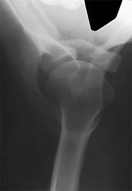
An 18-year-old high school football player sustains a thigh injury that results in the findings shown in Figure 1. Initial management should consist of


Explanation

The radiograph shows myositis ossificans within the quadriceps muscle. This condition occurs as a complication of muscle injury. Initial treatment should include rest, ice, compression, and elevation. While gentle active range of motion is encouraged in the functional recovery from this injury, passive stretching is contraindicated as it can enhance hemorrhage and accentuate the development of myositis ossificans. Ultrasound is similarly contraindicated because it can enhance the development of myositis ossificans and has no proven efficacy in this patient; electrical stimulation also has no proven benefits. Massage is contraindicated in the initial management of this injury because of its influence on increasing local blood flow. Anderson JE (ed): Grant's Atlas of Anatomy. Baltimore, MD, Williams & Wilkins, 1978, pp 4.39-4.49. Brumet ME, Hontas RB: The thigh, in DeLee JC, Drez D Jr (eds): Orthopaedic Sports Medicine. Philadelphia, PA, WB Saunders, 1994, pp 1086-1112. Antao NA: Myositis of the hip in a professional soccer player: A case report. Am J Sports Med 1988;16:82-83.

Question 2

What is the function of the rotator cuff during throwing?


Explanation

The coupled action of the rotator cuff prevents superior migration and controls anterior and posterior translation by depressing the humeral head. Poppen NK, Walker PS: Normal and abnormal motion of the shoulder. J Bone Joint Surg Am 1976;58:195-201.


Question 3

A 24-year-old female soccer player has had lateral joint line pain and a recurrent effusion in the left knee after sustaining a twisting injury 6 weeks ago. She reports that symptoms worsen with athletic activities. MRI scans are shown in Figures 2a through 2c. What is the most likely diagnosis?


Explanation

The MRI scans show the typical findings of a torn discoid lateral meniscus. The average transverse diameter of the lateral meniscus is 11 or 12 mm. A discoid lateral meniscus is suggested when three or more contiguous 5-mm sagittal sections on the MRI scan show continuity of the menicus between the anterior and posterior horns, or when two adjacent peripheral sagittal 5-mm sections show equal meniscal height. Normally the black "bow tie" would be seen on two contiguous sagittal sections. The presence of a discoid meniscus can be further confirmed if coronal views reveal increased width. Jordan MR: Lateral meniscal variants: Evaluation and treatment. J Am Acad Orthop Surg 1996;4:191-200.


Question 4

A 29-year-old woman who underwent an anterior cruciate ligament (ACL) reconstruction 6 months ago now reports difficulty achieving full knee extension, and physical therapy fails to provide relief. The knee is stable on ligament testing. Figure 3 shows the findings at a repeat arthroscopy. Treatment should now include


Explanation

The patient has a cyclops lesion. This is a nodule of fibroproliferative tissue that originates from either drilling debris from the tibial tunnel or remnants of the ACL stump; more rarely it is the result of broken graft fibers. The treatment of choice is excision of the nodule and, if needed, additional notchplasty. Marked improvements in function and symptoms have been noted after removal of the extension block and resumption of a rehabilitation program. Delince P, Krallis P, Descamps PY, et al: Different aspects of the cyclops lesion following anterior cruciate ligament reconstruction: A multifactorial etiopathogenesis. Arthroscopy 1998;14:869-876.


Question 5

The major blood supply to the cruciate ligaments arises from which of the following structures?


Explanation

The major blood supply to the cruciate ligaments arises from the ligamentous branches of the middle genicular artery. Few terminal branches of the inferior genicular artery contribute to the blood supply. The synovial plexus and sheath covering the cruciate ligaments are also supplied by branches of the middle genicular artery. The blood supply to the cruciate ligaments is predominately of soft-tissue origin. There is no significant osseous vascular contribution to the ligaments. Arnoczky SP: Anatomy of the anterior cruciate ligament. Clin Orthop 1983;172:19-25.


Question 6

In the anterior cruciate ligament (ACL)-deficient knee, which of the following variables has the highest correlation with the development of arthritis?


Explanation

Ample evidence supports an increased rate of degenerative arthritis in the ACL-deficient knee. Several variables play a role in the development of the arthritis, but the integrity of the meniscus has been shown to be the single most important factor. O'Brien WR: Degenerative arthritis of the knee following anterior cruciate ligament injury: Role of the meniscus. Sports Med Arthroscopy Rev 1993;1:114-118. Fetto JF, Marshall JL: The natural history and diagnosis of anterior cruciate ligament insufficiency. Clin Orthop 1980;147:29-38.


Question 7

A 20-year-old football player has immediate pain in the midfoot and is unable to bear weight after an opposing player lands on the back of his plantar flexed foot. AP and lateral radiographs are shown in Figures 4a and 4b. Management should consist of


Explanation

The history and radiographs indicate a Lisfranc fracture-dislocation of the foot. The radiographs show the classic "fleck sign," which is an avulsion of the Lisfranc ligament from the base of the second metatarsal. Most authors recommend open reduction and internal fixation of this injury. Closed reduction can be attempted, but anatomic reduction is unlikely because of the interposed bone fragments and soft tissues. Standard radiographs are not reliable in identifying 1 to 2 mm of subluxation of the tarsometatarsal joint. The tarsometatarsal joint has a poor tolerance to even mild subluxation, and the resulting decrease in joint contact area increases the likelihood of posttraumatic arthritis. Open reduction with the joint visible allows more anatomic reduction and internal fixation of larger osteochondral fragments or excision of smaller interposed fragments. Bellabarba C, Sanders R: Dislocations of the foot, in Coughlin MJ, Mann RA (eds): Surgery of the Foot and Ankle, ed 7. St Louis, MO, Mosby, 1999, vol 2, pp 1539-1558.


Question 8

What effect does deep freezing have on allograft tissue?


Explanation

Deep freezing is the simplest and most widely used method of ligament allograft storage. All cells in the tissue are destroyed with the freezing. However, for this reason, it is not a preferred storage method for menisci or cartilage allografts. Although this method may enhance success because it removes potential antigens located on the cells, it cannot guarantee elimination of HIV transmission. The advantage of cryopreservation storage is that a significant number of cells will survive the process, a factor important in meniscal allograft survival after implantation. No deleterious effects are noted clinically because of the acellularity of the tissue. Shelton WR, Treacy SH, Dukes AD, Bomboy AL: Use of allografts in knee reconstruction: I. Basic science aspects and current status. J Am Acad Orthop Surg 1998;6:165-168.


Question 9

A 32-year-old man who works as a laborer has had left trapezius wasting and lateral scapular winging after injuring his shoulder when a cargo box fell onto his neck 8 months ago. He now reports posterior shoulder pain and fatigue, and he has difficulty shrugging his shoulder. Examination reveals marked scapular winging, impingement signs, and an asymmetrical appearance when the patient attempts a shoulder shrug. Primary scapular-trapezius winging is the result of damage to the


Explanation

The patient has primary scapular-trapezius winging. This condition can be caused by blunt trauma to the relatively superficial spinal accessory nerve that is located in the floor of the posterior cervical triangle in the subcutaneous tissue. Other causes of injury include penetrating trauma, traction, or surgical injury. With trapezius winging, the shoulder appears depressed and laterally translated because of an unopposed serratus anterior. This contrasts with primary serratus anterior winging, which is caused by injury to the long thoracic nerve. In this condition, the scapula assumes a position of superior elevation and medial translation, and the inferior angle is rotated medially. The thoracodorsal nerve supplies the latissimus dorsi and is not involved in primary scapular winging. Kuhn JE, Plancher KD, Hawkins RJ: Scapular winging. J Am Acad Orthop Surg 1995;3:319-325.


Question 10

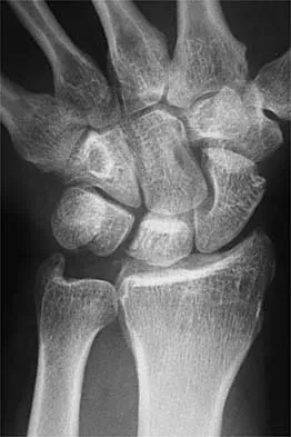
A 32-year-old football coach has had a 4-month history of increasing right wrist pain, particularly during blocking exercises, and he reports significant pain with range of motion and gripping activities. He denies any history of trauma. Examination reveals dorsal wrist tenderness and boggy fullness over the dorsum of the wrist. No erythema is noted. Grip strength is 60% compared with the opposite side. Radiographs are shown in Figures 5a and 5b. What is the most likely diagnosis?


Explanation

The patient has Kienbock's disease (osteonecrosis of the lunate), which presents with boggy synovitis of the wrist, decreased range of motion, and often normal radiographs. The patient's radiographs reveal small fragments from the lunate, with increased density in the lunate body. While a traumatic event may precede the patient's pain, often an insidious increase in pain is found. Repetitive trauma has been suggested as a possible cause. This disease process is classically associated with an ulnar-negative variant. An MRI scan, revealing a low-intensity signal in the lunate, is the best diagnostic tool for early Kienbock's disease. Green DP, Hotchkiss RN, Pederson WC: Green's Operative Hand Surgery, ed 4. Philadelphia, PA, Churchill Livingstone, 1999, pp 837-848.


Question 11

Which of the following properties apply to the human meniscus when compared with articular cartilage?


Explanation

The meniscal cartilage, like articular cartilage, possesses viscoelastic properties. The extracellular matrix is a biphasic structure composed of a solid phase (collagen, proteoglycan) that acts as a fiber-reinforced porous-permeable composite, and a fluid phase that may be forced through the solid matrix by a hydraulic pressure gradient. Although these properties are shared with articular cartilage, the meniscus is more elastic and less permeable than articular cartilage. Beaty JH (ed): Orthopaedic Knowledge Update 6. Rosemont, IL, American Academy of Orthopaedic Surgeons, 1999, pp 3-23.


Question 12

An 18-year-old football player lands on a flexed knee and ankle after being tackled. Examination reveals increased external rotation and posterior translation and varus at 30 degrees of flexion, which decreases as the knee is flexed to 90 degrees. What is the most likely diagnosis?


Explanation

The flexed knee and ankle mechanism of injury can result in a PCL and/or posterolateral corner injury. The examination reveals an isolated injury to the posterolateral corner (arcuate, popliteus, posterolateral capsule). This results in increased posterior translation and external rotation, as well as varus that is most notable at 30 degrees of flexion and decreases as the knee is further flexed to 90 degrees. Combined PCL and posterolateral corner injuries are characterized by increasing instability as the knee is flexed to 90 degrees from 30 degrees, while isolated PCL tears show the greatest degree of instability at 90 degrees of flexion. A rupture of the quadriceps tendon would not affect anterior or posterior stability, whereas an isolated rupture of the lateral collateral ligament, which is a rare injury, is characterized by varus instability at 30 degrees of knee flexion without posterior translation. Harner CD, Hoher J: Evaluation and treatment of posterior cruciate ligament injuries. Am J Sports Med 1998;26:471-482.


Question 13

Figure 6 shows the radiograph of a 14-year-old baseball player who felt a pop and had an immediate onset of pain in his elbow after a hard throw from the outfield. The best course of action should be to


Explanation

The valgus stress at the elbow caused by throwing strains the medial collateral ligament. The medial epicondyle, on which the ligament inserts, is the last ossification center to fuse to the distal humerus, and acute avulsion of the medial epicondyle can occur in adolescents. If the elbow is allowed to heal in a displaced position, valgus instability and loss of elbow extension may result. Valgus instability is especially problematic for the throwing athlete. Surgical treatment with rigid internal fixation is the treatment of choice for displaced medial epicondyle avulsion fractures. Valgus instability is prevented, and the rigid fixation allows for early range of motion. Case SL, Hennrikus WL: Surgical treatment of displaced medial epicondyle fractures in adolescent athletes. Am J Sports Med 1997;25:682-686.


Question 14

Osteophyte formation at the posteromedial olecranon and olecranon articulation in high-caliber throwing athletes is most often the result of underlying


Explanation

During the late acceleration phase of throwing, the triceps forcibly contracts, extending the elbow as the ball is released. Normally, this force is absorbed by the anterior capsule and the brachialis and biceps muscles. However, if the ulnar collateral ligament is insufficient, the elbow will be in a subluxated position during extension and cause impaction of the olecranon and the olecranon fossa posteromedially. Over time, osteophyte formation is likely to occur. Conway JE, Jobe FW, Glousman RE, Pink M: Medial instability of the elbow in throwing athletes: Treatment by repair or reconstruction of the ulnar collateral ligament. J Bone Joint Surg Am 1992;74:67-83.


Question 15

Sudden cardiac death in the young athlete is most frequently caused by


Explanation

Hypertrophic cardiomyopathy is the leading cause of sudden cardiac death in athletes, accounting for 40% of reported cases. Most athletes have no previous symptoms, and sudden death may be the first clinical manifestation. The prevalence of hypertrophic cardiomyopathy in the general population is 1 in 500, with a mortality rate of 2% to 4% in young adults. Athletes with active myocarditis should not engage in sports for up to 6 months, and although they may be at risk for the development of chronic cardiomyopathy, it is rarely a cause of sudden cardiac death. Mitral valve prolapse with an accompanying systolic murmur is common in the general population, but infrequently a cause of sudden cardiac death. Weakening of the aortic wall associated with Marfan syndrome can result in abrupt rupture of the aorta. This accounts for 3% of sudden cardiac deaths in young athletes. Marfan syndrome usually can be detected on preparticipation screenings by its skeletal and ocular manifestations. Atherosclerotic coronary artery disease is the most common cause of sudden cardiac death in older athletes, accounting for 75% of reported cases. However, it is much less common in the young competitive athlete. Burke AP, Farb A, Virmani R, Goodin J, Smialek JE: Sports-related and non-sports-related sudden cardiac death in young adults. Am Heart J 1991;121:568-575.


Question 16

A 14-year-old football player has had right knee pain for the past 2 months; however, he denies any history of trauma. Examination shows an abductor lurch and increased external rotation of the right lower extremity. The best course of action should be to


Explanation

Slipped capital femoral epiphysis is the most common pathology involving the hip in adolescents. While patients with acute slips may report severe pain and are unable to ambulate, those with chronic slips often have pain during ambulation, a limp, and increased external rotation of the hip. While 60% of the patients specifically report hip pain, the remainder have pain in the thigh or knee. The initial diagnostic study of choice is AP and frog-lateral radiographs of the pelvis; bilateral involvement is frequently seen. Boyer DW, Mickelson MR, Ponseti IV: Slipped capital femoral epiphysis: Long-term follow-up study of one hundred and twenty-one patients. J Bone Joint Surg Am 1981;63:85-95.


Question 17

Which of the following is considered the appropriate initial management protocol for an unconscious football player without spontaneous respirations?


Explanation

The on-field evaluation and management of the seriously injured athlete requires advance preparation and planning. It is imperative that the health care team have a game plan in place and the proper equipment readily available. The initial step consists of stabilizing the head and neck by manually holding the head and neck in a neutral position. Then, in the following order, check for breathing, pulses, and level of consciousness. If the athlete is breathing, simply remove the mouth guard and maintain the airway. If the athlete is not breathing, the face mask must be removed and the chin strap left in place. An open airway must be established, followed by assisted breathing. CPR is only instituted when breathing and circulation are compromised. If the athlete is unconcious or has a suspected cervical spine injury, the helmet must not be removed until the athlete has been transported to an appropriate facility and the cervical spine has been completely evaluated. McSwain NE, Garnelli RL: Helmet removal from injured patients. Bull Am Coll Surg 1997;82:42-44. Vegso JJ, Lehman RC: Field evaluation and management of head and neck injuries. Clin Sports Med 1987;6:1-15.


Question 18

Figure 7 shows the radiograph of an 18-year-old hockey player who sustained a shoulder injury during a fall into the side boards. Examination reveals a significant prominence at the acromioclavicular joint. Management should consist of


Explanation

The radiograph shows a type V acromioclavicular separation with greater than 100% superior elevation of the clavicle. This finding implies detachment of the deltoid and trapezius from the distal clavicle. Because of severe compromise of function and potential compromise to the overlying skin, surgery is the treatment of choice for type V acromioclavicular separations. During reduction and repair, meticulous repair of the deltotrapezial fascia will also aid in securing the repair. Nuber GW, Bowen MK: Acromioclavicular joint injuries and distal clavicle fractures. J Am Acad Orthop Surg 1997;5:11-18.


Question 19

A 22-year-old professional ballet dancer reports a 3-month history of posterior ankle pain that occurs when she changes from a flat foot to pointe (hyperplantar flexed position). Examination does not elicit the pain with forced passive plantar flexion. A radiograph is shown in Figure 8. What is the most likely cause of the pain?


Explanation

The most common causes of posterior ankle pain in ballet dancers are flexor hallucis longus tenosynovitis and os trigonum syndrome. Flexor hallucis longus tenosynovitis differs from a symptomatic os trigonum by the absence of pain with forced plantar flexion and the presence of pain with resisted plantar flexion of the great toe. The pain is often felt in the posterior ankle and can be associated with a snapping or triggering sensation. Os trigonum syndrome commonly occurs in ballet dancers who perform in a position of extreme plantar flexion. The pain occurs from entrapment of the os trigonum between the posterior portion of the talus and calcaneus. Hamilton WG, Geppert MJ, Thompson FM: Pain in the posterior aspect of the ankle in dancers: Differential diagnosis and operative treatment. J Bone Joint Surg Am 1996;78:1491-1500.


Question 20

An 18-year-old man recently underwent an uncomplicated arthroscopic partial medial meniscectomy that was complicated by reflex sympathetic dystrophy (RSD), also termed "sympathetically maintained pain" (SMP). What is the most common finding of this condition?


Explanation

The hallmark for RSD or SMP is the presence of pain that is out of proportion to that expected for the degree of the injury. SMP often extends well beyond the involved area and is present in a nonanatomic distribution. The pain is frequently described as a burning sensation, with extreme sensitivity to light touch. Joint stiffness can be present but is a nonspecific finding. There may be cold intolerance, but this is not a cardinal symptom. Sweating actually may be increased. Osteopenia, if present, is a late finding. Lindenfeld TN, Bach BR Jr, Wojtys EM: Reflex sympathetic dystrophy and pain dysfunction in the lower extremity. Instr Course Lect 1997;46:261-268.


Question 21

What is the main function of collagen found within articular cartilage?


Explanation

The main function of collagen in articular cartilage is to provide the tissue's tensile strength. It also immobilizes proteoglycans within the extracellular matrix. Compressive properties are maintained by proteoglycans. Cartilage metabolism is maintained by the indwelling chondrocytes. The flow of water through the tissue promotes transport of nutrients and provides a source of lubricant for the joint. Simon SR (ed): Orthopaedic Basic Science. Rosemont, IL, American Academy of Orthopaedic Surgeons, 1994, pp 3-44.


Question 22

A 15-year-old girl who competes in gymnastics has immediate pain and giving way of the left elbow after falling from the uneven parallel bars and landing on her outstretched arms. Examination reveals swelling and tenderness about the elbow, especially over the medial side. Measurement of elbow motion shows 0 degrees to 125 degrees of flexion, and valgus stress at the elbow is painful. AP, lateral, and stress radiographs are shown in Figures 9a through 9c. Management should consist of


Explanation

While many low-demand patients with injuries to the ulnar collateral ligament can be treated nonsurgically, Jobe and associates described two situations in which ulnar collateral ligament reconstruction is indicated: (1) an acute complete rupture in a competitive athlete who uses the upper extremities extensively and who wishes to remain active; and (2) chronic pain or instability that does not improve after at least 3 months of nonsurgical management. Rarely is direct surgical repair of the ligament possible or able to withstand the valgus stresses applied to the elbow. Most authors recommend surgical reconstruction of the ulnar collateral ligament using a palmaris longus, plantaris, or fourth toe extensor tendon from the fourth autograft. Andrews JR, Jelsma RD, Joyce ME, et al: Open surgical procedures for injuries to the elbow in throwers. Oper Tech Sports Med 1994;4:109-133. Jobe FW, Kvitne RS: Elbow instability in the athlete. Instr Course Lect 1991;40:17-23.


Question 23

A 15-year-old boy who participates in track reports acute pain along the left iliac crest during a sprint. Examination reveals that the anterior superior iliac spine is nontender. The most likely diagnosis is an injury to the


Explanation

The patient has iliac apophysitis. The radiographic findings are easily overlooked but usually reveal slight asymmetric widening of the iliac crest apophysis. The apophysis is the most vulnerable structure, as it is three to five times weaker than the tendon. This is not an epiphyseal site, and injury to the muscle or the tendinous insertion to bone (enthesis) is unlikely. Clancy WG Jr, Foltz AS: Iliac apophysitis and stress fractures in adolescent runners. Am J Sports Med 1976;4:214-218. Waters PM, Millis MB: Hip and pelvic injuries in the young athlete, in Stanitski CL, DeLee JC, Drez D Jr (eds): Pediatric and Adolescent Sports Medicine. Philadelphia, PA, WB Saunders, 1994, pp 279-293. Lombardo SJ, Retting AC, Kerlan RK: Radiographic abnormalities of the iliac apophysis in adolescent athletes. J Bone Joint Surg Am 1983;65:444-446.


Question 24

A 40-year-old woman who is an avid tennis player reports the insidious onset of progressive left shoulder pain for the past 2 months. Examination reveals full range of motion with a positive impingement sign. Strength in the supraspinatus and infraspinatus muscles is normal, although stress testing is painful. An earlier subacromial cortisone injection provided good, but only temporary relief. An AP radiograph of the left shoulder is shown in Figure 10. Management should now consist of


Explanation

The radiograph shows calcific deposits within the substance of the supraspinatus tendon. Patients with this condition are prone to recurrent bouts of acute inflammation in the shoulder. While the response to cortisone injection is often dramatic, repeated injections are not recommended because of injury to the collagen fibers. Good results have been obtained with arthroscopic evacuation of the calcium deposits. In one study, the addition of a subacromial decompression did not improve the results. Jerosch J, Strauss JM, Schmiel S: Arthroscopic treatment of calcific tendinitis of the shoulder. J Shoulder Elbow Surg 1998;7:30-37.


Question 25

Which of the following nerves is susceptible to entrapment near the calcaneal attachment site of the plantar fascia and can mimic or co-exist with plantar fasciitis?


Explanation

The first branch of the lateral plantar nerve is susceptible to entrapment beneath the deep fascia of the adductor hallucis muscle adjacent to the calcaneal attachment of the plantar fascia. This can be a cause of chronic heel pain. Additionally, the nerve is vulnerable to injury by a blind dissection in releasing the plantar fascia. The dorsal cutaneous branch of the superficial peroneal nerve supplies sensation to the dorsum of the foot. The medial calcaneal branch of the posterior tibial nerve lies in the subcutaneous tissues and innervates the skin of the heel. It is vulnerable to injury from skin incisions on the medial side of the heel. The lateral branch of the medial plantar nerve forms the second and third common digital nerves. Entrapment of the proper medial plantar nerve can occur at the master knot of Henry. This is well distal to the calcaneal attachment of the plantar fascia, and the pain usually radiates more distally in the arch, separate from heel pain. The communicating branch of the fourth common digital nerve crosses to the third common digital nerve. Therefore, the third common digital nerve receives supply from both the lateral and medial plantar nerves. This dual supply has been implicated in the increased incidence of digital neuroma of the third common digital nerve. Bordelon RL: Heel pain, in Mann RA, Coughlin MJ (eds): Surgery of the Foot and Ankle, ed 6. St Louis, MO, CV Mosby, 1993, pp 837-857. Mann RA, Baxter DE: Diseases of the nerves, in Mann RA, Coughlin MJ (eds): Surgery of the Foot and Ankle, ed 6. St Louis, MO, CV Mosby, 1993, pp 543-574.


Question 26

A 20-year-old soccer player sustains a non-contact twisting knee injury. Radiographs show a small avulsion fracture of the anterolateral proximal tibia.

What is the most likely associated ligamentous injury, and what is its primary biomechanical function?


Explanation

A Segond fracture is an avulsion of the anterolateral proximal tibia and is pathognomonic for an ACL tear. The primary function of the ACL is to resist anterior tibial translation.

Question 27

A 25-year-old professional baseball pitcher complains of posterior shoulder pain during the late cocking phase of throwing. Examination reveals a positive peel-back sign and increased external rotation with a deficit in internal rotation. What is the most likely diagnosis?


Explanation

Pain in the late cocking phase with increased external rotation and a positive peel-back sign is classic for internal impingement. This condition is often associated with a type II SLAP tear due to the greater tuberosity abutting the posterosuperior glenoid.

Question 28

During an anterior cruciate ligament (ACL) reconstruction using an anteromedial portal technique, the surgeon aims to place the femoral tunnel in the anatomic footprint. Compared to the traditional transtibial technique, the anteromedial portal technique is associated with which of the following tunnel characteristics?


Explanation

The anteromedial portal technique allows for independent drilling of the femoral tunnel, enabling a more anatomic, horizontal placement in the ACL footprint compared to the transtibial technique, which restricts femoral tunnel position based on the tibial tunnel trajectory.

Question 29

A 19-year-old collegiate hockey player presents with recurrent anterior shoulder instability. Preoperative evaluation determines an Instability Severity Index Score (ISIS) of 8. Which of the following surgical procedures is most appropriate to minimize his risk of recurrence?


Explanation

The Instability Severity Index Score (ISIS) identifies patients at high risk for recurrent instability after arthroscopic Bankart repair. A score >6 is an indication for a bone-block procedure, such as the Latarjet, to ensure adequate stability in high-risk contact athletes.

Question 30

A 35-year-old recreational basketball player sustains an acute Achilles tendon rupture. He opts for non-operative management. To optimize his clinical outcome and minimize the risk of re-rupture, which of the following is the most critical component of his non-operative protocol?


Explanation

Recent literature demonstrates that non-operative management of acute Achilles tendon ruptures using an early functional rehabilitation protocol (early weight-bearing in equinus bracing) yields re-rupture rates comparable to surgical repair, while avoiding surgical complications.

Question 31

A 24-year-old male sustains a traumatic knee injury. On physical examination, the dial test reveals 15 degrees of increased external rotation compared to the contralateral side at 30 degrees of knee flexion, but symmetrical external rotation at 90 degrees of knee flexion. What is the most likely diagnosis?


Explanation

The dial test evaluates posterolateral rotatory instability. Increased external rotation (>10 degrees compared to the normal side) at 30 degrees only indicates an isolated PLC injury. Increased external rotation at both 30 and 90 degrees indicates a combined PLC and PCL injury.

Question 32

A 45-year-old active female presents with sudden-onset posterior knee pain and a popping sensation while squatting. MRI reveals a medial meniscus posterior root tear with 4 mm of meniscal extrusion. Which of the following is the most appropriate management?


Explanation

Medial meniscus posterior root tears result in the loss of hoop stresses, leading to meniscal extrusion and rapid progression of osteoarthritis. Arthroscopic anatomic root repair (e.g., transtibial pull-out technique) is the treatment of choice in active patients without advanced arthritis.

Question 33

A 21-year-old wide receiver sustains a forceful external rotation injury to his ankle. Examination reveals tenderness over the anterior inferior tibiofibular ligament (AITFL) and a positive squeeze test. Radiographs show a tibiofibular clear space of 7 mm. What is the most appropriate treatment?


Explanation

A tibiofibular clear space >5 mm on AP or mortise radiographs indicates a syndesmotic injury with frank diastasis. Operative stabilization (using screws or suture-button constructs) is required to restore the mortise and prevent post-traumatic arthritis.

Question 34

A 28-year-old professional baseball pitcher presents with posterior shoulder pain during the late cocking phase of throwing. Physical examination reveals a glenohumeral internal rotation deficit (GIRD) of 25 degrees. What is the primary pathophysiological mechanism contributing to this condition?


Explanation

GIRD in throwing athletes is primarily caused by contracture of the posterior capsule, specifically the posterior band of the inferior glenohumeral ligament (IGHL). This alters glenohumeral kinematics, leading to superior and posterior translation of the humeral head during throwing and resulting in internal impingement.

Question 35

A 16-year-old female presents with recurrent lateral patellar dislocations. The decision is made to perform a medial patellofemoral ligament (MPFL) reconstruction. To prevent overloading the medial compartment and restricted knee flexion, the femoral attachment of the graft must be accurately placed. Where is the anatomic origin of the MPFL on the femur?


Explanation

The anatomic femoral origin of the MPFL (often localized radiographically via Schottle's point) is situated in a saddle-shaped depression between the adductor tubercle (proximally) and the medial epicondyle (distally). Non-anatomic placement, particularly too proximal, leads to increased graft tension in flexion.

Question 36

A 42-year-old heavy laborer sustains an acromioclavicular (AC) joint injury. Radiographs reveal a 150% superior displacement of the clavicle relative to the acromion with significant posterior displacement into the trapezius fascia. Based on the Rockwood classification, what is the injury type and optimal management?


Explanation

A Rockwood Type IV AC joint separation is characterized by posterior displacement of the distal clavicle into or through the trapezius fascia. Unlike many Type III injuries, Type IV injuries typically require surgical reconstruction due to significant functional impairment and pain.

Question 37

A 55-year-old male presents with shoulder pain and weakness after a fall. On examination, he has a positive "bear-hug" test and increased external rotation compared to the contralateral side. Which structure is most likely injured?


Explanation

The bear-hug test, belly-press test, and lift-off test evaluate the subscapularis tendon. Increased passive external rotation is a classic finding of a full-thickness subscapularis tear due to the loss of the anterior restraint.

Question 38

A 14-year-old male presents with vaguely localized knee pain and occasional catching. Radiographs reveal an osteochondritis dissecans (OCD) lesion. What is the most common anatomical location for this lesion in the knee?


Explanation

The most common location for an osteochondritis dissecans (OCD) lesion in the knee is the lateral aspect of the medial femoral condyle (classic location), accounting for approximately 70-80% of cases.

Question 39

A 40-year-old water skier sustains a complete proximal hamstring avulsion. He undergoes acute surgical repair. During the surgical approach to the ischial tuberosity, which of the following neurovascular structures is at the highest risk of iatrogenic injury, particularly with retraction?


Explanation

The sciatic nerve lies just lateral to the ischial tuberosity and the proximal hamstring origin. It is at significant risk of injury during proximal hamstring repair, especially from vigorous retraction or scar tissue dissection in delayed cases.

Question 40

A 25-year-old professional tennis player presents with shoulder pain and weakness in external rotation. An MRI demonstrates a massive, irreparable posterosuperior rotator cuff tear with severe fatty infiltration. The patient has preserved forward elevation but a positive external rotation lag sign. Which tendon transfer is most appropriate to restore active external rotation?


Explanation

The lower trapezius tendon transfer (often with an Achilles allograft) is highly effective for restoring active external rotation in patients with irreparable posterosuperior cuff tears. It has an in-phase line of pull that mimics the infraspinatus better than a latissimus dorsi transfer.

Question 41

A 19-year-old collegiate soccer player sustains a high ankle sprain. On evaluation 3 weeks post-injury, he is pain-free with walking but unable to run. Weight-bearing CT is utilized to assess the syndesmosis. Which finding would most strongly indicate the need for surgical stabilization?


Explanation

Weight-bearing CT is highly sensitive for detecting subtle syndesmotic instability. Asymmetry in the syndesmotic clear space, volume, or area (e.g., >2 mm side-to-side difference) under weight-bearing loads indicates a dynamic instability that warrants surgical fixation.

Question 42

A 22-year-old athlete sustains an isolated Grade II posterior cruciate ligament (PCL) injury. Which of the following non-operative rehabilitation strategies is most effective for promoting intrinsic ligament healing and restoring knee stability?


Explanation

For acute isolated Grade I and II PCL tears, bracing the knee in extension or using a dynamic PCL brace applies an anterior directed force to the proximal tibia. This prevents posterior sag, allowing the PCL to heal in an anatomic, non-elongated position.

Question 43

A 25-year-old male sustains a direct blow to the proximal tibia with his knee flexed during a rugby match. On physical examination, he has a positive posterior drawer test. A dial test is performed, demonstrating greater than 10 degrees of increased external rotation compared to the contralateral knee at both 30 degrees and 90 degrees of flexion. What is the most likely diagnosis?


Explanation

The dial test evaluates for excessive external rotation. Asymmetry of >10 degrees at 30 degrees of flexion only indicates an isolated PLC injury, whereas asymmetry at both 30 and 90 degrees of flexion indicates a combined PCL and PLC injury.

Question 44

A 22-year-old collegiate baseball pitcher presents with vague posterior shoulder pain during the late cocking phase of throwing. Examination reveals a 25-degree loss of internal rotation (GIRD) compared to the non-throwing shoulder. Which of the following is the primary pathoanatomy responsible for this clinical presentation?


Explanation

Glenohumeral internal rotation deficit (GIRD) in overhead athletes is primarily driven by contracture of the posteroinferior capsule. This tightness alters glenohumeral kinematics, leading to a posterosuperior shift of the humeral head during the throwing motion.

Question 45

An 18-year-old female soccer player undergoes primary anterior cruciate ligament (ACL) reconstruction. When comparing bone-patellar tendon-bone (BPTB) autograft to hamstring autograft, which of the following is a recognized disadvantage specific to the BPTB autograft?


Explanation

BPTB autografts have historically shown equivalent or slightly better objective stability compared to hamstring grafts but are consistently associated with a higher incidence of anterior knee pain and difficulty kneeling.

Question 46

A 24-year-old football player sustains an external rotation injury to his right ankle. Radiographs demonstrate a widened medial clear space and decreased tibiofibular overlap. In a syndesmotic injury, which ligament serves as the primary restraint to anterior translation of the distal fibula?


Explanation

The anterior inferior tibiofibular ligament (AITFL) is the primary restraint to anterior translation of the distal fibula and is typically the first ligament to tear in a syndesmotic injury.

Question 47

A 21-year-old rugby player presents with recurrent anterior shoulder instability. A CT scan of the shoulder reveals an engaging Hill-Sachs lesion and a 25% anterior glenoid bone defect.

What is the most appropriate definitive surgical management?


Explanation

In the setting of critical glenoid bone loss (typically >20-25%) and an engaging Hill-Sachs lesion, soft tissue stabilization (Bankart repair) has unacceptably high failure rates. A bony augmentation procedure like the Latarjet is the gold standard.

Question 48

A 16-year-old female presents with recurrent lateral patellar dislocations. She is scheduled for a medial patellofemoral ligament (MPFL) reconstruction. To ensure proper graft isometry, the femoral tunnel must be placed at Schöttle's point. Where is this anatomic location relative to the osseous landmarks?


Explanation

The anatomic femoral origin of the MPFL (Schöttle's point) is located in a saddle-like depression between the medial epicondyle and the adductor tubercle.

Question 49

A 35-year-old male "weekend warrior" suffers an acute Achilles tendon rupture. When discussing operative repair versus non-operative management with an early functional rehabilitation protocol, what should the patient be counseled regarding outcomes?


Explanation

Recent high-level evidence shows that when early functional rehabilitation is utilized, re-rupture rates are similar between operative and non-operative groups. However, operative management carries a higher risk of wound complications and sural nerve injury.

Question 50

A 45-year-old recreational tennis player is diagnosed with an isolated Type II SLAP tear that has failed 6 months of conservative management. Which surgical intervention has been shown to yield the highest rates of return to sport and patient satisfaction in patients of this age?


Explanation

In patients older than 40 years with a Type II SLAP tear, primary SLAP repair is associated with higher rates of stiffness and revision. Biceps tenodesis provides superior clinical outcomes and return to play in this demographic.

Question 51

A 48-year-old female feels a "pop" in her posterior knee while squatting to garden. An MRI reveals an isolated complete tear of the medial meniscus posterior root. Biomechanically, this injury is equivalent to which of the following?


Explanation

A complete tear of the meniscal root disrupts hoop stresses within the meniscus, causing it to extrude under load. Biomechanically, this alters tibiofemoral contact pressures to a degree equivalent to a total meniscectomy.

Question 52

A 26-year-old runner presents with chronic, deep ankle pain. MRI reveals a 1.8 square centimeter osteochondral lesion (OCL) on the medial talar dome without subchondral cysts. He has failed 6 months of conservative care. What is the most appropriate surgical treatment?


Explanation

Microfracture is generally recommended for talar OCLs smaller than 1.5 cm^2. For larger lesions (>1.5 cm^2), structural grafting with techniques like OATS provides superior outcomes and better hyaline-like cartilage restoration.

Question 53

A 30-year-old weightlifter feels a tearing sensation in his anterior axilla while performing a heavy bench press. Examination reveals loss of the anterior axillary fold and weakness in internal rotation. Which portion of the pectoralis major is most commonly ruptured in this scenario?


Explanation

Pectoralis major ruptures typically occur during eccentric contraction (like bench pressing). The sternal head at its humeral insertion is the most commonly torn portion due to its mechanical disadvantage at maximum extension.

Question 54

An 11-year-old male with wide-open physes sustains a complete anterior cruciate ligament (ACL) tear. He is highly active and wishes to return to pivoting sports. To minimize the risk of growth arrest, which of the following surgical techniques is recommended?


Explanation

In prepubescent patients with significant growth remaining (Tanner stage 1 or 2), physeal-sparing techniques like the modified MacIntosh/Kocher IT band reconstruction are recommended to avoid crossing the physes and causing growth arrest.

Question 55

A 23-year-old alpine skier presents with lateral ankle pain and a snapping sensation behind the lateral malleolus when dorsiflexing and everting the foot against resistance. What is the primary structure injured in this clinical presentation?


Explanation

The clinical presentation is classic for peroneal tendon subluxation. The primary restraint to peroneal tendon subluxation is the superior peroneal retinaculum (SPR), which is typically avulsed from the lateral malleolus.

Question 56

A 28-year-old male falls directly onto the point of his shoulder while cycling. Radiographs demonstrate a Type III acromioclavicular (AC) joint separation. Based on the Rockwood classification, what is the status of the supporting ligaments?


Explanation

A Rockwood Type III AC joint injury involves complete rupture of both the acromioclavicular (AC) and coracoclavicular (CC) ligaments, resulting in 25% to 100% superior translation of the distal clavicle.

Question 57

A 30-year-old male presents to the emergency department following an acute knee dislocation (Schenck KD-III). Vascular exam is normal, but he exhibits a complete foot drop and inability to extend his toes. Which nerve is injured, and at what anatomic site is it most commonly tethered during this injury?


Explanation

The common peroneal nerve is uniquely susceptible to traction injury during multi-ligament knee dislocations due to its rigid tethering at the fibular neck as it passes into the anterior compartment.

Question 58

A 19-year-old female gymnast with chronic lateral ankle instability fails non-operative management and is indicated for a modified Broström procedure. The Gould modification of this procedure involves advancing which structure to reinforce the repair?


Explanation

The standard Broström procedure involves anatomic repair of the ATFL and CFL. The Gould modification reinforces this repair by advancing the extensor retinaculum (specifically the inferior portion) over the repaired ligaments.

Question 59

A 29-year-old professional volleyball player presents with isolated weakness in shoulder external rotation. MRI reveals a paralabral cyst compressing a nerve. At what anatomic location is this cyst most likely situated?


Explanation

Compression of the suprascapular nerve at the spinoglenoid notch affects only the infraspinatus muscle, leading to isolated external rotation weakness. Compression at the suprascapular notch would affect both the supraspinatus and infraspinatus.

Question 60

A 14-year-old male complains of vague, activity-related knee pain. Radiographs demonstrate an osteochondritis dissecans (OCD) lesion.

What is the most common anatomic location for an OCD lesion in the knee?


Explanation

The most common location for osteochondritis dissecans (OCD) of the knee is the lateral aspect of the medial femoral condyle, accounting for over 70% of cases.

Question 61

A 22-year-old college basketball player sustains an acute fracture at the metaphyseal-diaphyseal junction of the 5th metatarsal (Jones fracture). To ensure the highest likelihood of union and rapid return to play, what is the treatment of choice?


Explanation

Due to the watershed blood supply at the metaphyseal-diaphyseal junction of the 5th metatarsal, Jones fractures in high-level athletes are best treated with intramedullary screw fixation to decrease nonunion risk and expedite return to play.

Question 62

A 16-year-old female soccer player sustains a torn anterior cruciate ligament (ACL) and is scheduled for reconstruction. What is the most significant disadvantage of utilizing a bone-patellar tendon-bone (BPTB) allograft compared to an autograft in this patient demographic?


Explanation

Allograft tissue has a significantly higher failure and rerupture rate compared to autograft when used for ACL reconstruction in young, highly active patients under the age of 25. This is largely attributed to delayed graft incorporation and slower biological remodeling.

Question 63

A 25-year-old football player complains of knee instability after a direct blow to the anteromedial tibia. The Dial test demonstrates 15 degrees of increased external rotation at 30 degrees of knee flexion, but symmetric rotation compared to the contralateral side at 90 degrees. Which structure is most likely injured?


Explanation

An isolated injury to the posterolateral corner (LCL and popliteus) typically presents with increased external rotation at 30 degrees of flexion. At 90 degrees, the intact posterior cruciate ligament acts as a secondary restraint, neutralizing the rotational asymmetry.

Question 64

A 22-year-old hockey player presents with severe lateral ankle pain after an eversion twisting injury. The squeeze test is positive, and radiographs reveal a widened tibiofibular clear space. Which ligament is the primary restraint to anterior translation of the distal fibula?


Explanation

The AITFL is the primary restraint to anterior translation of the distal fibula relative to the tibia. It is the most commonly disrupted structure in syndesmotic (high ankle) sprains, which are typically caused by an external rotation force.

Question 65

A 19-year-old collegiate baseball pitcher complains of posterior shoulder pain strictly during the late cocking phase of throwing. Examination reveals a 20-degree loss of internal rotation (GIRD) compared to the contralateral shoulder. What is the most likely pathomechanism of his pain?


Explanation

Internal impingement in overhead throwers involves pathological contact between the articular side of the posterior rotator cuff and the posterosuperior glenoid labrum. This occurs in the late cocking phase due to excessive external rotation and anterior capsular laxity coupled with posterior capsular contracture.

Question 66

A 30-year-old marathon runner presents with persistent deep ankle pain. MRI reveals a 1.8 square cm osteochondral lesion on the medial talar dome. Non-operative management has failed. What is the most appropriate surgical treatment algorithm for this specific defect size?


Explanation

Osteochondral lesions of the talus larger than 1.5 square cm have unacceptably high failure rates with marrow stimulation (microfracture). Osteochondral autograft transfer (OATS) restores the articular surface with structural hyaline cartilage and is indicated for these larger or cystic lesions.

Question 67

An 18-year-old female has recurrent lateral patellar instability. Advanced imaging demonstrates a tibial tubercle-trochlear groove (TT-TG) distance of 24 mm and a normal Caton-Deschamps ratio. What is the most appropriate surgical procedure?


Explanation

A TT-TG distance greater than 20 mm is pathognomonic for severe lateralizing patellar forces and requires a tibial tubercle osteotomy (anteromedialization) to normalize the extensor mechanism. Isolated MPFL reconstruction in this setting has a high failure rate due to uncorrected bony malalignment.

Question 68

A 28-year-old weightlifter feels a sharp pop in his anterior chest while performing a heavy bench press. Examination reveals a loss of the anterior axillary fold and significant weakness with resisted internal rotation. Which anatomical portion of the pectoralis major is most commonly ruptured in this specific scenario?


Explanation

The sternal head of the pectoralis major is under maximum mechanical tension when the arm is extended and externally rotated, such as at the bottom of a bench press. Consequently, ruptures most frequently occur at the sternal head humeral insertion site.

Question 69

A 21-year-old rugby player suffers from recurrent anterior shoulder instability. A pre-operative 3D CT scan demonstrates 28 percent anterior glenoid bone loss. What is the most appropriate definitive surgical management?


Explanation

Anterior glenoid bone loss greater than 20 to 25 percent creates an inverted pear glenoid, resulting in a high failure rate for isolated soft-tissue Bankart repairs. A Latarjet procedure (coracoid transfer) is the gold standard for restoring bony stability in these patients.

Question 70

A 45-year-old female experiences a painful pop in the posterior aspect of her knee while squatting to garden. MRI shows a radial tear adjacent to the posterior horn medial meniscus attachment. What is the primary biomechanical consequence of leaving this specific tear untreated?


Explanation

A posterior medial meniscal root tear completely disrupts the circumferential hoop stresses of the meniscus, leading to load transmission equivalent to a complete meniscectomy. This results in rapid articular cartilage wear and accelerated medial compartment osteoarthritis if left uncorrected.

Question 71

A 35-year-old male weekend warrior sustains an acute Achilles tendon rupture while playing basketball. He elects to pursue non-operative management. What rehabilitation protocol has been shown in Level I studies to optimize functional outcomes and minimize re-rupture rates?


Explanation

Recent Level I evidence demonstrates that dynamic functional rehabilitation with early weight-bearing provides equivalent functional outcomes and re-rupture rates to operative repair for acute Achilles tendon ruptures. This approach significantly reduces the risk of surgical wound complications.

Question 72

A 24-year-old motorcyclist sustains a traumatic knee dislocation (KD-IV). Upon closed reduction in the emergency department, the ipsilateral foot remains cool and pulseless. What is the immediate next step in management?


Explanation

The presence of hard signs of vascular injury, such as an absent pulse or active ischemia after a knee dislocation reduction, mandates immediate surgical exploration. Delaying treatment to obtain an ABI or advanced imaging can lead to irreversible ischemic limb loss.

Question 73

During diagnostic arthroscopy of a 22-year-old elite baseball pitcher, a Type II SLAP tear is identified. The dynamic peel-back mechanism is clearly demonstrated on the monitor. In what arm position is this specific pathomechanism most pronounced?


Explanation

The peel-back mechanism occurs during the late cocking phase of throwing, which corresponds to maximum abduction and external rotation. This specific position shifts the biceps vector posteriorly, twisting the superior labrum and actively peeling it off the underlying glenoid rim.

Question 74

A 14-year-old male presents with vague, aching knee pain. Radiographs demonstrate an osteochondritis dissecans (OCD) lesion. What is the classic and most common anatomic location for an OCD lesion in the knee?


Explanation

The classic and most common location for an osteochondritis dissecans (OCD) lesion in the knee is the lateral aspect of the medial femoral condyle. This location accounts for approximately 70 percent of all knee OCD lesions seen in clinical practice.

Question 75

A 26-year-old skier presents with a significantly swollen knee after a twisting fall. An anteroposterior radiograph reveals a small elliptical bone avulsion fragment from the proximal lateral tibia, known as a Segond fracture. What is the most commonly associated intra-articular injury?


Explanation

A Segond fracture is a cortical avulsion of the anterolateral ligament (ALL) and lateral capsule from the proximal lateral tibia. It is considered highly pathognomonic for an underlying anterior cruciate ligament (ACL) tear, occurring in up to 75 to 100 percent of these cases.

Question 76

A 19-year-old female soccer player sustains a noncontact anterior cruciate ligament (ACL) tear. Which of the following anatomic factors is most strongly associated with an increased risk of this injury?


Explanation

An increased posterior tibial slope increases anterior tibial translation under axial loads, elevating the risk of ACL rupture. Other anatomic risk factors include a narrow intercondylar notch and increased generalized joint laxity.

Question 77

A 24-year-old baseball pitcher presents with deep shoulder pain and decreased throwing velocity. The 'peel-back' mechanism during late cocking is suspected. Which physical examination test is most classically associated with diagnosing the resulting pathology?


Explanation

The O'Brien (active compression) test evaluates for Superior Labrum Anterior to Posterior (SLAP) tears. The peel-back mechanism of the biceps anchor during the late cocking phase is a primary cause of SLAP tears in overhead athletes.

Question 78

A 30-year-old male sustains a knee dislocation during a fall from a height. After closed reduction, his Ankle-Brachial Index (ABI) is calculated to be 0.7. He has palpable distal pulses and no active bleeding. What is the most appropriate next step in management?


Explanation

An ABI less than 0.9 after a knee dislocation raises high suspicion for a popliteal artery injury. CT angiography is the diagnostic modality of choice when hard signs of ischemia are absent but the ABI is abnormal.

Question 79

Compared to surgical repair, traditional nonoperative management with prolonged cast immobilization for an acute Achilles tendon rupture is historically associated with which of the following?


Explanation

Traditional nonoperative treatment with casting historically has a higher re-rupture rate compared to operative repair. However, surgical repair carries inherent risks of wound complications and sural nerve injury.

Question 80

A 22-year-old collegiate volleyball player complains of posterior shoulder pain when spiking the ball. Examination reveals a positive apprehension test and a relocation test that relieves her posterior shoulder pain. She demonstrates GIRD (glenohumeral internal rotation deficit). What is the most likely diagnosis?


Explanation

Internal impingement is characterized by posterior shoulder pain during late cocking (abduction and external rotation) due to the undersurface of the cuff impinging on the posterosuperior labrum. The relocation test typically relieves this posterior pain.

Question 81

A 55-year-old female presents with acute medial knee pain after feeling a 'pop' while descending stairs. MRI reveals a radial tear at the posterior horn attachment of the medial meniscus. If left untreated, what is the primary biomechanical consequence?


Explanation

A posterior medial meniscus root tear functionally unanchors the meniscus, disrupting hoop stresses and causing meniscal extrusion. This results in increased peak contact pressures and accelerated medial compartment osteoarthritis.

Question 82

A 60-year-old man undergoes arthroscopic evaluation for an irreparable rotator cuff tear and undergoes a biceps tenotomy. Compared to a biceps tenodesis, tenotomy is associated with a higher rate of which of the following?


Explanation

Biceps tenotomy carries a higher rate of cosmetic deformity (the 'Popeye' muscle appearance) and subjective muscle cramping compared to tenodesis. Both procedures provide similar levels of pain relief.

Question 83

A 26-year-old male presents with a posterior sag sign after a dashboard injury. Which of the following physical examination findings most suggests a combined Posterior Cruciate Ligament (PCL) and Posterolateral Corner (PLC) injury?


Explanation

An isolated PLC injury typically presents with increased external rotation only at 30 degrees of knee flexion. If external tibial rotation is increased at both 30 and 90 degrees, it indicates a combined PLC and PCL injury.

Question 84

A 21-year-old athlete sustains an external rotation injury to his ankle. Radiographs show a widened medial clear space and tibiofibular clear space. He is tender along the proximal fibula. What is the standard operative management?


Explanation

An external rotation force causing syndesmotic injury with medial clear space widening indicates an unstable syndesmosis (often a Maisonneuve variant). This requires operative reduction and syndesmotic fixation.

Question 85

A 16-year-old girl dislocates her patella laterally. The primary soft-tissue restraint to lateral patellar translation is ruptured. Where is the femoral footprint of this structure located?


Explanation

The medial patellofemoral ligament (MPFL) is the primary restraint to lateral patellar dislocation. Its femoral attachment (Schöttle's point) is located in the saddle between the medial epicondyle and the adductor tubercle.

Question 86

A 28-year-old cyclist falls directly onto his shoulder. Radiographs demonstrate superior displacement of the distal clavicle by 150% relative to the acromion. Which ligaments must be completely disrupted in this injury pattern?


Explanation

A Type III or greater acromioclavicular (AC) joint separation features complete disruption of both the AC ligaments and the coracoclavicular (CC) ligaments, leading to significant superior clavicle displacement.

Question 87

A 14-year-old boy presents with mechanical catching in his knee. Radiographs confirm Osteochondritis Dissecans (OCD). What is the most common anatomic location for an OCD lesion in the knee?


Explanation

The classic and most common location for osteochondritis dissecans (OCD) of the knee is the lateral aspect of the medial femoral condyle (often remembered by the mnemonic LAME).

Question 88

A 22-year-old elite soccer player sustains an acute fracture at the metaphyseal-diaphyseal junction of the fifth metatarsal. He desires to return to play as quickly as possible. What is the most appropriate management?


Explanation

A Jones fracture occurs in a vascular watershed area, predisposing it to nonunion. In elite athletes, early intramedullary screw fixation is the standard of care to expedite healing and return to play.

Question 89

A 72-year-old female presents with severe shoulder pseudoparalysis. Radiographs show advanced glenohumeral osteoarthritis with superior migration of the humeral head abutting the acromion. What is the most appropriate surgical intervention?


Explanation

Reverse total shoulder arthroplasty is the definitive treatment for rotator cuff tear arthropathy with pseudoparalysis. It medializes and distalizes the center of rotation, allowing the deltoid to elevate the arm without a functioning cuff.

Question 90

During ACL reconstruction in a patient with a high-grade pivot shift, an anterolateral ligament (ALL) reconstruction is considered. The ALL primarily acts to control which specific knee motion?


Explanation

The anterolateral ligament (ALL) functions as an important secondary stabilizer to the ACL. It specifically resists internal tibial rotation and helps eliminate the pivot shift phenomenon.

Question 91

A 40-year-old marathon runner complains of chronic, localized pain and thickening of the Achilles tendon 4 cm proximal to its calcaneal insertion. What is the most widely supported initial treatment?


Explanation

Noninsertional Achilles tendinopathy typically occurs in the hypovascular zone 2-6 cm proximal to the insertion. The gold standard first-line treatment is physical therapy emphasizing heavy eccentric strengthening.

Question 92

A 50-year-old female with poorly controlled diabetes presents with severe shoulder stiffness and night pain. Passive and active external rotation are equally and severely restricted. What is the primary pathophysiologic hallmark of her condition?


Explanation

Adhesive capsulitis (frozen shoulder) is characterized by fibroblastic proliferation, profound capsular thickening, and fibrosis, particularly involving the coracohumeral ligament and rotator interval.

Question 93

A 35-year-old man feels a pop in his knee while jumping. He cannot actively extend his knee, and radiographs reveal patella alta. What is the optimal timing and rationale for surgical repair?


Explanation

Acute patellar tendon ruptures should ideally be repaired within 2 weeks. Delayed repair increases the risk of severe quadriceps contracture and proximal patellar migration, severely complicating surgical reduction.

Question 94

A 20-year-old collegiate rugby player suffers from recurrent anterior shoulder instability. A pre-operative CT scan demonstrates 25% anterior glenoid bone loss. Which surgical stabilization procedure is most appropriate?


Explanation

In cases of recurrent anterior shoulder instability with critical anterior glenoid bone loss (>20-25%), isolated soft tissue repairs have unacceptably high failure rates. A bony augmentation such as the Latarjet procedure is required.

Question 95

A 24-year-old presents with deep ankle pain after an inversion injury 6 months ago. MRI shows a 1.2 cm osteochondral lesion on the medial talar dome. After failing conservative management, what is the standard first-line surgical intervention?


Explanation

For symptomatic osteochondral lesions of the talus smaller than 1.5 cm that fail nonoperative treatment, arthroscopic debridement and bone marrow stimulation (microfracture) is the primary surgical treatment to promote fibrocartilage healing.

Question 96

A 25-year-old athlete reports restricted knee flexion and persistent anterior knee instability in extension 8 months after an ACL reconstruction. Radiographs reveal an improperly placed femoral tunnel. Which of the following best describes the likely tunnel position and resulting biomechanics?


Explanation

An anteriorly placed femoral tunnel causes the ACL graft to tighten significantly during knee flexion (restricting motion) and become lax in extension (causing instability). Conversely, posterior placement results in a graft that is tight in extension and loose in flexion.

Question 97

A 21-year-old rugby player presents with recurrent anterior shoulder dislocations. A CT scan demonstrates 25% anterior glenoid bone loss. He undergoes a Latarjet procedure. Which of the following structures primarily creates the dynamic "sling effect" that stabilizes the anterior shoulder postoperatively?


Explanation

The Latarjet procedure provides stability via a triple effect: the osseous block, the capsule-coracoacromial ligament repair, and the dynamic "sling effect" of the conjoint tendon passing through the split lower subscapularis muscle when the arm is abducted and externally rotated.

Question 98

A 28-year-old professional soccer player sustains an external rotation injury to the right ankle. Radiographs show a widening of the medial clear space to 6 mm and a tibiofibular clear space of 8 mm on the AP view. Stress radiographs demonstrate further dynamic widening. Which of the following is the most appropriate surgical management?


Explanation

This scenario describes an unstable, purely ligamentous syndesmotic injury (high ankle sprain) without a fibular fracture. Operative stabilization with syndesmotic screws or a flexible suture-button device is indicated to restore the ankle mortise and prevent chronic instability.

Question 99

A 32-year-old male sustains a dashboard injury to his knee. On examination, the tibia rests posteriorly relative to the femoral condyles. A posterior drawer test reveals significantly increased posterior translation of the tibia at 90 degrees of flexion. Which of the following bundles of the affected ligament is the primary restraint to posterior tibial translation at 90 degrees of knee flexion?


Explanation

The posterior cruciate ligament (PCL) consists of two main bundles. The anterolateral bundle is the primary restraint to posterior tibial translation at 90 degrees of knee flexion, whereas the posteromedial bundle is tightest in full extension.

Question 100

A 45-year-old recreational overhead athlete presents with deep shoulder pain and mechanical catching. An MRI arthrogram reveals an isolated Type II SLAP tear. Six months of physical therapy and corticosteroid injections have failed to provide relief. Given the patient's age and pathology, what is the most reliable surgical intervention to provide pain relief while minimizing postoperative stiffness?


Explanation

In patients over 40 years old with Type II SLAP tears, primary biceps tenodesis has been shown to yield more reliable pain relief, lower complication rates, and a lower risk of postoperative stiffness compared to SLAP repair.

None

Clinic OS
Medically Verified Content by
Prof. Clinic OS
Consultant Orthopedic & Spine Surgeon
Chapter Index