العربية
Part of the Master Guide

100 High-Yield Orthopedic MCQs: Shoulder & Elbow | Mock Exam 251

AAOS Shoulder Board Review MCQs (Set 2): Rotator Cuff, Instability & Proximal Humerus Fractures

27 Apr 2026 62 min read 100 Views
Shoulder 2002 MCQs - Part 2

Key Takeaway

This high-yield question set (Set 2) for the AAOS and ABOS orthopedic exams focuses on critical shoulder pathology. It covers rotator cuff tears, including diagnosis and surgical management, various forms of shoulder instability and associated treatments, and the classification and treatment principles for proximal humerus fractures.

AAOS Shoulder Board Review MCQs (Set 2): Rotator Cuff, Instability & Proximal Humerus Fractures

Comprehensive 100-Question Exam


00:00

Start Quiz

Question 1

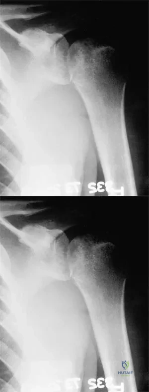
A 37-year-old electrician is diagnosed with a frozen shoulder after sustaining an electrical injury at work 2 weeks ago. Examination reveals that he cannot actively or passively externally rotate or abduct the arm. The glenohumeral joint and scapula move in a 1:1 ratio. Radiographs are shown in Figures 15a and 15b. The best course of action should be


Explanation

The patient's history, examination, and radiographs are classic for locked posterior dislocation of the glenohumeral joint. Posterior dislocation of the shoulder remains the most commonly missed dislocation of a major joint. Up to 80% are missed on initial presentation. The primary cause for failure to accurately diagnose this injury is inadequate radiographic evaluation. The typical presentation is a shoulder locked in internal rotation with loss of abduction. An axillary view not only will make the definitive diagnosis but will help assess the size of the articular surface defect and help plan treatment. This view can be done expediently as part of every trauma series. The AP view is suspicious for a posteriorly dislocated humerus with loss of the humeral neck profile, a vacant glenoid sign, and an anterior humeral head compression fracture (reverse Hill-Sachs lesion). Hawkins RJ, Neer CS II, Pianta RM, Mendoza FX: Locked posterior dislocation of the shoulder. J Bone Joint Surg Am 1987;69:9-18. Norris TR (ed): Orthopaedic Knowledge Update: Shoulder and Elbow. Rosement, IL, American Academy of Orthopaedic Surgeons, 1997, pp 181-189.

Question 2

An 80-year-old man has had increasing shoulder pain for the past 4 months. He reports that it began with soreness and stiffness after chopping some wood. A coronal MRI scan is shown in Figure 16. Initial management should consist of


Explanation

The MRI scan shows a massive tear of the supraspinatus tendon with medial retraction to the level of the glenoid. This is most likely an attritional tear with a high risk of failure of the repair. The preferred treatment is nonsurgical management for pain and stiffness. Acromioplasty and coracoacromial ligament release in this setting are controversial, as they can result in the devastating complication of anterosuperior subluxation of the humerus. Rockwood CA Jr, Williams GR Jr, Burkhead WZ Jr: Debridement of degenerative, irreparable lesions of the rotator cuff. J Bone Joint Surg Am 1995;77:857-866.


Question 3

Figure 17 shows the radiograph of a 25-year-old professional football player who has superior shoulder pain that prevents him from sports participation. History reveals that he sustained a shoulder injury that was treated with closed reduction and temporary pinning 3 years ago. The best course of action should be


Explanation

The radiograph shows a complete acromioclavicular separation. Because the patient is a professional athlete who is unable to participate, surgery is indicated. Chronic separations, especially those with previous trauma from joint pinning, should be treated with resection of the distal clavicle and stabilization to the coracoid. Some type of biologic reconstruction of the coracoclavicular ligaments is generally recommended. Open repair of the ligaments is generally not possible in such a delayed fashion. Screw fixation alone will not provide a lasting solution as the screws usually need to be removed, leaving no fixation in place. Reconstruction using the coracoacromial ligament is generally recommended with coracoclavicular fixation to protect the repair while it heals. Nuber GW, Bowen MK: Disorders of the acromioclavicular joint: Pathophysiology, diagnosis and management, in Iannotti JP, Williams GR (eds): Disorders of the Shoulder: Diagnosis and Management. Philadelphia, PA, Lippincott Williams and Wilkins, 1999.


Question 4

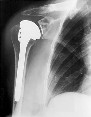
A 54-year-old man undergoes total shoulder arthroplasty for osteoarthritis. Despite compliance with an early passive range-of-motion exercise program, he does not regain more than 90 degrees of elevation, 10 degrees of external rotation, and has internal rotation to the fifth lumbar vertebra. At 6 months, his motion fails to improve. Radiographs are shown in Figures 18a and 18b. What is the best course of action?


Explanation

The patient has a global loss of motion that has failed to improve with 6 months of nonsurgical treatment; because he has reached a plateau, further nonsurgical management will likely be ineffective. Revision in the form of an open release is indicated to lyse intra- and extra-articular adhesions; subscapularis lengthening may be done concurrently as needed. Revising to a smaller head can be considered if adequate motion is not achieved. The radiographs reveal an adequate neck cut with appropriate seating of the component. Removing the glenoid component will decrease capsular tension but will probably increase pain because of the lack of glenoid resurfacing. Increasing humeral retroversion will not improve motion. Cuomo F, Checroun A: Avoiding pitfalls and complication in total shoulder arthroplasty. Orthop Clin North Am 1998;29:507-518.


Question 5

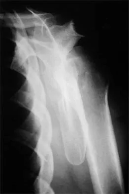
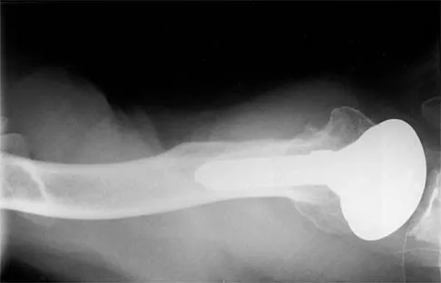
A 47-year-old patient has had persistent pain and weakness after undergoing a reamed intramedullary nailing for a midshaft humerus fracture 8 months ago. There is no evidence of infection. Radiographs are shown in Figures 19a and 19b. Management should consist of


Explanation

Compression plating remains the treatment of choice for most established humeral nonunions. Autograft is felt to be superior to allograft. Electrical stimulation has not been found to improve healing rates in patients with nonunion after intramedullary nailing. Retrograde nailing with flexible nails gives inadequate rotational control to promote healing in this patient. Adding cancellous graft alone will not stabilize the nonunion site. Dynamic locking has been successful only in the lower extremity because the bone can be loaded axially. McKee MD, Miranda MA, Riemer BL, et al: Management of humeral nonunion after the failure of locking intramedullary nails. J Orthop Trauma 1996;10:492-499.


Question 6

An 18-year-old man sustained closed humeral shaft and forearm fractures of his dominant arm in a motor vehicle accident. Neurovascular examination is intact, and his condition is stable. The best course of action for management of the injuries should be


Explanation

Fractures above and below the elbow constitute floating elbow injuries and are best treated with internal fixation to allow early range of motion and to prevent elbow stiffness. Use of a long arm cast would promote elbow stiffness. External fixation is indicated primarily for open injuries. Beaty JH (ed): Orthopaedic Knowledge Update 6. Rosemont, IL, American Academy of Orthopaedic Surgeons, 1999, pp 271-286.


Question 7

A 32-year-old woman has had pain and a visibly growing mass in the shoulder for 3 years but denies any history of trauma. Examination reveals a swollen, boggy shoulder mass. The AP radiograph and MRI scan are shown in Figures 20a and 20b. Figures 20c through 20e show a portion of the excised mass and the photomicrographs of the biopsy specimen. What is the most likely diagnosis?


Explanation

The radiographic findings are classic for synovial chondromatosis because of the small calcified opacities within the joint surrounding the synovium. The histologic findings show cartilaginous foci of metaplasia, which may be markedly cellular. However, unlike low-grade chondrosarcoma, it lacks cellular and nuclear pleomorphism. Murphy FP, Dahlin DC, Sullivan CR: Articular synovial chondromatosis. J Bone Joint Surg Am 1982;44:77-86.


Question 8

What is the most important factor regarding the risk of recurrent instability in a patient with an acute anterior dislocation of the shoulder?


Explanation

The recurrence rate of anterior dislocation of the shoulder after the first episode in athletes younger than age 20 years is thought to be as high as 90%, making surgery after the initial episode a consideration. The rate drops from 50% to 75% in the 20- to 25-year age group and down to 15% in patients older than age 40 years. An excellent prospective study of 257 patients in Sweden showed that there was no difference in those who did or did not complete 3 weeks of immobilization. The study also showed variability among different age groups in the importance of athletic participation; athletes in the 12- to 22-year age group had a higher recurrence rate, whereas the more sedentary patients in the 23- to 29-year age group had a higher rate. Hovelius L: The natural history of primary anterior dislocation of the shoulder in the young. J Orthop Sci 1999;4:307-317.


Question 9

A 25-year-old man injured his dominant shoulder after falling on his outstretched arm 4 months ago. Examination reveals that he cannot lift his arm above 90 degrees, and he has pronounced medial scapular winging. Management should consist of


Explanation

Serratus anterior palsy or long thoracic nerve palsy is usually caused by traction injury to the nerve, blunt injury, or iatrogenic injury. The palsy results in winging of the scapula and medial rotation of the inferior pole of the scapula. A patient with this injury will usually recover in 12 to 18 months. Initial treatment should include observation and shoulder strengthening exercises. Nerve exploration with repair has not proven beneficial in changing the outcome. Many orthopaedic surgeons favor using a split pectoralis major transfer for symptomatic patients. Electrodiagnostic studies are helpful in confirming the diagnosis. Post M: Pectoralis major transfer for winging of the scapula. J Shoulder Elbow Surg 1995;4:1-9.


Question 10

Treatment of adhesive capsulitis has a high failure rate when the underlying cause is


Explanation

Diabetes mellitus has been associated with resistant cases of adhesive capsulitis. With other causes of onset, adhesive capsulitis frequently responds to nonsurgical management such as stretching exercises or, when this fails, manipulation under anesthesia and/or arthroscopic release. Manipulation is rarely successful for the treatment of adhesive capsulitis associated with diabetes mellitus, and arthroscopic release may be preferred. Fisher L, Kurtz A, Shipley M: Association between cheiroarthropathy and frozen shoulder in patients with insulin-dependent diabetes mellitus. Br J Rheumatol 1986;25:141-146. Janda DH, Hawkins RJ: Shoulder manipulation in patients with adhesive capsulitis and diabetes mellitus: A clinical note. J Shoulder Elbow Surg 1993;2:36-38.


Question 11

Figure 21 shows the AP radiograph of a 41-year-old patient who sustained a closed bicolumnar fracture of the distal humerus that resulted in a painful nonunion. What is the best initial construct for rigid stabilization of this fracture pattern?


Explanation

The dual plate fixation construct is significantly stronger than single plate or "Y" plate fixation. Two-plate constructs at right angles, the ulnar plate medially and the lateral plate posteriorly, would appear to be biomechanically optimal. This approach usually is feasible at the time of surgery. Clinically, dual 3.5-mm reconstruction or dynamic compression plates are superior to one third tubular plate fixation. Supplementary external fixation is not considered a better treatment option. Failure of fixation and nonunion are often the result of inadequate fixation and osteoporosis. Helfet DL, Hotchkiss RN: Internal fixation of the distal humerus: A biomechanical comparison of methods. J Orthop Trauma 1990;4:260-264.


Question 12

Figure 22 shows the radiographs of a 16-year-old boy who injured his elbow in a fall 1 year ago. Although he has no pain, he reports restricted forearm rotation and elbow flexion. What is the most likely diagnosis?


Explanation

Congenital dislocation of the radial head is often confused with posttraumatic dislocation. The distinguishing feature here is the dome-shaped radial head. Some patients with congenital anomalies fail to recognize their limitations until an injury occurs. Soft-tissue contractures do not cause radial head dislocation nor do they usually cause this pattern of motion restriction (mainly flexion and rotation without significant loss of extension). There is no deformity of the ulna to suggest an old Monteggia lesion. Morrey BF (ed): The Elbow and Its Disorders, ed 2. Philadelphia, PA, WB Saunders, 1993, p 196.


Question 13

A 55-year-old man has had progressive right shoulder pain for the past 2 years. Examination reveals active elevation to 120 degrees, external rotation to 20 degrees, and internal rotation to the sacrum. AP and axillary radiographs are shown in Figures 23a and 23b. Which of the following procedures would result in the most predictable long-term pain relief?


Explanation

Total shoulder arthroplasty yields excellent pain relief and function in patients with osteoarthritis. It is favored over humeral arthroplasty, especially when there is asymmetric posterior glenoid wear and posterior humeral subluxation as shown on the axillary radiograph. Arthroscopic debridement of the glenohumeral joint may be helpful in delaying the need for arthroplasty when the arthritic changes are mild to moderate but is not indicated for advanced osteoarthritis. Cofield RH, Frankle MA, Zuckerman JD: Humeral head replacement for glenohumeral arthritis. Semin Arthroplasty 1995;6:214-221. Levine WN, Djurasovic M, Glasson JM, Pollock RG, Flatow EL, Bigliani LU: Hemiarthroplasty for glenohumeral osteoarthritis: Results correlated to degree of glenoid wear. J Shoulder Elbow Surg 1997;6:449-454.


Question 14

A 20-year-old professional baseball pitcher has had a 3-year history of increased aching in his shoulder that is associated with pitching, and he is now seeking a second opinion. Nonsurgical management consisting of rest, anti-inflammatory drugs, ice, heat, and cortisone injections has failed to provide relief. A previous work-up that included radiographs and gadolinium-enhanced MRI arthrography was negative. Results of an arteriogram suggest quadrilateral space syndrome. Assuming that this is the correct diagnosis, what nerve needs to be decompressed?


Explanation

Quadrilateral space syndrome is a rare condition and is the result of compression of the contents of the quadrilateral space. The contents of the quadrilateral space include the posterior circumflex vessels and the axillary nerve. Cahill BR, Palmer RE: Quadrilateral space syndrome. J Hand Surg 1983;8:65-69.


Question 15

A right-handed 24-year-old woman underwent an arthroscopic Bankart repair for recurrent anterior dislocations 9 months ago. Despite extensive physical therapy for 8 months, the patient has very limited range of motion (elevation to 130 degrees and external rotation to 10 degrees with the arm at the side). Shoulder radiographs are normal. The next step in management should consist of


Explanation

Arthroscopic capsular release is an effective means of treating stiffness that is the result of capsular contractures, such as in the case of a tight Bankart repair. Open release allows lengthening of a surgically shortened subscapularis, such as after a tight Putti-Platt repair. Additional physical therapy is unlikely to be effective because 8 months of treatment has failed to result in improvement. Accepting this degree of asymptomatic limited motion is not advisable because of the functional limitations for the patient and the increased risk of postoperative degenerative arthritis. Warner JJ, Allen AA, Marks PH, Wong P: Arthroscopic release of postoperative capsular contracture of the shoulder. J Bone Joint Surg Am 1997;79:1151-1158.


Question 16

A patient with deficient anteroinferior bone stock undergoes a Latarjet procedure that transfers a portion of the coracoid to the glenoid rim and secures it with two screws. After surgery, the patient reports numbness on the anterolateral forearm. To verify the diagnosis, what muscle should be tested for strength?


Explanation

A Latarjet procedure is similar to a Bristow procedure, but with the Latarjet procedure a larger portion of the coracoid is transferred to the scapular neck at the anteroinferior glenoid. As in a Bristow procedure, if the fragment is pulled or twisted during the dissection or during fixation, the musculocutaneous nerve can be injured. With loss of biceps function, elbow flexion and forearm supination will be weaker. Ho E, Cofield RH, Balm MR, Hattrup SJ, Rowland CM: Neurologic complications of surgery for anterior shoulder instability. J Shoulder Elbow Surg 1999;8:266-270. Boardman ND 3rd, Cofield RH: Neurologic complications of shoulder surgery. Clin Orthop 1999;368:44-53.


Question 17

A 34-year-old woman has had painful snapping and popping in the elbow since falling while in-line skating 6 months ago. The popping also occurs when she pushes off with her hands to rise from a seated position. Initial radiographs were normal, and she was told that she had sprained her elbow. Examination reveals few findings except that she is very apprehensive when the forearm is forcefully supinated with the elbow extended or partially flexed. A radiograph taken in that position is shown in Figure 24. Treatment should consist of


Explanation

The radiograph reveals posterolateral rotatory subluxation of the radiohumeral and ulnohumeral joints. The space between the ulna and trochlea is enlarged, particularly posteriorly at the olecranon. These findings are diagnostic of posterolateral rotatory instability, which causes recurrent subluxation and reduction as the elbow is flexed from an extended and supinated position with valgus load. The posterolateral rotatory instability apprehension test was performed on this patient and the result was positive. The lateral pivot-shift test causes a clunk as the elbow reduces but is more difficult to perform, even under general anesthesia. The patient does not have isolated subluxation of the radial head, although these findings can be mistakenly diagnosed as such. The radial head is normally shaped and does not represent a congenital dislocation. There are no findings here to suggest osteochondritis dissecans or loose bodies. O'Driscoll SW, Bell DF, Morrey BF: Posterolateral rotatory instability of the elbow. J Bone Joint Surg Am 1991;73:440-446. Burgess RC, Sprague HH: Post-traumatic posterior radial head subluxation: Two case reports. Clin Orthop 1984;186:192-194.


Question 18

A 49-year-old woman noted pain in her right axilla 1 day after moving heavy furniture. Two weeks later, she now reports persistent numbness and paresthesias along the inner aspect of her upper arm radiating into the ulnar digits. Examination reveals full shoulder motion, tenderness over the first rib, and a decreased radial pulse with the shoulder placed overhead. What is the most likely diagnosis?


Explanation

Thoracic outlet syndrome is thought to be caused by compression of the neurovascular supply to the upper limb in the supraclavicular and axillary regions of the shoulder. While typically progressive in onset, thoracic outlet syndrome may develop after acute injury. Injury or weakness of the scapular muscles, especially the trapezius, may result in descent of the scapula and cause compression of the thoracic outlet. In general, most symptoms are the result of neural compression. Typical symptoms include pain in the neck or shoulder and numbness or tingling that predominantly involves the ulnar side of the arm and hand. Exacerbation of these symptoms is typical when the arm is abducted. Initial management should consist of postural exercises aimed at restoring proper scapular stability. Severe recalcitrant symptoms may warrant surgical decompression. Leffert RD: Thoracic outlet syndrome. J Am Acad Orthop Surg 1994;2:317-325.


Question 19

A patient has had a locked posterior dislocation of the shoulder for the past 6 months. After undergoing total shoulder arthroplasty that includes adequate anterior releases and posterior capsulorrhaphy, the patient still exhibits posterior instability intraoperatively. The postoperative rehabilitation regimen should include


Explanation

Achieving stability in chronic locked posterior dislocations of the shoulder remains a difficult challenge. Intraoperative measures include decreased humeral retroversion, anterior releases, and posterior capsular tightening. Postoperative rehabilitation is of equal importance. Immobilization in an external rotation brace (10 degrees to 15 degrees) with the arm at the side for 4 to 6 weeks is recommended to decrease tension in the posterior capsule. When passive range-of-motion exercises are instituted, they should be performed in the plane of the scapula to avoid stress posteriorly. Internal rotation and supine elevation should be avoided for similar reasons. Hawkins RJ, Neer CS II, Pianta RM, Mendoza FX: Locked posterior dislocation of the shoulder. J Bone Joint Surg Am 1987;69:9-18.


Question 20

Which of the following factors is associated with failure of arthroscopic excision of the distal clavicle?


Explanation

Uneven resection of bone, typically leaving a retained posterolateral corner of the distal clavicle, can lead to failure of arthroscopic distal clavicle excision. The amount of bone resected, the gender of the patient, or the diagnosis (osteoarthritis versus osteolysis) does not appear to affect the results.


Question 21

Anterior subluxation in a throwing athlete is most commonly the result of


Explanation

Subtle anterior subluxation in the throwing athlete most frequently results from excessive capsular laxity because of repetitive microtrauma. Avulsion of the inferior glenohumeral ligament from the glenoid, or more rarely from the humerus, occurs with macrotrauma. A large Hill-Sachs lesion and a glenoid rim fracture also may result from a traumatic anterior dislocation. Kvitne RS, Jobe FW: The diagnosis and treatment of anterior instability in the throwing athlete. Clin Orthop 1993;291:107-123.


Question 22

What is the most significant prognostic factor in nontraumatic osteonecrosis of the humeral head?


Explanation

Use of systemic steroids has been implicated in the development of nontraumatic osteonecrosis of the humeral head. Staging of the disease is most relevant to prognosis and treatment. Cruess has described a widely accepted staging system. Several authors have shown that patients who have a lower stage of disease (ie, stage I or II) have a much less likely chance of progression compared with those who are in the later stages (IV and V). Cruess RL: Osteonecrosis of bone: Current concepts as to etiology and pathogenesis. Clin Orthop 1986;208:30-39. Cruess RL: Steroid-induced avascular necrosis of the humeral head: Natural history and management. J Bone Joint Surg Br 1976;58:313-317. Rutherford CS, Cofield RH: Osteonecrosis of the shoulder. Orthop Trans 1987;11:239.


Question 23

A 43-year-old former professional hockey player reports severe pain in his chest after being checked from the side in a pick-up hockey game. An MRI scan and plain radiographs are shown in Figures 25a through 25c. What is the most likely diagnosis?


Explanation

Anterior dislocation is the most common type of sternoclavicular dislocation. The medial end of the clavicle is displaced anterior or anterosuperior to the anterior margin of the sternum. In a study by Omer, 31% of athletic injuries have been known to cause a dislocation of the sternoclavicular joint. The serendipity view can show this dislocation, as will CT of the chest. This view requires the x-ray beam to be aimed at the manubrium with 40 degrees of cephalic tilt. An anterior sternoclavicular joint dislocation will appear superiorly displaced, while a posterior sternoclavicular joint dislocation is inferiorly displaced on the serendipity view. Rockwood CA Jr, Matsen FA III (eds): The Shoulder. Philadelphia, PA, WB Saunders, 1998, vol 1, pp 566-572.


Question 24

Which of the following is considered a contraindication to functional bracing for the treatment of humeral shaft fractures?


Explanation

Most closed humeral shaft fractures and fractures caused by a low-velocity hand gun can be managed nonsurgically with closed reduction and application of a coaptation splint followed by a functional brace. Contraindications to use of the functional brace include: 1) massive soft-tissue or bone loss; 2) an unreliable or noncompliant patient; and 3) an inability to maintain acceptable fracture alignment of up to 20 degrees of anterior or posterior angulation, 30 degrees of varus or valgus angulation, and greater than 3 cm of shortening. Beaty JH (ed): Orthopaedic Knowledge Update 6. Rosemont, IL, American Academy of Orthopaedic Surgeons, 1999, pp 271-286. Pollock FH, Drake D, Bovill EG, Day L, Trafton PG: Treatment of radial neuropathy associated with fractures of the humerus. J Bone Joint Surg Am 1981;63:239-243.


Question 25

A 20-year-old man with fascioscapulohumeral dystrophy has severe scapular winging of both shoulders. He can no longer abduct above 80 degrees, and it affects his activities of daily living. A clinical photograph is shown in Figure 26. Definitive management should consist of


Explanation

The patient's history is typical of patients with severe fascioscapulohumeral dystrophy. The scapular winging can be so pronounced that there is significant loss of function of the upper extremity. The surgical options include transfer of the pectoralis major muscle with a tendon graft or scapulothoracic fusion. The latter is a technically demanding procedure but can provide a very stable platform for the upper extremity. Most patients will see increased elevation of the extremity once the scapula is stabilized. Pectoralis minor transfer has not been described and would not be effective. Shapiro F, Specht L: The diagnosis and orthopaedic treatment of inherited muscular diseases of childhood. J Bone Joint Surg Am 1993;75:439-454.


Question 26

A 22-year-old collegiate rugby player presents with his fourth anterior shoulder dislocation. A 3D CT scan reveals 26% anterior glenoid bone loss. What is the most appropriate surgical management?


Explanation

The Latarjet procedure is indicated for recurrent anterior shoulder instability in the presence of critical glenoid bone loss (typically >20-25%). Soft tissue stabilization alone in this setting has an unacceptably high failure rate.

Question 27

A 55-year-old man falls on an outstretched arm and experiences a popping sensation in his right shoulder. On exam, he has increased passive external rotation compared to the contralateral side and weakness on the belly-press test. MRI confirms an isolated, complete tear of the subscapularis tendon. What is the most appropriate initial management?


Explanation

Acute, traumatic subscapularis tears in active patients should be treated with early surgical repair to prevent rapid tendon retraction and muscle atrophy. Delaying surgery significantly reduces the chances of a successful primary repair.

Question 28

A 65-year-old female presents with a 4-part proximal humerus fracture.

According to the Hertel criteria, which radiographic feature is most predictive of humeral head ischemia?


Explanation

Hertel identified specific predictors of humeral head ischemia, the strongest of which are a short calcar segment (<8 mm), a disrupted medial hinge, and an anatomic neck fracture pattern.

Question 29

A 28-year-old man has recurrent anterior shoulder instability. Diagnostic arthroscopy reveals an engaging Hill-Sachs lesion and <10% glenoid bone loss. Which of the following is the most appropriate surgical treatment?


Explanation

An engaging Hill-Sachs lesion in the setting of subcritical glenoid bone loss is best treated with a Bankart repair and Remplissage (infraspinatus tenodesis into the defect) to prevent engagement and recurrent dislocation.

Question 30

A 70-year-old woman presents with chronic, severe right shoulder pain and an inability to actively elevate her arm past 60 degrees (pseudoparalysis). Radiographs demonstrate superior migration of the humeral head with an acromiohumeral interval of 2 mm. MRI confirms massive, retracted tears of the supraspinatus and infraspinatus with fatty infiltration. What is the treatment of choice?


Explanation

Reverse total shoulder arthroplasty is the treatment of choice for elderly patients with rotator cuff tear arthropathy and pseudoparalysis, as it relies on the deltoid to restore active elevation.

Question 31

A 35-year-old man presents to the emergency department with shoulder pain after a generalized seizure. His arm is locked in adduction and internal rotation, with essentially no passive external rotation possible. An AP radiograph shows a 'lightbulb' sign. Which imaging view is most critical to confirm the diagnosis?


Explanation

The clinical presentation strongly suggests a missed posterior shoulder dislocation. An axillary lateral radiograph is the most reliable conventional view to confirm the posterior relationship of the humeral head to the glenoid.

Question 32

A 30-year-old elite volleyball player presents with painless weakness in external rotation of the dominant shoulder. Physical exam reveals isolated atrophy of the infraspinatus fossa. MRI shows a paralabral cyst in the spinoglenoid notch. What is the most likely associated intra-articular pathology?


Explanation

Cysts at the spinoglenoid notch are highly associated with posterior or posterosuperior SLAP tears. They selectively compress the suprascapular nerve branch to the infraspinatus, causing isolated external rotation weakness and atrophy.

Question 33

The biomechanical 'suspension bridge' concept of the rotator cuff relies on force couples to maintain the humeral head centered on the glenoid. Which two muscles form the primary force couple in the transverse plane?


Explanation

The transverse plane force couple of the shoulder is formed by the subscapularis anteriorly and the infraspinatus and teres minor posteriorly. This couple is crucial for dynamic glenohumeral stability.

Question 34

A 75-year-old woman sustains a minimally displaced 2-part surgical neck fracture of the proximal humerus. She is treated nonoperatively in a sling. To optimize functional outcomes and prevent adhesive capsulitis, when should gentle pendulum exercises and passive range of motion begin?


Explanation

For minimally displaced proximal humerus fractures treated nonoperatively, early progressive motion (pendulums/passive ROM) should ideally begin within 7-14 days to prevent severe post-traumatic shoulder stiffness.

Question 35

An 18-year-old female gymnast complains of bilateral shoulder pain and a sensation of 'slipping'. On exam, she has a positive sulcus sign and apprehension in multiple positions. Beighton score is 6/9. What is the most appropriate initial management?


Explanation

Multidirectional instability (MDI) typically presents in young, hyperlax patients. The first-line treatment is always an extended course of dedicated physical therapy focusing on rotator cuff and periscapular strengthening.

Question 36

A 24-year-old throwing athlete is diagnosed with a Type II SLAP tear via MRI arthrogram. Diagnostic arthroscopy confirms a 'peel-back' lesion of the superior labrum. When placing suture anchors for repair, where is the optimal location to restore biomechanics and prevent internal impingement?


Explanation

For Type II SLAP tears involving a peel-back mechanism, repairing the labrum slightly posterior to the biceps anchor is critical to neutralize the posterior peel-back forces generated during the late cocking phase of throwing.

Question 37

A 31-year-old man suffers a traumatic anterior shoulder dislocation. MRI reveals a 'J-sign' involving the inferior glenohumeral ligament (IGHL) complex. This indicates a Humeral Avulsion of the Glenohumeral Ligament (HAGL) lesion. Which of the following is true regarding this condition?


Explanation

A HAGL lesion involves avulsion of the IGHL from its humeral attachment, leading to anterior instability. It is an important cause of recurrent instability that occurs without a traditional anterior labral (Bankart) tear.

Question 38

During open reduction and internal fixation of a proximal humerus fracture, a deltopectoral approach is utilized. Which two nerves supply the muscles that form the internervous plane for this approach?


Explanation

The deltopectoral approach uses the internervous plane between the deltoid (innervated by the axillary nerve) and the pectoralis major (innervated by the medial and lateral pectoral nerves).

Question 39

A patient presents with anterior shoulder pain exacerbated by internal rotation and forward elevation. MRI demonstrates a narrowed coracohumeral interval (<6 mm). Subcoracoid impingement syndrome is suspected. This condition is most strongly associated with pathology of which structure?


Explanation

Subcoracoid impingement involves compression of the tissues between the coracoid process and the lesser tuberosity, which most directly affects and damages the subscapularis tendon.

Question 40

The Latarjet procedure provides stability through a 'triple effect'. Which structure provides the dynamic 'sling effect' when the arm is abducted and externally rotated?


Explanation

The Latarjet procedure transfers the coracoid and its attached conjoined tendon. In abduction and external rotation, the conjoined tendon acts as a dynamic sling across the anterior-inferior capsule, providing critical stability.

Question 41

A 45-year-old heavy laborer has a massive, retracted, and irreparable subscapularis tendon tear. He complains of severe weakness with lifting objects in front of his body. Which tendon transfer is the most appropriate primary option to restore function?


Explanation

The pectoralis major tendon transfer (routed either anterior or posterior to the conjoined tendon) is the preferred procedure to restore active internal rotation and stability in the setting of an irreparable subscapularis tear.

Question 42

A 55-year-old physically active man presents with an irreparable posterosuperior rotator cuff tear (supraspinatus and infraspinatus). The subscapularis is completely intact, and there is no glenohumeral arthritis. He has a severe external rotation lag. Which tendon transfer is most appropriate?


Explanation

The latissimus dorsi (or lower trapezius) transfer is indicated for irreparable posterosuperior rotator cuff tears to restore external rotation and elevation in young, active patients without significant glenohumeral arthritis.

Question 43

A 72-year-old female undergoes open reduction and internal fixation with a locking plate for a displaced 3-part proximal humerus fracture. Osteoporotic bone is noted during surgery. What is the most common hardware-related complication postoperatively?


Explanation

In the setting of osteoporotic proximal humerus fractures fixed with a locking plate, the most common hardware complication is intra-articular screw penetration. This typically occurs as the fracture settles or collapses, causing the fixed-angle screws to protrude into the joint.

Question 44

The 'terrible triad' of the shoulder is a distinct clinical entity commonly seen in middle-aged or older patients following high-energy trauma. Which three injuries comprise this triad?


Explanation

The terrible triad of the shoulder consists of an anterior glenohumeral dislocation, a rotator cuff tear (often massive), and a neurologic injury (most commonly the axillary nerve or brachial plexus).

Question 45

A 32-year-old man sustains a displaced 3-part proximal humerus fracture after a high-speed motorcycle accident. An axillary nerve injury is suspected. Which clinical finding is the most reliable indicator of axillary nerve dysfunction?


Explanation

The axillary nerve provides motor innervation to the deltoid and teres minor, and sensory innervation to the lateral shoulder (regimental badge area). Sensation loss here combined with lack of palpable deltoid contraction confirms its dysfunction.

Question 46

A 21-year-old collegiate rugby player presents with his fourth anterior shoulder dislocation. A 3D CT scan is obtained to evaluate bone stock.

The imaging reveals 25% anterior glenoid bone loss. What is the most appropriate definitive surgical management?


Explanation

Critical glenoid bone loss (>20-25%) in a young collision athlete is a strong indication for a bony augmentation procedure like the Latarjet to restore anterior stability. Soft tissue repairs alone in this setting have an unacceptably high failure rate.

Question 47

A 65-year-old female sustains a proximal humerus fracture after a mechanical fall. According to Hertel's criteria, which of the following radiographic findings is the most reliable predictor of humeral head ischemia?


Explanation

Hertel's criteria for predicting humeral head ischemia include a disrupted medial hinge, short calcar length (metaphyseal extension < 8 mm), and basicervical/anatomic neck fracture patterns. An intact medial hinge and valgus impaction are protective of the blood supply.

Question 48

A 72-year-old male presents with long-standing shoulder pain and an inability to actively elevate his arm above 45 degrees. Radiographs demonstrate an acromiohumeral distance of 3 mm.

MRI reveals massive, retracted supraspinatus and infraspinatus tears with grade 4 fatty infiltration. What is the most appropriate surgical treatment?


Explanation

Reverse total shoulder arthroplasty (RTSA) is the treatment of choice for elderly patients with massive, irreparable rotator cuff tears and pseudoparalysis or cuff tear arthropathy. RTSA relies on the deltoid to restore active elevation.

Question 49

A 28-year-old offensive lineman presents with deep posterior shoulder pain and clicking during bench pressing. Examination reveals a positive jerk test. If an MRI arthrogram is obtained, it is most likely to show a labral tear at which clock-face location on the right glenoid?


Explanation

A positive jerk test indicates posterior shoulder instability, which is highly associated with a reverse Bankart lesion. On a right shoulder, this corresponds to the posteroinferior labrum between 7 and 9 o'clock.

Question 50

A 45-year-old male presents with acute shoulder pain and weakness after falling on an outstretched arm while skiing. Physical examination demonstrates increased passive external rotation compared to the contralateral side and a positive belly-press test. Which of the following structures is most likely injured?


Explanation

The subscapularis is the primary internal rotator of the shoulder. A tear results in increased passive external rotation and weakness on specific testing, such as a positive lift-off, belly-press, or bear-hug test.

Question 51

A 24-year-old male is undergoing arthroscopic evaluation for recurrent anterior shoulder instability. Diagnostic arthroscopy reveals an 'engaging' Hill-Sachs lesion, but pre-operative CT scan showed minimal glenoid bone loss (<10%). Which of the following is the most appropriate management strategy?


Explanation

An engaging Hill-Sachs lesion (off-track lesion) without critical glenoid bone loss is effectively treated by combining an arthroscopic Bankart repair with a Remplissage. Remplissage involves tenodesing the infraspinatus into the humeral defect to prevent engagement.

Question 52

When performing a reverse total shoulder arthroplasty for a 4-part proximal humerus fracture in an elderly patient, anatomic healing of the greater tuberosity to the proximal humerus shaft is most critical for restoring which of the following active motions?


Explanation

In RTSA for proximal humerus fractures, healing of the greater tuberosity (containing the infraspinatus and teres minor insertions) is strongly correlated with improved active external rotation and overall functional outcomes.

Question 53

A 62-year-old woman undergoes open reduction and internal fixation with a locked plate for a 3-part proximal humerus fracture.

Which of the following is the most common complication associated with this specific procedure?


Explanation

Intra-articular screw penetration is the most frequent complication following locked plating of proximal humerus fractures. This often occurs secondary to fracture settling or varus collapse over rigid fixed-angle screws.

Question 54

A 19-year-old gymnast presents with bilateral shoulder pain and a sensation of her shoulders 'slipping' during routines. She has a positive sulcus sign bilaterally and generalized ligamentous laxity. Supervised physical therapy has failed to improve her symptoms after 6 weeks. What is the next best step in management?


Explanation

Multidirectional instability (MDI) is primarily managed non-operatively. An extended course (at least 6 months) of physical therapy focusing on periscapular and rotator cuff strengthening is indicated before considering surgical intervention like a capsular shift.

Question 55

During an arthroscopic rotator cuff repair, the surgeon evaluates the normal anatomical footprint on the greater tuberosity. Which of the following best describes the normal insertion site of the supraspinatus tendon?


Explanation

The supraspinatus tendon inserts onto the superior facet of the greater tuberosity. The infraspinatus inserts onto the middle facet, and the teres minor inserts onto the inferior facet.

Question 56

A 22-year-old rugby player has recurrent anterior shoulder instability. CT scan with 3D reconstruction demonstrates 25% anterior glenoid bone loss. What is the most appropriate definitive management?


Explanation

Critical glenoid bone loss (>20-25%) in a collision athlete is an absolute indication for a bony augmentation procedure like the Latarjet. Soft tissue repairs alone in this setting have an unacceptably high failure rate.

Question 57

A 70-year-old man presents with an inability to actively elevate his right arm above 45 degrees, though passive elevation is full. Radiographs show superior migration of the humeral head with an acromiohumeral interval of 3 mm. MRI confirms a massive, retracted rotator cuff tear with Goutallier grade 4 fatty infiltration of the supraspinatus and infraspinatus. What is the best treatment option?


Explanation

Reverse total shoulder arthroplasty is the treatment of choice for elderly patients with cuff tear arthropathy and pseudoparalysis. It provides a stable fulcrum, allowing the deltoid to effectively restore active forward elevation.

Question 58

A 65-year-old woman sustains a 3-part proximal humerus fracture. According to Hertel's criteria, which of the following radiographic findings is most strongly predictive of humeral head ischemia?


Explanation

Hertel's criteria for high risk of humeral head ischemia include a posteromedial metaphyseal head extension <8 mm, disruption of the medial hinge >2 mm, and an anatomic neck fracture pattern. Disruption of the medial hinge removes the critical blood supply from the ascending branch of the anterior circumflex humeral artery.

Question 59

A 34-year-old man has chronic posterior shoulder pain and limited external rotation following a seizure 3 months ago. Radiographs confirm a missed posterior dislocation with an associated reverse Hill-Sachs defect involving 35% of the articular surface. The joint is reducible but unstable in internal rotation. What is the most appropriate management?


Explanation

For reverse Hill-Sachs defects involving >30-40% of the articular surface, an osteoarticular allograft is recommended to restore joint congruity and stability. Smaller defects (<20%) can be treated with a modified McLaughlin procedure.

Question 60

A 45-year-old man presents with anterior shoulder pain and weakness after a forceful external rotation injury. He demonstrates increased passive external rotation compared to the contralateral side. Which physical examination test is most sensitive and specific for his likely injury?


Explanation

The clinical presentation suggests a subscapularis tendon tear, indicated by increased passive external rotation. The bear hug test is highly sensitive and specific for evaluating the integrity of the upper subscapularis.

Question 61

A 55-year-old woman undergoes locking plate osteosynthesis for a 4-part proximal humerus fracture. Six months later, she presents with severe pain and a mechanical block to motion. Radiographs show fracture healing but the superior-most locking screws are protruding through the articular surface. What intraoperative step is most critical to prevent this complication?


Explanation

Primary varus collapse of the humeral head is the most common reason for secondary screw cutout in proximal humerus plating. An inferomedial calcar screw provides structural support to resist this varus displacement.

Question 62

In the setting of recurrent anterior shoulder instability, the addition of an arthroscopic remplissage (infraspinatus tenodesis) to a Bankart repair is most appropriately indicated for which of the following scenarios?


Explanation

Remplissage is indicated for engaging or "off-track" Hill-Sachs lesions in the setting of subcritical glenoid bone loss. It prevents the defect from engaging the anterior glenoid rim during external rotation.

Question 63

A 50-year-old laborer undergoes a latissimus dorsi tendon transfer for an irreparable posterosuperior rotator cuff tear. To function effectively, the transferred latissimus dorsi primarily replicates the function of which native muscle?


Explanation

Latissimus dorsi transfer is used for irreparable massive posterosuperior tears to restore external rotation and provide a head-depressing effect. It primarily replicates the function of the infraspinatus and teres minor.

Question 64

A 72-year-old right-hand-dominant woman sustains a minimally displaced 2-part surgical neck fracture of the proximal humerus. She lives alone and has well-controlled diabetes. What is the most appropriate initial management?


Explanation

Minimally displaced 2-part proximal humerus fractures in elderly patients have excellent outcomes with nonoperative management. Treatment consists of brief sling immobilization followed by early motion to prevent shoulder stiffness.

Question 65

A 28-year-old volleyball player presents with posterior shoulder pain and weakness in external rotation. MRI reveals a paralabral cyst in the spinoglenoid notch and a superior labral tear. Which physical examination finding is most likely to be present?


Explanation

A cyst at the spinoglenoid notch compresses the suprascapular nerve after it has provided motor branches to the supraspinatus. This results in isolated denervation, weakness, and atrophy of the infraspinatus.

Question 66

The concept of the "suspension bridge" model of the rotator cuff, described by Burkhart, emphasizes the importance of which intact structures in maintaining normal glenohumeral kinematics despite a supraspinatus tear?


Explanation

The rotator cable acts as a suspension bridge to transfer forces. As long as the anterior (subscapularis) and posterior (infraspinatus) transverse force couples remain intact, shoulder kinematics can remain balanced and functional.

Question 67

A 45-year-old man sustains a severe fracture-dislocation of the proximal humerus. During an open reduction through a deltopectoral approach, brisk arterial bleeding is encountered near the inferior border of the subscapularis. Which vessel is most likely injured?


Explanation

The anterior circumflex humeral artery, often referred to as the "three sisters" along with its venae comitantes, runs laterally across the inferior border of the subscapularis. It must be carefully identified and ligated or cauterized during the deltopectoral approach.

Question 68

A 24-year-old swimmer presents with anterior shoulder pain and apprehension. An MR arthrogram demonstrates a "J-sign" with extravasation of contrast inferiorly. What is the most likely diagnosis?


Explanation

The "J-sign" on MR arthrogram represents a disrupted inferior glenohumeral ligament at its humeral attachment (HAGL lesion). This allows contrast to extravasate inferiorly forming the characteristic J shape.

Question 69

In performing a superior capsular reconstruction (SCR) for an irreparable supraspinatus tear, the graft is typically attached medially to the superior glenoid and laterally to the greater tuberosity. What is the primary biomechanical goal of this procedure?


Explanation

SCR utilizes a dermal or fascial graft to statically replace the absent superior capsule. This provides a restraint that prevents superior migration of the humeral head, restoring the anatomic fulcrum for the deltoid.

Question 70

In the treatment of a comminuted 4-part proximal humerus fracture with a reverse total shoulder arthroplasty, anatomic healing of the greater tuberosity is most strongly associated with which functional outcome?


Explanation

In RSA for proximal humerus fractures, healing of the greater tuberosity (which houses the infraspinatus and teres minor attachments) is critical for restoring active external rotation and maximizing overall functional outcomes.

Question 71

During a Latarjet procedure, the coracoid process is osteotomized and transferred to the anterior glenoid. Which muscle tendon units are transferred with the coracoid to provide a dynamic "sling" effect?


Explanation

The conjoined tendon, consisting of the short head of the biceps and coracobrachialis, remains attached to the transferred coracoid process. When the arm is abducted and externally rotated, it acts as a dynamic sling across the anteroinferior capsule.

Question 72

A 22-year-old rugby player presents with recurrent anterior shoulder dislocations. Advanced imaging demonstrates 25% glenoid bone loss and an engaging Hill-Sachs lesion. What is the most appropriate definitive surgical management?


Explanation

For anterior shoulder instability with >20-25% glenoid bone loss and an engaging Hill-Sachs lesion, a Latarjet procedure (coracoid transfer) is the standard of care to restore joint stability.

Question 73

A 65-year-old female sustains a comminuted 4-part proximal humerus fracture. Which of the following radiographic findings is most predictive of avascular necrosis of the humeral head?


Explanation

Hertel's radiographic criteria for predicting humeral head ischemia include a posteromedial metaphyseal extension of less than 8 mm and disruption of the medial hinge greater than 2 mm.

Question 74

A 55-year-old laborer suffers from chronic shoulder pain and pseudoparalysis of external rotation. MRI reveals a massive, retracted, and fatty-infiltrated tear of the supraspinatus and infraspinatus with an intact subscapularis. Which of the following tendon transfers is most appropriate for restoring active external rotation?


Explanation

Latissimus dorsi tendon transfer is classically indicated to restore active external rotation and forward elevation in patients with massive, irreparable posterosuperior rotator cuff tears and an intact subscapularis.

Question 75

A 45-year-old male presents with shoulder pain and weakness in internal rotation after a fall. Which of the following physical examination tests is most sensitive and specific for evaluating a tear of the upper border of the subscapularis tendon?


Explanation

The bear hug test is highly sensitive and specific for evaluating tears of the upper portion of the subscapularis tendon, whereas the lift-off test primarily isolates the lower portion.

Question 76

A 30-year-old weightlifter presents with vague posterior shoulder pain and a positive jerk test. Nonoperative management has failed. Imaging shows a posterior labral tear without significant glenoid bone loss. What is the most appropriate surgical intervention?


Explanation

Arthroscopic posterior labral repair is the surgical treatment of choice for recurrent posterior shoulder instability in patients who have failed conservative management and lack significant bone loss.

Question 77

A 24-year-old professional baseball pitcher is diagnosed with an isolated Type II SLAP tear after failing 6 months of physical therapy. What is the recommended surgical management?


Explanation

In young, overhead athletes (typically <30 years old), arthroscopic repair of a type II SLAP tear is the preferred treatment to restore throwing mechanics, whereas older patients often undergo biceps tenodesis.

Question 78

A 78-year-old female undergoes a reverse total shoulder arthroplasty for a severely comminuted 4-part proximal humerus fracture. What is the most critical surgical step for ensuring optimal functional external rotation and joint stability?


Explanation

Anatomic healing of the greater tuberosity is the most critical factor for restoring external rotation and providing dynamic stability following a reverse total shoulder arthroplasty for fracture.

Question 79

A 22-year-old throwing athlete experiences posterior shoulder pain during the late cocking phase. An MRI arthrogram reveals a partial articular-sided supraspinatus tendon tear and posterosuperior labral fraying. This condition is primarily associated with which of the following pathomechanics?


Explanation

Internal impingement in overhead throwers is heavily associated with glenohumeral internal rotation deficit (GIRD), which is caused by a contracted posteroinferior capsule.

Question 80

A 32-year-old athlete presents with isolated weakness in external rotation of the shoulder. MRI reveals a paralabral cyst causing nerve compression. Where is the cyst most likely located and which muscle will demonstrate isolated atrophy?


Explanation

The suprascapular nerve innervates the supraspinatus before passing through the spinoglenoid notch. A cyst at this notch compresses the nerve distally, resulting in isolated infraspinatus atrophy.

Question 81

A 72-year-old man presents with progressive pseudoparalysis and severe glenohumeral osteoarthritis secondary to a massive rotator cuff tear. Which of the following is an absolute contraindication to performing a reverse total shoulder arthroplasty in this patient?


Explanation

Reverse total shoulder arthroplasty relies on a functioning deltoid muscle to elevate the arm. A non-functioning deltoid or paralyzed axillary nerve is an absolute contraindication.

Question 82

A 60-year-old female undergoes open reduction and internal fixation of a 2-part surgical neck humerus fracture with a locking plate. Postoperatively, radiographs show that the fracture has collapsed into varus. Which of the following technical errors most likely contributed to this complication?


Explanation

The placement of medial calcar screws provides vital medial column support, which is critical to preventing varus collapse and subsequent screw cut-out in proximal humerus fracture plating.

Question 83

A 65-year-old male undergoes an arthroscopic biceps tenotomy for a symptomatic superior labral tear and associated biceps tendinopathy. Compared to biceps tenodesis, biceps tenotomy is associated with a significantly higher incidence of which of the following?


Explanation

Biceps tenotomy has a significantly higher rate of a cosmetic "Popeye" deformity and subjective muscle cramping compared to biceps tenodesis, though clinical shoulder outcome scores are similar.

Question 84

A 19-year-old gymnast presents with bilateral shoulder pain and multidirectional instability. On examination, she has a positive sulcus sign that does not diminish when the arm is placed in external rotation. This finding specifically indicates incompetence of which of the following structures?


Explanation

A positive sulcus sign that persists in external rotation indicates incompetence of the rotator interval, specifically the coracohumeral ligament and superior glenohumeral ligament.

Question 85

A 28-year-old male bodybuilder feels a pop in his anterior chest while performing a heavy bench press. Examination reveals an asymmetric chest wall contour and weakness in internal rotation. Which of the following describes the most common anatomic location of a pectoralis major rupture in this demographic?


Explanation

Pectoralis major ruptures in weightlifters most commonly occur as avulsions of the sternocostal tendon from its insertion on the humerus, typically requiring surgical repair.

Question 86

A 35-year-old skier sustains an acute shoulder dislocation and presents with continued apprehension and instability. An MRI arthrogram reveals extravasation of contrast inferiorly into the axillary pouch, demonstrating a classic "J-sign". What is the most likely diagnosis?


Explanation

A Humeral Avulsion of the Glenohumeral Ligament (HAGL) presents on an MRI arthrogram as extravasation of contrast into the axillary pouch due to capsular avulsion, classically described as the "J-sign".

Question 87

A 68-year-old female sustains a 4-part proximal humerus fracture. Which of the following radiographic features is the most reliable predictor of humeral head ischemia?


Explanation

Hertel identified that a metaphyseal head extension (calcar length) of less than 8 mm attached to the articular segment and disruption of the medial hinge are the most reliable predictors of humeral head ischemia.

Question 88

A 22-year-old rugby player presents with recurrent anterior shoulder instability. CT evaluation demonstrates a 25% anteroinferior glenoid bone loss (an "inverted pear" glenoid). What is the most appropriate surgical management?


Explanation

In patients with significant glenoid bone loss (typically >20-25%), soft tissue stabilization (Bankart repair) has an unacceptably high failure rate. A coracoid transfer (Latarjet procedure) is the gold standard to restore the glenoid arc and provide a dynamic sling effect.

Question 89

A 72-year-old man presents with chronic shoulder pain and inability to actively elevate his arm above 45 degrees, though passive ROM is full. MRI shows massive, retracted tears of the supraspinatus and infraspinatus with Goutallier grade 4 fatty infiltration. What is the best treatment option?


Explanation

For an elderly patient with an irreparable massive rotator cuff tear (indicated by high-grade fatty infiltration) and pseudoparalysis, reverse total shoulder arthroplasty is the most reliable option. It restores elevation by medializing the center of rotation and utilizing the intact deltoid.

Question 90

During an arthroscopic stabilization for recurrent shoulder instability, an engaging Hill-Sachs lesion is noted with minimal glenoid bone loss. Which of the following procedures is indicated to address the humeral defect?


Explanation

An engaging Hill-Sachs lesion without critical glenoid bone loss is effectively treated with a remplissage procedure. This involves tenodesis of the infraspinatus and posterior capsule into the defect, preventing it from engaging the anterior glenoid rim during external rotation and abduction.

Question 91

A 60-year-old woman undergoes open reduction and internal fixation with a locking plate for a 3-part proximal humerus fracture. What is the most common complication specifically associated with this fixation method?


Explanation

The most common complication following locked plating of proximal humerus fractures is intra-articular screw penetration. This often occurs due to osteoporotic bone settling and varus collapse, causing the rigid locking screws to cut out through the articular surface.

Question 92

A 28-year-old overhead athlete presents with persistent shoulder pain. MRI arthrogram reveals a partial articular-sided supraspinatus tendon avulsion (PASTA) involving 60% of the tendon footprint. Conservative management has failed. What is the recommended surgical approach?


Explanation

Partial articular-sided rotator cuff tears involving greater than 50% of the tendon footprint typically require surgical repair. This can be accomplished via tear completion and formal repair, or a transtendon in situ repair to restore the footprint without violating the intact bursal fibers.

Question 93

A 35-year-old man presents with shoulder pain and a locked arm in internal rotation after a first-time seizure. Radiographs reveal a posterior shoulder dislocation with an anteromedial humeral head defect (reverse Hill-Sachs lesion) involving 35% of the articular surface. What is the most appropriate surgical treatment?


Explanation

Posterior dislocations with an anteromedial humeral head defect (reverse Hill-Sachs) between 20% and 40% are typically treated with a McLaughlin or modified McLaughlin procedure. This involves transferring the subscapularis or lesser tuberosity into the defect to provide dynamic stability.

Question 94

When treating a 4-part proximal humerus fracture in an elderly patient with a reverse total shoulder arthroplasty, which of the following factors is most critical for achieving active external rotation postoperatively?


Explanation

In reverse total shoulder arthroplasty for acute fractures, restoration of active external rotation is highly dependent on tuberosity healing. Stable anatomic healing of the greater tuberosity (the attachment of the infraspinatus and teres minor) to the shaft is essential for external rotation function.

Question 95

A 55-year-old male sustains an acute traumatic anterior shoulder dislocation. After successful reduction, he complains of profound weakness in internal rotation. Which of the following physical examination tests is most sensitive for diagnosing an upper subscapularis tendon tear?


Explanation

The Bear hug test is highly sensitive and specific for diagnosing tears of the upper subscapularis tendon. The lift-off test is also used but primarily isolates the lower subscapularis and requires the patient to be able to internally rotate the arm behind the back.

Question 96

During an arthroscopy for recurrent anterior shoulder instability, the surgeon identifies an Anterior Labroligamentous Periosteal Sleeve Avulsion (ALPSA) lesion. How does this pathology differ anatomically from a classic Bankart lesion?


Explanation

An ALPSA lesion involves an intact anterior scapular periosteum that strips off the bone, allowing the avulsed labroligamentous complex to displace medially. It often heals in a non-anatomic position on the glenoid neck, unlike a classic Bankart which has a ruptured periosteum.

Question 97

Which of the following best describes the fundamental biomechanical alteration achieved by a classic Grammont-style reverse total shoulder arthroplasty in a patient with advanced rotator cuff arthropathy?


Explanation

The Grammont-style reverse total shoulder arthroplasty shifts the joint's center of rotation medially and inferiorly. This medialization increases the deltoid moment arm, while inferiorization tensions the deltoid fibers, allowing it to efficiently compensate for the deficient rotator cuff.

Question 98

During an open reduction and internal fixation of a proximal humerus fracture using an extended deltopectoral approach, which nerve is at greatest risk of iatrogenic injury when mobilizing the inferior capsule and placing retractors inferior to the humeral head?


Explanation

The axillary nerve travels anteriorly to posteriorly through the quadrilateral space, passing just inferior to the shoulder capsule. It is at significant risk of injury during inferior capsular release, excessive traction, or from misplaced retractors positioned inferior to the humeral head.

Question 99

A 30-year-old professional volleyball player presents with posterior shoulder pain and isolated weakness in external rotation. MRI reveals a paralabral cyst located in the spinoglenoid notch. This finding is most commonly associated with which of the following concomitant labral pathologies?


Explanation

Paralabral cysts in the spinoglenoid notch typically compress the infraspinatus branch of the suprascapular nerve, causing isolated external rotation weakness. They are highly associated with posterior or superior labral tears (often Type II SLAP tears), which act as a one-way valve for joint fluid to escape.

Question 100

A 28-year-old male sustains a shoulder dislocation and subsequent recurrent instability. An MRI arthrogram demonstrates extravasation of contrast into the axillary pouch in a U-shape, replacing the normal V-shaped axillary recess. What is the most likely diagnosis?


Explanation

A HAGL lesion involves avulsion of the inferior glenohumeral ligament from the anatomic neck of the humerus. On MRI arthrography, the normal V-shaped axillary pouch becomes U-shaped (the classic "J-sign") as contrast leaks inferiorly through the avulsed humeral attachment.

None

Clinic OS
Medically Verified Content by
Prof. Clinic OS
Consultant Orthopedic & Spine Surgeon
Chapter Index