العربية
Part of the Master Guide

100 High-Yield Orthopedic MCQs: Shoulder & Elbow | Mock Exam 251

AAOS Sports Medicine MCQs (Set 3): Knee & Shoulder Ligament Trauma | OITE & ABOS Review

27 Apr 2026 50 min read 99 Views
Sports Medicine 2001 MCQs - Part 3

Key Takeaway

This high-yield Sports Medicine MCQ set (Set 3) for AAOS/ABOS exams covers critical topics in knee and shoulder pathology. Questions delve into the diagnosis, imaging, and management of ACL tears, meniscal injuries, shoulder instability, and rotator cuff pathology, aiding comprehensive board preparation.

AAOS Sports Medicine MCQs (Set 3): Knee & Shoulder Ligament Trauma | OITE & ABOS Review

Comprehensive 100-Question Exam


00:00

Start Quiz

Question 1

Figure 24 shows the radiograph of a 10-year-old boy who sustained a valgus injury to the knee. Examination reveals grade III medial laxity. Initial management should consist of


Explanation

Based on the mechanism of injury and findings of medial laxity, the most likely diagnosis is injury to either the growth plate or the medial collateral ligament. With the open physeal plate, this area of injury is presumed present until proven otherwise; therefore, stress radiographs should be obtained before implementing any treatment or ordering more extensive and expensive tests. DeLee JC: Ligamentous injury of the knee, in Stanitski CL, DeLee JC, Drez D Jr (eds): Pediatric and Adolescent Sports Medicine. Philadelphia, PA, WB Saunders, 1994, vol 3, pp 406-432. Clanton TO, DeLee JC, Sanders B, Neidre A: Knee ligament injuries in children. J Bone Joint Surg Am 1979;61:1195-1201.

Question 2

A right-handed 14-year-old pitcher has had a 3-month history of shoulder pain while pitching. Examination reveals full range of motion, a mildly positive impingement sign, pain with rotational movement, and no instability. Plain AP radiographs of both shoulders are shown in Figures 25a and 25b. Management should consist of


Explanation

The patient has the classic signs of Little Leaguer's shoulder, with findings that include pain localized to the proximal humerus during the act of throwing and radiographic evidence of widening of the proximal humeral physis. Examination usually reveals tenderness to palpation over the proximal humerus, but the presence of any swelling, weakness, atrophy, or loss of motion is unlikely. The treatment of choice is rest from throwing for at least 3 months, followed by a gradual return to pitching once the shoulder is asymptomatic. Carson WG Jr, Gasser SI: Little Leaguer's shoulder: A report of 23 cases. Am J Sports Med 1998;26:575-580.


Question 3

A 38-year-old man sustains a complete avulsion with retraction of the ischial attachment of the hamstring muscles in a fall while water skiing. He indicates that he is an aggressive athlete who participates regularly in multiple running and cutting-type sports, and he strongly desires to continue his athletic competition. Management should consist of


Explanation

Several studies have identified a complete proximal avulsion of the hamstring muscles as an injury that leads to significant long-term disability, with a high percentage of athletes who must permanently restrict their activities following nonsurgical management. Early surgical repair and prolonged rehabilitation have yielded consistently better results than nonsurgical management. Orava S, Kujala UM: Rupture of the ischial origin of the hamstring muscles. Am J Sports Med 1995;23:702-705.

Question 4

What mechanism contributes to strength gains during conditioning of the preadolescent athlete?


Explanation

Prepubescent athletes gain strength through neurogenic adaptations, including recruitment of motor units, reduced inhibition, and learned motor skills. Myogenic adaptations (muscle hypertrophy) occur after puberty and include increased contractile proteins, thickening of the connective tissue, and increased short-term energy sources such as creatine phosphate. Grana WA: Strength training, in Stanitski CL, DeLee JC, Drez D Jr (eds): Pediatric and Adolescent Sports Medicine. Philadelphia, PA, WB Saunders, 1994, pp 520-526.

Question 5

Following an episode of transient quadriplegia in contact sports, an athlete's return to play is absolutely contraindicated when


Explanation

Return to play decisions after traumatic spinal or spinal cord injury are not always clear-cut and often must be made on a patient-by-patient basis. The Torg ratio has been found to have low sensitivity in patients with large vertebral bodies. Abnormal electromyographic studies can persist in the face of normal function and do not define spinal injury. Duration of quadriplegia is not related to anatomic pathology. Findings on MRI scans or contrast-enhanced CT scans consistent with stenosis include lack of a significant cerebrospinal fluid signal around the cord, bony or ligament hypertrophy, or disk encroachment. Based on these findings, return to play should be avoided. Cantu RC, Bailes JE, Wilberger JE Jr: Guidelines for return to contact or collision sport after a cervical spine injury. Clin Sports Med 1998;17:137-146. Herzog RJ, Wiens JJ, Dillingham MF, Sontag MJ: Normal cervical spine morphometry and cervical stenosis in asymptomatic professional football players: Plain film radiography, multiplanar computer tomography, and magnetic resonance imaging. Spine 1991;16:178-186.

Question 6

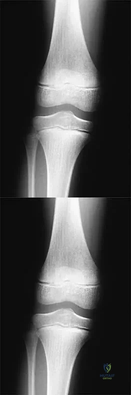
A 16-year-old snowboarder has significant pain and is still unable to bear weight after sustaining a lateral ankle injury in a fall 1 week ago. Examination reveals swelling and tenderness in the sinus tarsi. AP, lateral, and mortise radiographs of the ankle are unremarkable. Management should consist of


Explanation

Because there is a significant possibility that the patient may have a fracture of the lateral process of the talus, there is some disagreement as to the best radiographic study to identify this injury. A CT scan is an appropriate diagnostic tool to visualize the fracture and identify any displacement. Displaced lateral process fractures are best treated surgically. Kirkpatrick DP, Hunter RE, Janes PC, Mastrangelo J, Nicholas RA: The snowboarder's foot and ankle. Am J Sports Med 1998;26:271-277.

Question 7

A 24-year-old man who plays golf noted the immediate onset of pain on the ulnar side of his hand and has been unable to swing a club for the past 6 weeks after striking a tree root with his club during his golf swing. Examination reveals full motion of the wrist, diminished grip strength, and tenderness over the hypothenar region. A CT scan of the hand and wrist is shown in Figure 26. Management should consist of


Explanation

Fractures of the hook of the hamate frequently are not identified in the acute phase. Because the fracture can be difficult to see on plain radiographs, the lack of findings can lead to a painful nonunion. A carpal tunnel view may show the fracture, but a CT scan will best detect the injury. Immobilization is the treatment of choice and will result in union in most patients unless the diagnosis is delayed. However, excision of the fragment may be necessary for patients who have nonunion, persistent pain, or ulnar nerve palsy. Carroll RE, Lakin JF: Fracture of the hook of the hamate: Acute treatment. J Trauma 1993;34:803-805.


Question 8

An 18-year-old football player sustains a contact injury to his right lower leg, and radiographs show a closed transverse fracture of the middle third of the tibia. Based on the clinical examination, a compartment syndrome is suspected. When measuring compartment pressures, the highest tissue pressure is recorded how many centimeters proximal or distal to the fracture site?


Explanation

Measurements of compartment pressures in patients with tibial fractures and compartment syndrome reveal that the highest tissue pressures are recorded at the level of the fracture or within 5 cm of the fracture. Tissue pressures show a statistically significant decrease when they are recorded at increasing distances proximal and distal to the site of the highest pressure recorded. To reliably determine the location of the highest tissue pressure in patients with tibial fractures, measurements should be obtained, at a minimum, in both the anterior and deep posterior compartments at the level of the fracture, as well as at locations proximal and distal. The highest tissue pressure recorded should serve as a basis for determining the need for fasciotomy. Heckman MM, Whitesides TE Jr, Grewe SR, Rooks MD: Compartment pressure in association with closed tibial fractures: The relationship between tissue pressure, compartment, and the distance from the site of the fracture. J Bone Joint Surg Am 1994;76:1285-1292.

Question 9

A 50-year-old patient who plays tennis sustained the deformity shown in Figure 27 following a high volley. Further diagnostic work-up should include


Explanation

The patient has a rupture of the long head of the biceps; however, patients older than age 45 years are at greater risk of having an associated rotator cuff tear. An MRI scan should be ordered to avoid missing concomitant rotator cuff pathology. While patients may report pain radiating down the arm at the time of the tendon rupture, an EMG is not indicated. The short head of the biceps is intact and needs no further work-up, even though the muscle descends in most cases. The anterior labrum can be injured but is not associated with this deformity. Neer CS II, Bigliani LU, Hawkins RJ: Rupture of the long head of the biceps related to the subacromial impingement. Orthop Trans 1977;1:114.


Question 10

A 16-year-old ice hockey player is struck on the chest by the puck. He skates a few strides and then collapses. What is the most likely diagnosis?


Explanation

Sudden cardiac arrest following a blow to the chest in young athletes has been termed "commotio cordis." It is most common in Little League and other youth projectile sports (eg, ice hockey, lacrosse). The cause, although not completely determined, is most likely an arrhythmia related to the impact in a vulnerable time in the cardiac cycle. Resuscitation has proven to be exceedingly difficult, resulting in a high mortality rate. Maron BJ, Strasburger JF, Kugler JD, Bell BM, Brodkey FD, Poliac LC: Survival following blunt chest impact-induced cardiac arrest during sports activities in young athletes. Am J Cardiol 1997;79:840-841.

Question 11

A 24-year-old dancer sustains the injury shown in Figure 28. Management should consist of


Explanation

The patient has a moderately displaced distal diaphyseal fracture of the fifth metatarsal, and the most appropriate treatment is brief immobilization and symptomatic management. Attempts at closed reduction are unlikely to appreciably alter the position of the fracture. Surgical techniques for either reduction of the fracture or fixation have not been shown to result in improved functional outcomes. O'Malley MJ, Hamilton WG, Munyak J: Fractures of the distal shaft of the fifth metatarsal: "Dancer's Fracture." Am J Sports Med 1996;24:240-243. DeLee JC: Fractures and dislocations of the foot, in Mann RA, Coughlin MJ (eds): Surgery of the Foot and Ankle, ed 6. St Louis, MO, CV Mosby, 1993, pp 1465-1703.


Question 12

A 22-year-old volleyball player has atrophy of the infraspinatus muscle. This deficit is the result of entrapment of what nerve?


Explanation

Suprascapular deficits, as the result of repetitive forceful internal rotation with overhead ball striking, occur in the spinoglenoid notch. Compression interferes with distal suprascapular nerve innervation to the infraspinatus, while allowing the supraspinatus to function normally. A scapular notch entrapment of this nerve would involve both the supraspinatus and the infraspinatus. The axillary, dorsal scapular, and subscapular nerves do not affect the infraspinatus. Ferretti A, Cerullo G, Russo G: Suprascapular neuropathy in volleyball players. J Bone Joint Surg Am 1987;69:260-263.

Question 13

Figure 29 shows the radiograph of a 25-year-old woman who has had a 3-month history of ankle pain after sustaining an inversion injury to the ankle. She reports occasional catching, but no sense of instability. Examination reveals ligament stability. Management should consist of


Explanation

Osteochondral lesions of the talar dome can have a traumatic or nontraumatic etiology. Most authors site a probable traumatic etiology for lateral lesions. Stage I and II lesions, which are composed of compressed subchondral bone or a partial detached osteochondral fragment, can be treated initially in a non-weight-bearing short leg cast for 6 weeks. Stage III medial lesions can also be treated in the same manner. If symptoms persist, the treament of choice is debridement of the fracture, curettage of the lesion, and drilling of the subchondral bone. This treatment also applies to lateral stage III and all stage IV lesions. If the fragment is at least one third of the size of the talar dome, management should consist of open reduction and internal fixation. In patients with more chronic lesions (4 to 6 months of persistent pain), the threshold to proceed with surgery is lower, even in a stage II lesion. Lutter LD, Mizel MS, Pfeffer GB (eds): Orthopaedic Knowledge Update: Foot and Ankle. Rosemont, IL, American Academy of Orthopaedic Surgeons, 1994, pp 205-226.


Question 14

A 19-year-old college cross-country runner is amenorrheic and has recurrent stress fractures. Long-term management should consist of


Explanation

The triad of menstrual dysfunction, disordered eating, and stress fracture is well recognized in women who participate in endurance sports. The best treatment remains to be determined, but at present, the combination of oral contraceptives to regulate menses, an increased intake of calcium and vitamin D, as well as nutritional counseling, is the recommended treatment for decreased bone mass related to exercise-induced amenorrhea. Nattiv A, Armsey TD Jr: Stress injury to bone in the female athlete. Clin Sports Med 1997;16:197-224.

Question 15

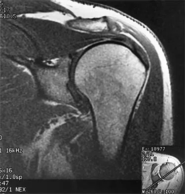
A 47-year-old male tennis player has pain in his nondominant shoulder that has failed to respond to 4 months of nonsurgical management. Examination reveals acromial tenderness and pain at the supraspinatus tendon insertion. He has a positive impingement sign, pain on forward elevation, and minimal cuff weakness. The MRI scans are shown in Figures 30a and 30b. To completely resolve his symptoms, treatment should consist of


Explanation

The MRI scans show a mesoacromion with tendonopathy of the supraspinatus. The history and physical findings indicate that the patient has a symptomatic os acromiale. Simple excision of the unstable os acromiale has not yielded consistently good results. Meticulous internal fixation using tension banding with cannulated screws and autologous bone grafting has shown good results for this problem. Hutchinson MR, Veenstra MA: Arthroscopic decompression of shoulder impingement secondary to os acromiale. Arthroscopy 1993;9:28-32.


Question 16

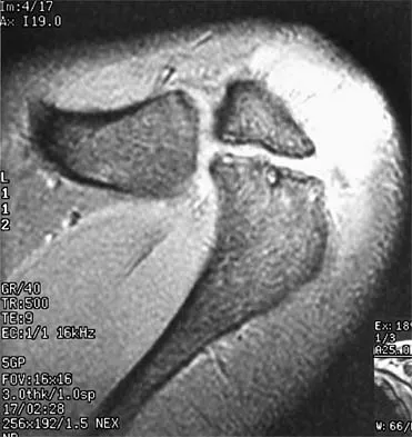
A 39-year-old competitive cyclist sustains an injury to her left hip in a fall. Gadolinium arthrography, with an accompanying MRI scan, is shown in Figure 31. A cleft, or defect, identified by the arrow, indicates a detachment of the


Explanation

The area indicated by the arrow represents gadolinium contrast extending into a separation between the lateral labrum and its acetabular attachment. This can be a traumatic detachment, but occasionally a cleft may be present as a normal variant of the labral morphology. The capsular attachment of the iliofemoral ligament is peripheral to the labrum. The pulvinar is the common name applied to the fat and overlying synovium contained within the acetabular fossa above the ligamentum teres. The zona orbicularis is a circumferential thickening of the capsule around the femoral neck, and the retinacular vessels travel within the capsular synovium up the femoral neck to supply the femoral head. Petersilge CA, Haque MA, Petersilge WJ, Lewin JS, Lieberman JM, Buly R: Acetabular labral tears: Evaluation with MR arthrography. Radiology 1996;200:231-235. Czerny C, Hofmann S, Neuhold A, et al: Lesions of the acetabular labrum: Accuracy of MR imaging and MR arthrography in detection and staging. Radiology 1996;200:225-230.


Question 17

A cortisone injection in the subacromial space will most likely result in


Explanation

A cortisone injection in the subacromial space will most likely result in elevated blood glucose levels in patients with type I diabetes mellitus. Patients should be warned of this potential complication. Cortisone does not have an effect on instability or proprioception, and a single injection would not affect osteoporosis. Repetitive injections or injection into the tendon itself could accelerate rupture of the biceps tendon. Matsen FA III, Arntz CT: Subacromial impingement, in Rockwood CA, Matsen FA III (eds): The Shoulder. Philadelphia, PA, WB Saunders, 1990, pp 623-646.

Question 18

A high school athlete reports the sudden onset of low back pain while performing a dead lift. Examination reveals a lumbar paraspinal spasm and a positive straight leg raising test. The deep tendon reflexes, motor strength, and sensation in the lower extremeties are normal. The radiographs are normal. If symptoms persist for more than a few weeks, management should consist of


Explanation

In adolescents, a lumbar herniated disk is characterized by a paucity of clinical findings; a positive straight leg raising test may be the only consistent positive finding. This may result in a long period of nonsurgical management that fails to provide relief. Activities that place a significant shear load on the lumbar spine, such as the dead lift, are associated with an increased risk of central disk herniation. When an adolescent who lifts weights has a history of low back pain that fails to respond to a short period of active rest, an MRI scan is the study of choice to evaluate for a lumbar herniated disk. Epstein JA, Epstein NE, Marc J, Rosenthal AD, Lavine LS: Lumbar intervertebral disk herniation in teenage children: Recognition and management of associated anomalies. Spine 1984;9:427-432.

Question 19

A 22-year-old skier reports painful range of motion in the left thumb after falling forward on his outstretched hand while holding his ski pole. Examination of the left thumb reveals increased AP laxity and 45 degrees of valgus laxity at the metacarpophalangeal (MCP) joint. Examination of the right thumb shows 25 degrees of valgus laxity at the MCP joint. Radiographs are normal. Management should consist of


Explanation

The patient has a complete tear of the ulnar collateral ligament as defined by MCP joint laxity of greater than 30 degrees (or 15 degrees greater laxity compared with the opposite side). Primary repair is the treatment of choice because displacement of the ligament superficial to the adductor aponeurosis (Stener lesion) must be corrected. Any volar plate injury can be addressed during repair of the ulnar collateral ligament.

Question 20

Which of the following structures is most commonly involved in lateral epicondylitis?


Explanation

The most common specific site of involvement is the origin of the extensor carpi radialis brevis. It is usually caused by overuse activities, such as the eccentric overload exhibited during a backhand in tennis. In most patients, the characteristic friable, grayish tissue described as angiofibroblastic hyperplasia or hyaline degeneration is seen at the extensor carpi radialis brevis origin. Nirschl RP: Elbow tendinosis/tennis elbow. Clin Sports Med 1992;11:851-870.

Question 21

When comparing surgical and nonsurgical extremities in patients who underwent anterior cruciate ligament (ACL) reconstruction using patellar tendon or hamstrings autografts, isokinetic strength measurements obtained 6 months after the surgery would most likely reveal


Explanation

Follow-up examination at 6 months revealed no statistically significant differences in quadricep or hamstring strength when comparing surgical versus nonsurgical extremities isokinetically. Therefore, the selection of autogenous hamstring or patellar tendon for ACL reconstruction should not be based solely on the assumption of the graft tissue source altering the recovery of quadricep and/or hamstring strength. Carter TR, Edinger S: Isokinetic evaluation of anterior cruciate ligament reconstruction: Hamstring versus patellar tendon. Arthroscopy 1999;15:169-172 Howell SM, Taylor MA: Brace-free rehabilitation, with early return to activity, for knees reconstructed with a double-looped semitendinosus and gracilis graft. J Bone Joint Surg Am 1996;78:814-825.

Question 22

A quarterback sustains a rough tackle after which he appears confused, has a dazed look on his face and an unsteady gait on standing. He denies loss of consciousness. Reexamination within 10 minutes is normal, the patient is lucid, and he wants to return to play. The coach and the player should be advised that he may


Explanation

The patient has a grade I (mild) concussion that can result in confusion and disorientation, without loss of consciousness. This concussion syndrome is completely reversible, with no long-term sequelae. Athletes who sustain a grade I concussion may return to play after 15 minutes if there are no lingering symptoms, such as headache or vertigo. A grade II concussion is characterized by loss of consciousness of less than 5 minutes. With this type of injury, the athlete can return to play in 1 week, if asymptomatic. If a grade III (severe) concussion is sustained, the athlete should avoid contact for a minimum of 1 month before considering a return to competition. A grade III concussion is characterized by a loss of consciousness of greater than 5 minutes or posttraumatic amnesia of greater than 24 hours. A CT scan is not indicated in a grade I injury. An athlete who sustains three grade I or grade II concussions, or two grade III concussions may not return to play for the season. Torg JS, Gennarelli TA: Head and cervical spine injuries, in DeLee JC, Drez D Jr (eds): Orthopaedic Sports Medicine Principles & Practice. Philadelphia, PA, WB Saunders, 1994, vol 1, pp 417-462.

Question 23

The bone avulsion shown in Figure 32 has a high correlation with tearing of the


Explanation

As described by Segond in 1987, an avulsion fracture of the lateral tibial plateau is commonly referred to as a Segond fracture. Subsequent to 1987, several authors have also found that the lateral capsular sign represents, but is not limited to, a disruption of the middle third of the lateral capsule and a tear of the anterior cruciate ligament. Bach BR, Warren RF: Radiographic indicators of anterior cruciate ligament injury, in Feagin JA (ed): The Crucial Ligaments. New York, NY, Churchill Livingston, 1988, pp 301-327. Segond P: Recherches cliniques et experimentales sur les epanchements sanguins du genou par entorse. Prog Med (Paris) 1987;7:297.


Question 24

A 21-year-old college defensive lineman sustains a minimally displaced (less than 1 mm) midthird scaphoid fracture during the first game of the season. Management should consist of


Explanation

The union rate for minimally displaced midthird scaphoid fractures is quite high with cast immobilization while allowing a return to sports. Inadequate immobilization results in a much higher nonunion rate. Early fixation and rehabilitation have been proposed for sports or positions that are not amenable to cast immobilization. While immobilization of a nondisplaced fracture results in an acceptably high union rate, there is no advantage to fixation in conjunction with immobilization in the course of healing. With adequate immobilization and protection, play restrictions until healing has occurred are unnecessary. Rettig AC, Kollias SC: Internal fixation of acute stable scaphoid fractures in the athlete. Am J Sports Med 1996;24:182-186. Rettig AC, Weidenbener EJ, Gloyeske R: Alternative management in midthird scaphoid fractures in the athlete. Am J Sports Med 1994;22:711-714.

Question 25

A 16-year-old football player sustains a direct blow to the anterior aspect of his flexed right knee. Examination reveals a contusion over the anterior tibial tubercle and a small effusion. MRI scans are shown in Figures 33a through 33c. What is the most likely diagnosis?


Explanation

The MRI scans show disruption of the fibers of the PCL. Patients sustaining an isolated acute PCL injury can present with only minimal discomfort and have full range of motion. When examination reveals a contusion over the tibial tubercle and discomfort with the posterior drawer examination, with or without instability, a possible injury to the PCL should be considered. In acute injuries, the reported accuracy of MRI imaging for diagnosing PCL tears ranges from 96% to 100%. Resnick D, Kang HS: Internal Derangement of Joints: Emphasis on MRI Imaging. Philadelphia, PA, WB Saunders, 1997, pp 699-700. Harner CD, Hoher J: Evaluation and treatment of posterior cruciate ligament injuries. Am J Sports Med 1998;26:471-482.


Question 26

A 25-year-old rugby player sustains a direct blow to the anteromedial aspect of his knee. Examination reveals increased external rotation of the tibia at 30 degrees of knee flexion, but symmetric external rotation at 90 degrees compared to the contralateral side. Which structure is the primary deficient restraint responsible for this examination finding?


Explanation

An isolated posterolateral corner (PLC) injury presents with increased external rotation at 30 degrees of flexion but not at 90 degrees. The fibular collateral ligament (FCL) is the primary static constraint to varus and external rotation at 30 degrees.

Question 27

A 22-year-old competitive rugby player presents with recurrent anterior shoulder dislocations. CT imaging demonstrates a 25% anterior glenoid bone loss and a deep, engaging Hill-Sachs lesion. What is the most appropriate surgical management?


Explanation

Anterior glenoid bone loss greater than 20-25% in a collision athlete is a critical indication for a bony augmentation procedure. The Latarjet procedure restores glenoid width and provides a dynamic sling effect via the conjoint tendon.

Question 28

A 16-year-old elite female soccer player sustains an acute ACL tear. She is considering graft options for reconstruction. Compared to hamstring autograft, bone-patellar tendon-bone (BTB) autograft has been consistently associated with which of the following outcomes?


Explanation

BTB autografts have historically shown a lower re-rupture rate in young, high-demand athletes compared to hamstring grafts. However, they carry a significantly higher risk of donor site morbidity, particularly anterior knee pain and pain with kneeling.

Question 29

A 25-year-old rugby player presents with a twisting knee injury. On examination, the dial test demonstrates 15 degrees of increased external rotation compared to the contralateral normal knee at 30 degrees of knee flexion, but symmetrical external rotation at 90 degrees of flexion. Which structure is most likely injured?


Explanation

An isolated posterolateral corner (PLC) injury presents with increased external rotation at 30 degrees but not 90 degrees of knee flexion. If increased external rotation is present at both 30 and 90 degrees, a combined PLC and PCL injury is present.

Question 30

A 34-year-old male is brought to the emergency department after a motorcycle accident. He has a visibly deformed knee that spontaneously reduces. Examination reveals a gross multiligamentous knee injury. His pedal pulses are palpable, but an ankle-brachial index (ABI) is measured at 0.85. What is the most appropriate next step in management?


Explanation

In the setting of a knee dislocation, an ABI less than 0.90 is highly suspicious for a popliteal artery injury, even if pulses are palpable. A CT angiogram is indicated to evaluate for vascular compromise requiring possible surgical intervention.

Question 31

A 22-year-old collegiate linebacker presents with recurrent anterior shoulder instability. Preoperative CT imaging reveals a 28% anterior glenoid bone loss and an engaging Hill-Sachs lesion. Which of the following is the most appropriate surgical management?


Explanation

Anterior glenoid bone loss exceeding critical thresholds (typically 20-25%) in a high-demand collision athlete is an absolute indication for a bony augmentation procedure like the Latarjet. Arthroscopic soft-tissue repairs have unacceptably high failure rates in the presence of such severe glenoid bone loss.

Question 32

A 19-year-old male presents to the trauma bay after a rugby tackle. He complains of severe medial clavicle pain, dysphagia, and a choking sensation. Clinical examination demonstrates a palpable depression at the medial end of the clavicle. What is the most appropriate definitive management?


Explanation

Posterior sternoclavicular dislocations can compress vital mediastinal structures, causing dysphagia, dyspnea, or vascular compromise. They require prompt reduction in the operating room with a cardiothoracic surgeon on standby due to the risk of catastrophic hemorrhage during reduction.

Question 33

During a medial patellofemoral ligament (MPFL) reconstruction for recurrent patellar instability, the surgeon uses fluoroscopy to identify the femoral attachment site. According to Schöttle's method, where is the anatomic femoral origin of the MPFL located?


Explanation

Schöttle's point is located 1 mm anterior to the posterior femoral cortical line and 2.5 mm distal to the posterior origin of the medial femoral condyle. Clinically, this lies in the saddle just proximal to the medial epicondyle and distal to the adductor tubercle.

Question 34

A 24-year-old athlete undergoes an anterior cruciate ligament (ACL) reconstruction. Postoperatively, he demonstrates an absent Lachman test but a persistently positive pivot shift test. What is the most likely technical error made during the procedure?


Explanation

A vertical femoral tunnel in ACL reconstruction controls anteroposterior translation (negative Lachman) but fails to adequately control rotatory loads, resulting in a persistent pivot shift. Anatomic tunnel placement is required for robust rotational stability.

Question 35

A 21-year-old collegiate rugby player presents with recurrent anterior shoulder instability. A 3D CT scan reveals 25% anterior glenoid bone loss. Which of the following is the most appropriate definitive management?


Explanation

In a young contact athlete with recurrent instability and >20-25% anterior glenoid bone loss, a Latarjet procedure (coracoid transfer) is the gold standard. Soft tissue procedures alone (Bankart) have unacceptably high failure rates in this setting.

Question 36

A 28-year-old man sustains a twisting injury to his knee. Examination reveals 15 degrees of increased external rotation on the dial test at 30 degrees of knee flexion compared to the contralateral side. At 90 degrees of knee flexion, the external rotation is equal bilaterally. Which structure is most likely injured?


Explanation

The dial test evaluates PLC and PCL integrity. Greater than 10 degrees of external rotation asymmetry at 30 degrees only indicates an isolated PLC injury. Asymmetry at both 30 and 90 degrees indicates a combined PLC and PCL injury.

Question 37

A 22-year-old collegiate football player sustains a non-contact pivoting knee injury. MRI confirms an isolated ACL tear. He elects for reconstruction using a bone-patellar tendon-bone autograft. During the procedure, the femoral tunnel is drilled too anteriorly. Which complication is most likely postoperatively?


Explanation

If the femoral tunnel is placed too anteriorly during ACL reconstruction, the graft will tighten excessively in flexion, leading to a flexion deficit. An anteriorly placed femoral tunnel is non-anatomic and typically fails to restore normal kinematics.

Question 38

A 28-year-old skier sustains an acute knee injury and presents with a positive dial test at 30 degrees of flexion, which normalizes to the contralateral side at 90 degrees of flexion. Which of the following structures is most likely injured?


Explanation

A positive dial test (increased external rotation of >10 degrees compared to the normal side) at 30 degrees of flexion that reduces at 90 degrees indicates an isolated injury to the posterolateral corner (PLC). If the test is positive at both 30 and 90 degrees, it suggests a combined PCL and PLC injury.

Question 39

A 25-year-old overhead athlete presents with anterior shoulder pain. He describes a "dead arm" sensation when throwing. Physical exam reveals a positive O'Brien's active compression test. Which of the following MRI arthrogram findings is most consistent with a Type II SLAP tear?


Explanation

A Type II SLAP tear is defined by the detachment of the superior labrum and the long head of the biceps anchor from the superior glenoid tubercle. This is the most common type of SLAP tear seen in overhead athletes.

Question 40

A 20-year-old male sustains a traumatic anterior shoulder dislocation. After closed reduction, CT scan demonstrates a 25% anterior glenoid bone loss. What is the most appropriate definitive surgical management?


Explanation

For critical anterior glenoid bone loss (>20-25%), isolated arthroscopic soft-tissue repair has unacceptably high recurrence rates. A bone block augmentation procedure, such as the Latarjet procedure (coracoid transfer), is the standard of care.

Question 41

A 35-year-old laborer falls directly on the lateral aspect of his shoulder and is diagnosed with a grade III acromioclavicular (AC) joint separation. Which ligaments are completely disrupted in this injury?


Explanation

A Rockwood grade III AC joint separation involves complete disruption of both the acromioclavicular (AC) and coracoclavicular (CC) ligaments. This results in superior displacement of the clavicle by 25-100%.

Question 42

A 16-year-old female experiences recurrent patellar dislocations. Physical exam reveals apprehension with lateral patellar translation. What is the primary soft-tissue restraint to lateral patellar translation at 20 degrees of knee flexion?


Explanation

The medial patellofemoral ligament (MPFL) provides approximately 50-60% of the restraining force against lateral patellar displacement, acting primarily in early flexion (0 to 30 degrees).

Question 43

A 30-year-old male sustains a posterior knee dislocation. After reduction, his ankle-brachial index (ABI) is measured at 0.85. What is the most appropriate next step in management?


Explanation

Following a knee dislocation, an ABI < 0.9 is highly suggestive of a vascular injury and mandates further advanced imaging, usually CT angiography, to evaluate the popliteal artery. Immediate surgical exploration is reserved for hard signs of vascular injury like absent pulses or expanding hematoma.

Question 44

Which bundle of the anterior cruciate ligament (ACL) is the primary restraint to anterior tibial translation when the knee is in 90 degrees of flexion?


Explanation

The anteromedial (AM) bundle of the ACL is tightest in knee flexion and acts as the primary restraint to anterior tibial translation at 90 degrees. Conversely, the posterolateral (PL) bundle is tight in extension and primarily controls rotational stability.

Question 45

A 24-year-old gymnast presents with a painful shoulder. Examination demonstrates generalized ligamentous laxity, positive sulcus sign, and apprehension with anterior and posterior translation. She has failed 6 months of physical therapy. What is the most appropriate surgical treatment for her multidirectional instability?


Explanation

For multidirectional shoulder instability (MDI) that has failed extensive conservative management, global capsular reduction via open or arthroscopic capsular plication (capsular shift) is the surgical treatment of choice. Isolated labral repairs do not address the primary pathology of capsular redundancy.

Question 46

During an ACL reconstruction using a hamstring autograft, the surgeon uses a suspensory fixation device on the femur and an interference screw on the tibia. What is the most common mechanism of graft failure in the first 3 months postoperatively?


Explanation

In the early postoperative period (less than 3 months), graft integration is incomplete. Failure during this period is most commonly due to loss of graft fixation at the bone-tunnel interface rather than a mid-substance rupture.

Question 47

A 19-year-old football player sustains a valgus blow to his knee. Exam reveals a grade III MCL injury and an ACL tear. What is the recommended treatment strategy?


Explanation

Combined ACL and grade III MCL injuries are typically managed with a period of bracing to allow the MCL to heal, followed by delayed ACL reconstruction. This approach minimizes the significant risk of postoperative arthrofibrosis seen with acute simultaneous surgeries.

Question 48

A 30-year-old baseball pitcher complains of posterior shoulder pain during the late cocking phase of throwing. Physical examination reveals a loss of 25 degrees of internal rotation compared to the contralateral side, while external rotation is increased. What is the most likely diagnosis?


Explanation

Glenohumeral internal rotation deficit (GIRD) is common in overhead athletes and is defined as a loss of >20 degrees of internal rotation compared to the non-throwing shoulder. It is associated with posterior capsular contracture and internal impingement.

Question 49

A patient presents with a chronic posterolateral corner (PLC) deficient knee resulting in a varus thrust during gait. Radiographs show mechanical axis falling medial to the knee center and no advanced osteoarthritis. What is the most appropriate initial surgical management?


Explanation

In the setting of chronic posterolateral corner deficiency with varus malalignment, correcting the bony alignment with a valgus-producing High Tibial Osteotomy (HTO) is essential. Soft-tissue reconstruction alone will likely fail due to the constant varus overload.

Question 50

In performing an arthroscopic repair of a Bankart lesion, the surgeon places suture anchors at the 3, 4, and 5 o'clock positions on the glenoid (in a right shoulder). Which nerve is most at risk if the drill penetrates the anteroinferior glenoid neck too deeply?


Explanation

The axillary nerve runs immediately inferior to the glenohumeral joint capsule, near the 6 o'clock position. Deep drilling or suture passage at the anteroinferior glenoid (5 to 6 o'clock) places the axillary nerve at risk of injury.

Question 51

A 21-year-old athlete undergoes revision ACL reconstruction. Preoperative CT scan shows an expanded tibial tunnel measuring 16 mm in diameter. What is the preferred surgical strategy in this setting?


Explanation

When significant tunnel widening is present (>14 mm), a two-stage revision strategy is recommended. The first stage involves filling the dilated tunnel with bone graft, followed by the definitive ACL reconstruction 4 to 6 months later once the graft has incorporated.

Question 52

A 22-year-old collegiate soccer player undergoes anterior cruciate ligament (ACL) reconstruction using a bone-patellar tendon-bone autograft. Compared to a quadruple-stranded hamstring autograft, what is the most likely long-term complication associated with this graft choice?


Explanation

Bone-patellar tendon-bone autografts are historically associated with a higher incidence of anterior knee pain and pain with kneeling compared to hamstring autografts. Hamstring grafts, on the other hand, are associated with decreased deep knee flexion strength.

Question 53

A 25-year-old man presents with knee pain after a twisting injury. Physical examination reveals an asymmetric increased external rotation of the tibia of 15 degrees at 30 degrees of knee flexion, but no difference compared to the contralateral knee at 90 degrees of flexion. Which of the following structures is most likely injured?


Explanation

The dial test evaluates the PCL and PLC. Greater than 10 degrees of asymmetric external rotation at 30 degrees but symmetric at 90 degrees indicates an isolated PLC injury. Asymmetry at both 30 and 90 degrees indicates a combined PLC and PCL injury.

Question 54

A 20-year-old rugby player undergoes a Latarjet procedure for recurrent anterior shoulder instability with significant glenoid bone loss. Postoperatively, he presents with weakness in elbow flexion and decreased sensation over the lateral forearm. Which nerve was most likely injured during the procedure?


Explanation

The musculocutaneous nerve is the most commonly injured nerve during the Latarjet procedure due to its proximity to the coracoid process. It innervates the biceps brachii and brachialis, and provides sensation to the lateral forearm via the lateral antebrachial cutaneous nerve.

Question 55

A 30-year-old male is evaluated in the trauma bay after a high-speed motorcycle accident. He has a grossly deformed knee which is quickly reduced and splinted. His ankle-brachial index (ABI) is 0.85. What is the most appropriate next step in management?


Explanation

An ABI less than 0.9 following a knee dislocation is a strong indicator of a potential vascular injury. The most appropriate next step to definitively evaluate the popliteal artery in the absence of hard signs of ischemia is a CT angiogram.

Question 56

A 24-year-old skier sustains an isolated Grade III injury to the medial collateral ligament (MCL). Magnetic resonance imaging reveals an avulsion of the MCL from its tibial insertion with the distal end flipped superficial to the pes anserinus. What is the most appropriate management?


Explanation

Distal MCL avulsions with the ligament displaced superficial to the pes anserinus (Stener-like lesion of the knee) lack the ability to heal properly due to soft tissue interposition. Unlike proximal tears which typically heal nonoperatively, these specific distal avulsions require primary surgical repair.

Question 57

A 45-year-old male manual laborer complains of deep shoulder pain and mechanical clicking. MRI demonstrates a Type II Superior Labrum Anterior to Posterior (SLAP) tear. Nonoperative management has failed. What is the most appropriate surgical treatment in this patient demographic?


Explanation

In patients older than 40 years, especially non-overhead athletes or manual laborers, biceps tenodesis provides superior outcomes and lower revision rates compared to SLAP repair for Type II SLAP lesions. SLAP repair in this age group has a higher rate of postoperative stiffness and persistent pain.

Question 58

A 35-year-old man presents to the emergency department complaining of shoulder pain after a first-time generalized tonic-clonic seizure. On examination, his arm is locked in adduction and internal rotation. Radiographs reveal a posterior shoulder dislocation with an anteromedial humeral head defect involving 20% of the articular surface. What is this defect termed?


Explanation

A reverse Hill-Sachs lesion (McLaughlin lesion) is an impaction fracture of the anteromedial humeral head commonly associated with posterior shoulder dislocations. A classic Hill-Sachs lesion is a posterolateral defect associated with anterior dislocations.

Question 59

During an anterior cruciate ligament (ACL) reconstruction, the surgeon evaluates the anatomy of the native ACL footprint. Which of the following statements correctly describes the biomechanics of the two main bundles of the ACL?


Explanation

The ACL consists of the anteromedial (AM) and posterolateral (PL) bundles. The AM bundle tightens in flexion to resist anterior tibial translation, while the PL bundle tightens in extension and is the primary restraint to rotatory loads.

Question 60

A 28-year-old dashboard-injury victim presents with a positive posterior drawer test and a posterior sag sign. MRI confirms an isolated Grade II posterior cruciate ligament (PCL) tear. What is the most appropriate initial management?


Explanation

Isolated Grade I and II PCL tears have a high intrinsic healing capacity. They are best managed nonoperatively with relative immobilization in extension (to prevent posterior tibial sag) and physical therapy focusing on quadriceps strengthening.

Question 61

A 35-year-old man presents with a locked shoulder after a generalized seizure. Radiographs demonstrate a posterior shoulder dislocation with an anterior humeral head defect involving 25% of the articular surface as shown in Figure 1.

What is the most appropriate surgical management?


Explanation

Posterior shoulder dislocations with an associated reverse Hill-Sachs lesion involving 20% to 40% of the articular surface are best treated with a lesser tuberosity transfer (McLaughlin procedure) into the defect. Defects >40% typically require arthroplasty.

Question 62

A 22-year-old football player undergoes an anterior cruciate ligament (ACL) reconstruction using a bone-patellar tendon-bone autograft. Six months postoperatively, he complains of anterior knee pain and a persistent 10-degree extension lag. A sagittal MRI reveals graft impingement against the intercondylar notch roof. Which of the following technical errors most likely caused this complication?


Explanation

A tibial tunnel placed too far anteriorly causes the ACL graft to impinge on the roof of the intercondylar notch in terminal extension. This leads to an extension deficit, anterior knee pain, and potentially a cyclops lesion.

Question 63

A 26-year-old male is brought to the emergency department after a high-speed motorcycle accident. Examination reveals a grossly unstable knee with a suspected multi-ligamentous injury (knee dislocation). Distal pulses are palpable, but the ankle-brachial index (ABI) is 0.85. What is the most appropriate next step in management?


Explanation

An ABI < 0.90 in the setting of a knee dislocation suggests a high risk of vascular injury and warrants an immediate CT angiogram. Immediate surgical exploration is indicated only if there are hard signs of vascular ischemia, such as absent pulses or an expanding hematoma.

Question 64

An 8-year-old girl (Tanner stage 1) sustains a midsubstance anterior cruciate ligament (ACL) tear. She experiences recurrent instability despite bracing and physical therapy. What is the most appropriate surgical technique to minimize the risk of physeal growth arrest?


Explanation

In prepubescent children with significant remaining growth (Tanner stage 1 or 2), physeal-sparing techniques like an all-epiphyseal reconstruction or extra-articular ITB tenodesis are recommended. Transphyseal drilling, especially with bone blocks, carries an unacceptably high risk of growth arrest.

Question 65

A 22-year-old collegiate soccer player undergoes anterior cruciate ligament (ACL) reconstruction using a bone-patellar tendon-bone (BPTB) autograft. Which of the following is the most common complication uniquely associated with this specific graft choice compared to hamstring autograft?


Explanation

Bone-patellar tendon-bone (BPTB) autografts are historically associated with a higher incidence of donor site morbidity, specifically anterior knee pain and pain with kneeling, compared to hamstring autografts. Rates of graft rupture and postoperative laxity are generally comparable between the two.

Question 66

A 19-year-old female gymnast presents with bilateral shoulder pain and a sensation of the shoulders "slipping" out of joint. Examination reveals positive sulcus signs bilaterally and generalized ligamentous laxity (Beighton score 7/9). Initial management should consist of:


Explanation

Multidirectional instability (MDI) often presents with generalized ligamentous laxity and bilateral symptoms. First-line treatment is strictly nonoperative, focusing on an extensive rehabilitation program to strengthen the dynamic stabilizers of the shoulder.

Question 67

A 26-year-old rugby player sustains a direct blow to the proximal tibia with the knee flexed at 90 degrees. Examination demonstrates a posterior sag sign and 8 mm of posterior translation of the tibia on the femur at 90 degrees of flexion, but normal translation at 30 degrees. The dial test is negative. What is the most appropriate initial management?


Explanation

The scenario describes an isolated Grade II posterior cruciate ligament (PCL) injury (5-10 mm translation). The standard of care for isolated Grade I and II PCL injuries is nonoperative management, emphasizing early range of motion and quadriceps strengthening to counteract posterior tibial translation.

Question 68

A 24-year-old throwing athlete undergoes an arthroscopic Type II SLAP repair. What is the most common postoperative complication that prevents a return to the previous level of overhead competition?


Explanation

The most common complication following SLAP repair in overhead athletes is postoperative stiffness, specifically a loss of external rotation. This loss of motion can severely impact throwing mechanics and prevent a successful return to elite play.

Question 69


A 35-year-old man sustains an ultra-low velocity knee dislocation after a misstep. Following closed reduction in the emergency department, his ankle-brachial index (ABI) is measured at 0.7. What is the most appropriate next step in management?


Explanation

An ABI less than 0.9 following a knee dislocation is highly suspicious for a popliteal artery injury. Urgent vascular evaluation, typically via CT angiography or formal arteriogram, is mandated to rule out limb-threatening ischemia before any definitive orthopedic intervention.

Question 70

A 29-year-old cyclist falls directly onto his shoulder point. Radiographs confirm a 150% superior displacement of the distal clavicle relative to the acromion, and the coracoclavicular distance is increased by 150% compared to the contralateral side. This represents which type of acromioclavicular (AC) joint injury according to the Rockwood classification?


Explanation

A Rockwood Type V AC joint injury is characterized by >100% to 300% superior displacement of the clavicle relative to the acromion. This involves severe disruption of the AC ligaments, coracoclavicular (CC) ligaments, and the deltotrapezial fascia.

Question 71

A 31-year-old soccer player sustains a hyperextension and varus injury to his knee. On examination, he has 15 degrees of increased external tibial rotation at 30 degrees of knee flexion compared to the contralateral side. This asymmetry completely resolves when tested at 90 degrees of flexion. Which structure is most likely injured?


Explanation

Increased external rotation of the tibia at 30 degrees of flexion that reduces to normal at 90 degrees indicates an isolated injury to the posterolateral corner (PLC). If the rotational asymmetry persisted at 90 degrees, it would suggest a combined PLC and PCL injury.

Question 72

A 22-year-old football player sustains a recurrent anterior shoulder dislocation.

Advanced imaging demonstrates an engaging Hill-Sachs lesion and anterior glenoid bone loss of 28%. What is the most appropriate surgical management to minimize the risk of recurrence?


Explanation

In the setting of significant anterior glenoid bone loss (subcritical typically >13.5%, critical >20-25%) and an engaging Hill-Sachs lesion, soft tissue procedures have unacceptably high failure rates. A bony augmentation procedure, such as the Latarjet procedure, is indicated to restore stability.

Question 73

A 40-year-old recreational skier presents with medial knee pain after catching an inside edge. Examination reveals 4 mm of medial opening to valgus stress at 30 degrees of flexion with a firm endpoint, but no opening at 0 degrees. MRI confirms a partial tear of the superficial MCL at its femoral attachment. What is the recommended treatment?


Explanation

An isolated Grade I or II medial collateral ligament (MCL) injury is optimally managed nonoperatively. A hinged knee brace allows early range of motion while protecting against valgus stress, leading to excellent ligamentous healing and return to sport.

None

Clinic OS
Medically Verified Content by
Prof. Clinic OS
Consultant Orthopedic & Spine Surgeon
Chapter Index