Shoulder Orthopedics MCQs (Set 3): Rotator Cuff, Instability & Proximal Humerus | ABOS Board Review
Key Takeaway
This high-yield Shoulder Orthopedics MCQ set (Set 3) is curated for AAOS, ABOS, and OITE exam preparation. It covers critical topics like the diagnosis and management of rotator cuff tears, treatment algorithms for shoulder instability, and classification of proximal humerus fractures. Enhance your understanding of key shoulder pathologies.
Shoulder Orthopedics MCQs (Set 3): Rotator Cuff, Instability & Proximal Humerus | ABOS Board Review
Comprehensive 100-Question Exam
00:00
Start Quiz
Question 1
Flexion and extension of the elbow occur about an axis of rotation that

Explanation
Question 2
Figure 27 shows the radiograph of a 26-year-old man who sustained a closed head injury and a closed elbow dislocation 6 weeks ago. Examination reveals 65 degrees to 115 degrees of flexion, and intensive physical therapy has resulted in no improvement. A decision regarding the timing of surgical correction of the contracture should be based on

Explanation
Question 3
A 70-year-old man who underwent an uncomplicated large rotator cuff repair 6 months ago is now seeking a second opinion regarding persistent pain and weakness in his shoulder. Examination reveals that his incision is well healed and unreactive. The surgical report suggests that the tendons were secured back to bone with sutures through the greater tuberosity. Figure 28 shows a radiograph that was obtained 1 week ago. What is the most likely diagnosis?

Explanation
Question 4
A 29-year-old man who lifts weights states that he injured his left shoulder while performing a bench press 2 days ago. The following morning he noted ecchymosis and swelling in the left chest wall. Examination reveals ecchymosis and tenderness and deformity in the left anterior chest wall and axillary fold that is accentuated with resisted adduction of the arm. Passive range of motion beyond 90 degrees of forward flexion and 45 degrees of external rotation is extremely painful. Glenohumeral stability is difficult to assess because of severe guarding. Figure 29 shows an MRI scan. Management should consist of

Explanation
Question 5
What range of motion parameters are required for a patient with posttraumatic elbow stiffness to accomplish all the normal activities of daily living?
Explanation
Question 6
A 24-year-old athlete has a painful right shoulder. Figure 30 shows an intra-articular photograph that was obtained through a posterior portal during arthroscopy; the labrum is indicated by the arrow. Based on these findings, management should consist of

Explanation
Question 7
The use of a screw between the clavicle and the coracoid process to maintain the clavicle and acromioclavicular (AC) joint in a reduced position is a treatment option for AC joint separations. Screw removal is generally recommended after soft-tissue healing. What effect does this rigid coracoclavicular fixation have on shoulder kinematics?
Explanation
Question 8
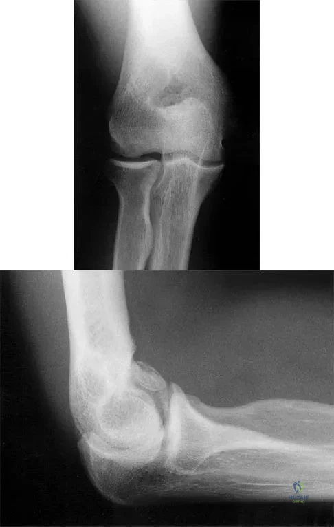
Figure 31 shows the AP and lateral radiographs of the elbow of a 56-year-old man with chronic polyarticular rheumatoid arthritis. His function continues to be limited by pain with activities of daily living. Examination shows that his total arc of motion is 110 degrees. Nonsurgical management has failed to provide relief. Treatment should now consist of

Explanation
Question 9
A 12-year-old pitcher has had a 2-month history of pain in his right dominant shoulder after throwing. He reports that the pain has gradually progressed to the point where he cannot throw without pain. He also notes that the pain now awakens him at night if he has been active. Anti-inflammatory drugs have failed to provide relief. Examination reveals no abnormalities except for some localized tenderness over the proximal humerus. Figures 32a and 32b show radiographs of both shoulders. What is the most likely diagnosis?
Explanation
Question 10
Which of the following ligaments is the primary static restraint against inferior translation of the arm when the shoulder is in 0 degrees of abduction?
Explanation
Question 11
A 16-year-old high school student undergoes a routine preparticipation physical examination at the beginning of the school year. Examination reveals marked laxity of both shoulders but only mild generalized laxity in other joints. The load and shift test allows for anterior humeral translation to the glenoid rim and posterior humeral translation beyond the glenoid rim. The sulcus sign is present. What is the next most appropriate step in management?
Explanation
Question 12
A 21-year-old professional baseball player has had painful catching and stiffness in his dominant right elbow for the past year. Examination reveals a flexion contracture of 2 degrees and mild pain with full elbow flexion. Radiographs are shown in Figures 33a and 33b. The most effective management should consist of
Explanation
Question 13
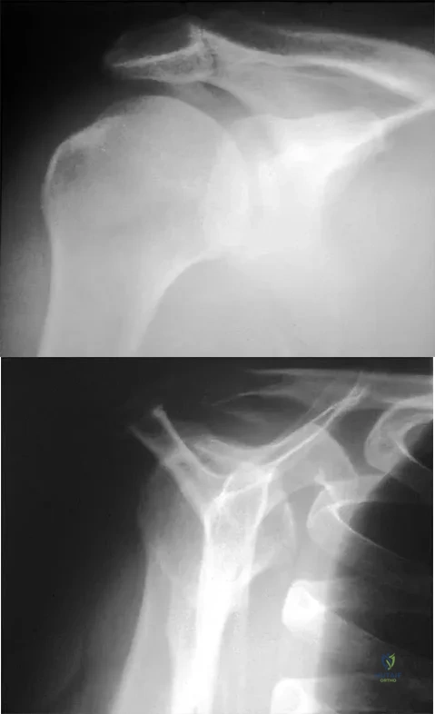
A 42-year-old patient has had painful inferior subluxation of the glenohumeral joint following a recent cerebrovascular accident (CVA). Figure 34 shows the AP radiograph of the shoulder. Management should consist of

Explanation
Question 14
A 50-year-old man who underwent an arthroscopic rotator cuff repair 5 days ago now returns for an early postoperative follow-up because of increasing pain in his shoulder. He reports increasing malaise and has a low-grade fever. Examination reveals no redness or swelling, but he has scant serous drainage from the posterior portal. An emergent Gram stain is positive for gram-positive cocci. The next most appropriate step in management should consist of
Explanation
Question 15
A 42-year-old man who is right-hand dominant injured his right shoulder when he fell from a ladder onto his outstretched arm 1 hour ago. Radiographs reveal a two-part greater tuberosity anterior fracture-dislocation. Initial management should consist of
Explanation
Question 16
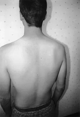
A 19-year-old man who plays college volleyball undergoes a routine preparticipation physical examination. Figure 35 shows a posterior view of his dominant shoulder. An electromyogram shows that this is a chronic injury, and an MRI scan shows no abnormalities. The best course of action should be
Explanation
Question 17
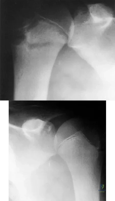
A 59-year-old construction worker who is right-hand dominant has had right shoulder pain for the past 9 months with no history of injury. Nonsurgical management consisting of two cortisone injections, physical therapy for 3 months, and nonsteroidal anti-inflammatory drugs has failed to provide lasting relief. Examination reveals tenderness over the acromioclavicular (AC) joint and over the subacromial bursa. He has positive Neer and Hawkins impingement signs and AC joint pain with adduction of the shoulder. Radiographs are shown in Figures 36a and 36b. An MRI scan reveals an intact rotator cuff. Management should now consist of
Explanation
Question 18
What three structures are considered the primary constraints necessary for elbow stability?
Explanation
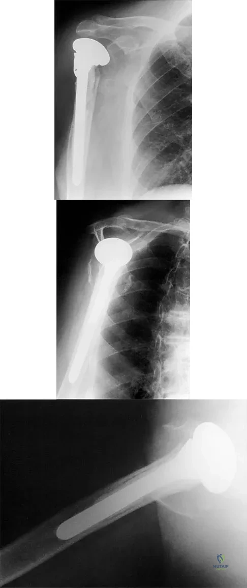
Question 19
A 68-year-old woman has been progressing slowly after undergoing humeral head replacement for a four-part fracture 3 months ago. She has not regained active elevation, she feels an audible clunk on attempting elevation, and she reports pain and weakness. She used a sling for 2 weeks in the immediate postoperative period. Radiographs are shown in Figure 37a through 37c. Management should consist of
Explanation
Question 20
What is the most important feature in choosing an outcome instrument to assess shoulder disorders?
Explanation
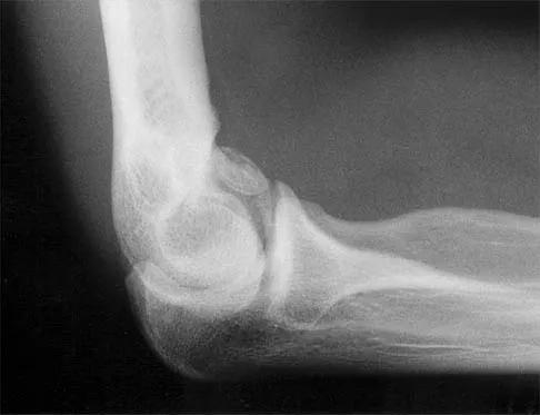
Question 21
Figure 38 shows the radiograph of a 16-year-old wrestler who injured his elbow when he was thrown to the mat by his opponent. To minimize additional trauma to the medial soft tissues, the elbow should be reduced in

Explanation
Question 22
In patients older than age 40 years who sustain a first-time anterior dislocation of the shoulder, prolonged morbidity is most commonly associated with
Explanation
Question 23
Figure 39 shows the AP radiograph of a 62-year-old man with degenerative osteoarthritis secondary to trauma. History reveals that he underwent total elbow arthroplasty 3 years ago. He continues to report instability and constant pain. A complete work-up, including aspiration and cultures, is negative. Treatment should consist of removal of the components and

Explanation
Question 24
A 67-year-old woman undergoes a revision total shoulder arthroplasty for replacement of a loose glenoid component. Examination in the recovery room reveals absent voluntary deltoid and triceps contraction, weakness of wrist and thumb extension, and absent sensation in the palmar aspect of all fingertips and the radial forearm. The next most appropriate step in management should consist of
Explanation
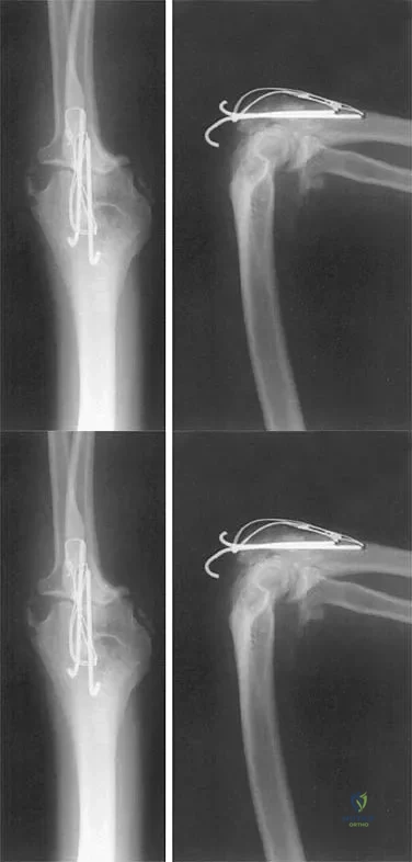
Question 25
Figure 40 shows the radiograph of a 16-year-old wrestler who injured his elbow when he was thrown to the mat by his opponent. Closed reduction is readily accomplished, and the elbow seems stable. Management should now consist of application of a splint for

Explanation
Question 26
A 75-year-old man presents with chronic right shoulder pain and an inability to actively elevate his arm above 45 degrees. Radiographs demonstrate an acromiohumeral distance of 3 mm. MRI reveals massive, retracted tears of the supraspinatus and infraspinatus with grade 4 fatty infiltration, while the subscapularis and deltoid are intact. What is the most appropriate surgical treatment?
Explanation
Question 27
A 22-year-old rugby player presents with recurrent anterior shoulder instability. A 3D CT scan reveals 26% anterior glenoid bone loss. Which of the following is the most appropriate definitive management?
Explanation
Question 28
Which of the following radiographic findings is the most reliable predictor of subsequent humeral head ischemia (avascular necrosis) following a severe proximal humerus fracture?
Explanation
Question 29
A 28-year-old professional baseball pitcher presents with posterior shoulder pain. MRI arthrogram shows undersurface fraying of the posterior supraspinatus and posterosuperior labrum. This pathology is primarily exacerbated by which of the following shoulder positions?
Explanation
Question 30
During the physical examination of a 55-year-old man with a suspected rotator cuff tear, the examiner asks the patient to place the palm of his hand on his contralateral shoulder and attempts to externally rotate the patient's hand while the patient resists. Which specific structure is best isolated by this test?
Explanation
Question 31
A 35-year-old man presents after a seizure with a locked posterior shoulder dislocation. CT scan demonstrates an anteromedial humeral head defect (reverse Hill-Sachs lesion) involving 35% of the articular surface. The dislocation is irreducible closed. What is the most appropriate surgical intervention?
Explanation
Question 32
A 45-year-old woman undergoes arthroscopy for persistent anterior shoulder pain. The surgeon notes medial subluxation of the long head of the biceps tendon and a tear of the coracohumeral ligament. Which of the following additional structures must be torn to allow this biceps subluxation?
Explanation
Question 33
During an open Latarjet procedure, the surgeon inadvertently places a self-retaining medial retractor deep to the conjoined tendon and applies excessive traction. Which of the following nerves is at greatest risk of injury from this maneuver?
Explanation
Question 34
A 68-year-old woman with severe osteoporosis sustains a 4-part proximal humerus fracture with a head-split component. She is functionally independent and active. What is the most appropriate treatment to optimize her functional outcome and minimize complications?
Explanation
Question 35
In evaluating a standard anteroposterior radiograph of the shoulder in a patient with chronic rotator cuff disease, what is the earliest radiographic sign indicative of rotator cuff tear arthropathy?
Explanation
Question 36
A 40-year-old construction worker presents with persistent shoulder pain despite 6 months of nonoperative management. MRI reveals an articular-sided partial tear of the supraspinatus tendon involving 60% of the tendon footprint depth (PASTA lesion). What is the recommended surgical management?
Explanation
Question 37
The concept of the 'glenoid track' is used to evaluate anterior shoulder instability. Which of the following statements correctly defines an 'off-track' Hill-Sachs lesion?
Explanation
Question 38
A 42-year-old man is undergoing an open subpectoral biceps tenodesis. The surgeon makes an incision in the axillary fold and exposes the intertubercular groove. Which nerve is most at risk of injury during medial retraction of the conjoint tendon in this approach?
Explanation
Question 39
A 25-year-old man presents with an acute anterior shoulder dislocation. After reduction, an MR arthrogram reveals disruption of the inferior glenohumeral ligament at its insertion onto the anatomic neck of the humerus, with contrast leaking into the axillary pouch. What is this lesion called?
Explanation
Question 40
A 58-year-old man with a massive, irreparable posterosuperior rotator cuff tear is scheduled for a latissimus dorsi tendon transfer. For this procedure to be successful in restoring active forward elevation, which of the following muscles MUST be functionally intact to provide a balanced force couple?
Explanation
Question 41
A 22-year-old elite collegiate baseball pitcher is diagnosed with an isolated Type II SLAP tear via MRI arthrogram. He has significant glenohumeral internal rotation deficit (GIRD) on exam. What is the most appropriate initial management?
Explanation
Question 42
A 28-year-old male volleyball player presents with isolated, painless weakness in external rotation of his dominant shoulder. MRI reveals a paralabral ganglion cyst. At which of the following anatomic locations is the cyst most likely causing nerve compression?
Explanation
Question 43
What is the most frequent hardware-related complication following open reduction and internal fixation of a proximal humerus fracture using a locking plate?
Explanation
Question 44
During biomechanical testing of the glenohumeral joint, which capsuloligamentous structure is the primary restraint to anterior translation when the arm is positioned in 90 degrees of abduction and maximum external rotation?
Explanation
Question 45
A 52-year-old woman sustains an acute anterior shoulder dislocation. After a successful closed reduction in the emergency department, she complains of persistent pain and is completely unable to actively abduct her arm. Axillary nerve sensation is intact. What is the most likely associated injury?
Explanation
Question 46
A 22-year-old competitive rugby player presents with recurrent anterior shoulder dislocations. CT scan reveals 25% anterior glenoid bone loss. What is the most appropriate surgical management?
Explanation
Question 47
A 65-year-old man presents with chronic shoulder weakness and a massive, retracted rotator cuff tear. Which of the following preoperative MRI findings is the strongest contraindication to a primary tendon repair?
Explanation
Question 48
A 55-year-old woman sustains a proximal humerus fracture. According to Hertel's criteria, which radiographic factor is the most reliable predictor of subsequent humeral head ischemia?
Explanation
Question 49
When evaluating a patient with recurrent anterior shoulder instability and bipolar bone loss, a Hill-Sachs lesion is considered 'off-track' (engaging) under which of the following conditions?
Explanation
Question 50
A 45-year-old laborer with an intact subscapularis presents with persistent pain and pseudoparalysis of external rotation due to a massive, irreparable posterosuperior rotator cuff tear. Which tendon transfer is most appropriate?
Explanation
Question 51
A 75-year-old female sustains a comminuted 4-part proximal humerus fracture with severe osteoporosis and poor tuberosity bone quality. What is the most reliable surgical option to restore active elevation?
Explanation
Question 52
A 30-year-old weightlifter presents with posterior shoulder pain during bench presses. Examination reveals a positive jerk test. MRI confirms a posterior labral tear. What is the most appropriate initial management?
Explanation
Question 53
A patient presents with isolated weakness of external rotation but normal active forward elevation. Examination reveals isolated atrophy in the infraspinatus fossa. A paralabral cyst is most likely compressing the suprascapular nerve at which anatomical location?
Explanation
Question 54
During a deltopectoral approach for a proximal humerus fracture, the axillary nerve must be protected. Which of the following describes its anatomical course relative to the shoulder joint?
Explanation
Question 55
A 28-year-old male with recurrent anterior instability undergoes an MRI arthrogram that shows no Bankart lesion, but reveals an avulsion of the inferior glenohumeral ligament from the humeral neck. What is this lesion termed?
Explanation
Question 56
In a patient with a massive, chronic rotator cuff tear, which structure becomes the primary static restraint to anterosuperior translation of the humeral head?
Explanation
Question 57
When performing open reduction and internal fixation of a surgical neck proximal humerus fracture with a locking plate, what is the primary biomechanical advantage of placing inferior medial calcar screws?
Explanation
Question 58
How does an Anterior Labroligamentous Periosteal Sleeve Avulsion (ALPSA) lesion classically differ from a standard Bankart lesion?
Explanation
Question 59
A 40-year-old overhead athlete presents with shoulder pain. MRI shows a partial articular-sided supraspinatus tendon avulsion (PASTA) involving 60% of the tendon footprint. If nonoperative management fails, what is the most appropriate surgical treatment?
Explanation
Question 60
The arthroscopic remplissage procedure, utilized for engaging Hill-Sachs lesions, involves capsulotenodesis of which structures into the humeral defect?
Explanation
Question 61
What is the most common complication following open reduction and internal fixation of a proximal humerus fracture using a fixed-angle locking plate?
Explanation
Question 62
During a Superior Capsular Reconstruction (SCR) for a massive irreparable rotator cuff tear, the graft is anchored to the superior glenoid medially. Where is it anchored laterally?
Explanation
Question 63
Following an acute anterior shoulder dislocation, a 24-year-old male is unable to actively elevate his arm and reports numbness over the lateral aspect of his shoulder. Which nerve is most likely injured?
Explanation
Question 64
A patient is evaluated for anterior shoulder pain. They demonstrate a positive belly-press test and increased passive external rotation. If an MRI confirms a full-thickness subscapularis tear, what associated pathology is most likely to be found?
Explanation
Question 65
Recent quantitative anatomic studies have demonstrated that the predominant arterial blood supply to the humeral head is derived from which of the following vessels?
Explanation
Question 66
A 34-year-old man presents with severe shoulder pain and inability to externally rotate the arm following a generalized seizure. An axillary lateral radiograph is obtained.
Advanced imaging reveals a locked posterior shoulder dislocation with an anteromedial humeral head defect (reverse Hill-Sachs lesion) involving 30% of the articular surface. What is the most appropriate surgical management?
Explanation
Question 67
A 55-year-old man presents with profound weakness in active internal rotation after a traumatic lifting injury. MRI confirms an isolated, full-thickness, retracted tear of the subscapularis tendon. Which of the following physical examination findings is most specific for this condition?
Explanation
Question 68
A 65-year-old woman sustains a displaced proximal humerus fracture after a fall.
According to the Hertel criteria, which of the following radiographic findings is most predictive of developing avascular necrosis of the humeral head?
Explanation
Question 69
A 22-year-old collegiate rugby player experiences recurrent anterior shoulder instability. A pre-operative 3D CT scan reveals 27% anterior glenoid bone loss with an "inverted pear" appearance. What is the most appropriate definitive management?
Explanation
Question 70
A 72-year-old woman presents with severe shoulder pain and pseudoparalysis. Radiographs demonstrate advanced cuff tear arthropathy with superior migration of the humeral head. She is scheduled for a reverse total shoulder arthroplasty (RTSA).
The stability and primary functional elevation of this implant rely most heavily on the tension and function of which muscle?
Explanation
Question 71
A 40-year-old man sustains a severe proximal humerus fracture and presents with decreased sensation over the lateral aspect of his shoulder. Assuming the nerve supplying this area is injured, which of the following muscles is most likely to exhibit denervation weakness?
Explanation
Question 72
A 28-year-old overhead athlete is diagnosed with a Type II SLAP (Superior Labrum Anterior and Posterior) tear on MR arthrogram. Which of the following best describes the anatomic pathology of a Type II SLAP lesion?
Explanation
Question 73
An MRI of a 45-year-old recreational tennis player reveals a partial articular-sided supraspinatus tendon avulsion (PASTA lesion). The tear is estimated to involve 65% of the tendon footprint thickness. Nonoperative management has failed. What is the most appropriate surgical intervention?
Explanation
Question 74
A 25-year-old man undergoes an arthroscopic anterior stabilization procedure. Intraoperatively, an engaging Hill-Sachs lesion is identified, and the surgeon decides to perform an arthroscopic remplissage. This procedure involves securing which of the following structures into the humeral head defect?
Explanation
Question 75
A 21-year-old collegiate baseball pitcher complains of posterior shoulder pain during the late cocking phase of throwing. Physical examination reveals a positive apprehension test, but pain is completely relieved with the relocation test. MR arthrogram demonstrates posterior/superior labral fraying and a partial articular-sided supraspinatus tear. What is the most likely diagnosis?
Explanation
Question 76
A 68-year-old man sustains a minimally displaced 2-part surgical neck fracture of the proximal humerus and is treated nonoperatively.
To minimize the risk of adhesive capsulitis while ensuring fracture stability, when is the optimal time to initiate early passive range of motion exercises?
Explanation
Question 77
A 19-year-old female competitive swimmer presents with bilateral shoulder pain and a sensation that her shoulders "slide out of place." Examination shows generalized hyperlaxity, a positive sulcus sign that does not reduce with external rotation, and normal rotator cuff strength. What is the most appropriate initial management?
Explanation
Question 78
A 50-year-old woman undergoes ORIF with a locking plate for a 3-part proximal humerus fracture.
Six weeks postoperatively, she reports a grinding sensation and mechanical block during elevation. Radiographs show varus settling of the humeral head. What is the most common hardware-related complication necessitating reoperation in this scenario?
Explanation
Question 79
A 38-year-old weightlifter presents with vague posterior shoulder pain and isolated weakness in external rotation. Abduction strength is fully preserved. MRI demonstrates a paralabral cyst located strictly within the spinoglenoid notch. Which of the following muscles is most likely selectively denervated?
Explanation
Question 80
A 30-year-old male powerlifter feels a "pop" in his anterior axilla while bench pressing. Examination reveals an asymmetric anterior axillary fold and weakness in internal rotation. MRI confirms a complete avulsion of the pectoralis major tendon from the humerus. Which of the following statements regarding the relevant anatomy and repair is TRUE?
Explanation
Question 81
A 60-year-old male with an irreparable massive rotator cuff tear (supraspinatus and infraspinatus) without advanced glenohumeral arthritis undergoes a superior capsular reconstruction (SCR) using dermal allograft. The primary biomechanical goal of this procedure is to:
Explanation
Question 82
During open repair of a massive rotator cuff tear, the surgeon meticulously decorticates the greater tuberosity to enhance biological healing. To fully restore native biomechanics, the healing tissue must eventually replicate the normal enthesis. The native, direct rotator cuff tendon insertion onto the bone consists of how many distinct histological zones?
Explanation
Question 83
A 68-year-old woman sustains a severely displaced 4-part proximal humerus fracture. Radiographic evaluation demonstrates a calcar segment of 4 mm and a completely disrupted medial hinge. According to the Hertel criteria, what is the most reliable management option to ensure a predictable functional recovery given her risk of avascular necrosis?
Explanation
Question 84
A 22-year-old collegiate rugby player presents with recurrent anterior shoulder instability. A 3D CT scan of the shoulder reveals an anteroinferior glenoid bone loss of 26%. Which of the following is the most appropriate surgical intervention?
Explanation
Question 85
A 74-year-old man presents with chronic shoulder pain and inability to actively elevate his arm above 60 degrees. MRI demonstrates a massive, retracted posterosuperior rotator cuff tear with Goutallier stage 4 fatty infiltration of the supraspinatus and infraspinatus. The subscapularis is intact. What is the most appropriate definitive surgical treatment?
Explanation
Question 86
A 35-year-old man with a seizure disorder presents with a locked posterior shoulder dislocation. CT scan demonstrates a reverse Hill-Sachs lesion involving 35% of the humeral articular surface. What is the most appropriate surgical management?
Explanation
Question 87
During a physical examination of a patient with suspected rotator cuff pathology, the examiner asks the patient to place the palm of their hand on the opposite shoulder with the elbow anterior to the body, and the examiner applies an upward force to the wrist. Which specific structure is primarily being evaluated?
Explanation
Question 88
A 28-year-old man sustains an anterior shoulder dislocation with a concomitant greater tuberosity fracture. Following closed reduction, the patient is unable to actively abduct the shoulder and has diminished sensation over the lateral aspect of the shoulder. Injury to which of the following nerves is most likely?
Explanation
Question 89
A 24-year-old man with recurrent anterior instability undergoes a preoperative MRI which reveals an "off-track" Hill-Sachs lesion and 10% anterior glenoid bone loss. What is the most appropriate surgical intervention?
Explanation
Question 90
Normal tendon-to-bone healing in the rotator cuff footprint is characterized by four distinct histological zones. What is the correct sequence of these zones from the tendon to the bone?
Explanation
Question 91
A 45-year-old manual laborer presents with deep shoulder pain and mechanical catching. He has a positive O'Brien test and dynamic labral shear test. MRI arthrogram demonstrates a Type II SLAP tear. After failing 6 months of nonoperative management, what is the most strongly supported surgical recommendation?
Explanation
Question 92
A 55-year-old man underwent open reduction and internal fixation with a locked plate for a 3-part proximal humerus fracture 6 months ago. He now presents with severe shoulder pain and a mechanical clicking sensation during elevation.
What is the most common hardware-related complication that leads to this presentation?
Explanation
Question 93
A 78-year-old woman presents with chronic right shoulder pain. Radiographs demonstrate superior migration of the humeral head with "acetabularization" of the coracoacromial arch and advanced glenohumeral osteoarthritis. Which classification system specifically stages these radiographic findings in the setting of rotator cuff arthropathy?
Explanation
Question 94
A 21-year-old collegiate baseball pitcher complains of posterior shoulder pain during the late cocking phase of throwing. Physical exam reveals a glenohumeral internal rotation deficit (GIRD) of 25 degrees. What is the primary pathoanatomy associated with internal impingement in this athlete?
Explanation
Question 95
A 30-year-old male weightlifter felt a "pop" in his anterior axilla while performing a heavy bench press. Exam shows loss of the normal anterior axillary fold contour. When considering the relevant surgical anatomy for repair, the sternal head of the pectoralis major tendon normally inserts in what position relative to the clavicular head?
Explanation
Question 96
A 19-year-old female gymnast presents with bilateral multidirectional shoulder instability. She has failed 9 months of dedicated physical therapy emphasizing periscapular stabilization. If surgical intervention is pursued, what is the most historically validated "gold standard" procedure?
Explanation
Question 97
A 29-year-old volleyball player presents with insidious onset right shoulder weakness. MRI demonstrates an isolated paralabral cyst located in the spinoglenoid notch causing nerve compression. Which of the following clinical findings is most expected?
Explanation
None
