العربية
Part of the Master Guide

100 High-Yield Orthopedic MCQs: Shoulder & Elbow | Mock Exam 251

Orthopedic Shoulder 2026 MCQs: Board Review Questions & Answers (Part 3)

23 Apr 2026 64 min read 60 Views
Figure for Shoulder 2002 MCQs - Part 3 - Question 51

Key Takeaway

We review everything you need to understand about Orthopedic Shoulder 2026 MCQs: Board Review Questions & Answers (Part 3). Top-rated Orthopedic Shoulder 2026 MCQs bank. Practice with clinical case questions, orthopedic surgery board review, and evidence-based answers updated for 2026.

Orthopedic Shoulder 2026 MCQs: Board Review Questions & Answers (Part 3)

Comprehensive 100-Question Exam


00:00

Start Quiz

Question 1

Flexion and extension of the elbow occur about an axis of rotation that


Explanation

The elbow mimics a true hinge and flexes and extends around an axis that is centered in the centers of the trochlea and capitellum. The medial epicondyle is not perfectly isometrically placed; rather the axis of rotation passes through a point on the anteroinferior aspect of the medial epicondyle. Application of a hinged external fixator is possible because of the fact that there is a single axis of rotation. Morrey BF (ed): The Elbow and Its Disorders, ed 2. Philadelphia, PA, WB Saunders, 1993, pp 53-54. London JT: Kinematics of the elbow. J Bone Joint Surg Am 1981;63:529-535.

Question 2

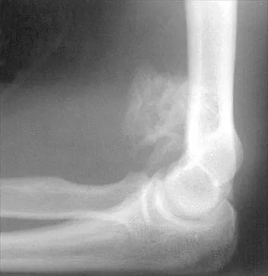
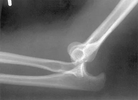
Figure 27 shows the radiograph of a 26-year-old man who sustained a closed head injury and a closed elbow dislocation 6 weeks ago. Examination reveals 65 degrees to 115 degrees of flexion, and intensive physical therapy has resulted in no improvement. A decision regarding the timing of surgical correction of the contracture should be based on


Explanation

The patient has heterotopic ossification, a more common finding in patients who have sustained head injuries. Treatment will require removal of the heterotopic bone and anterior and posterior capsulectomies. The main concern about timing is the possible recurrence of heterotopic bone. While an extended wait was once thought necessary, this is no longer true. The timing is based on the time since injury and evidence of bone maturation on plain radiographs. A sharp marginal demarcation of the new bone and a trabecular pattern within it are usually present 3 to 6 months after onset, indicating that it is safe to proceed with surgical excision. It is not necessary to wait more than 6 months. Bone scan results are not good indicators because they may remain "hot" for long periods of time. The levels of alkaline phosphatase and serum calcium-phosphorus product do not need to be measured.

Question 3

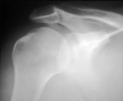
A 70-year-old man who underwent an uncomplicated large rotator cuff repair 6 months ago is now seeking a second opinion regarding persistent pain and weakness in his shoulder. Examination reveals that his incision is well healed and unreactive. The surgical report suggests that the tendons were secured back to bone with sutures through the greater tuberosity. Figure 28 shows a radiograph that was obtained 1 week ago. What is the most likely diagnosis?


Explanation

Symptoms can persist following a rotator cuff repair for a variety of reasons. In the early postoperative period, infection is the primary concern. Stiffness and loss of motion can occur because of postoperative scarring. Complex regional pain syndrome can occur but is rare, and the diagnosis is not made with a plain radiograph. This radiograph shows a superiorly migrated humeral head that articulates with the acromion, indicating that the repair has failed. While large to massive tears may fail more commonly than once thought, the clinical outcome may be satisfactory in many patients. Mansat P, Cofield RH, Kersten TE, Rowland CM: Complications of rotator cuff repair. Orthop Clin North Am 1997;28:205-213.

Question 4

A 29-year-old man who lifts weights states that he injured his left shoulder while performing a bench press 2 days ago. The following morning he noted ecchymosis and swelling in the left chest wall. Examination reveals ecchymosis and tenderness and deformity in the left anterior chest wall and axillary fold that is accentuated with resisted adduction of the arm. Passive range of motion beyond 90 degrees of forward flexion and 45 degrees of external rotation is extremely painful. Glenohumeral stability is difficult to assess because of severe guarding. Figure 29 shows an MRI scan. Management should consist of


Explanation

Rupture of the pectoralis major tendon most commonly occurs during bench pressing. Wolfe and associates have shown that the most inferiorly located fibers of the sternal head lengthen disproportionately during the final 30 degrees of humeral extension during the bench press. This creates a mechanical disadvantage in the final portion of the eccentric phase of the lift; with forceful flexion of the shoulder these maximally stretched fibers may rupture. In most patients, particularly in young athletes, the treatment of choice is anatomic repair of the ruptured tendon to its insertion in the proximal humerus either with suture anchors or transosseous sutures. Following surgery, most patients experience a near normal return of strength and significant improvement in the cosmetic appearance of the deformity. While more technically challenging, repair of chronic rupture is possible and is indicated in some patients. Wolfe SW, Wickiewicz TL, Cavanaugh JT: Ruptures of the pectoralis major muscle: An anatomic and clinical analysis. Am J Sports Med 1992;20:587-593.

Question 5

What range of motion parameters are required for a patient with posttraumatic elbow stiffness to accomplish all the normal activities of daily living?


Explanation

Activities of daily living such as dressing, eating, and bathing can all be performed with elbow motion through a 100 degrees arc of flexion and extension (30 degrees to 130 degrees) and a 100 degrees arc of forearm rotation (50 degrees pronation, 50 degrees supination). Some patients can accomplish these activities of daily living with 10 degrees less motion at each end point. This is referred to as the functional arc of motion. Kasser JR (ed): Orthopaedic Knowledge Update 5. Rosemont, IL, American Academy of Orthopaedic Surgeons, 1996, pp 283-294.

Question 6

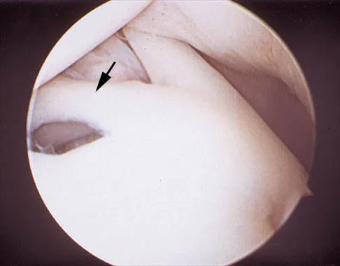
A 24-year-old athlete has a painful right shoulder. Figure 30 shows an intra-articular photograph that was obtained through a posterior portal during arthroscopy; the labrum is indicated by the arrow. Based on these findings, management should consist of


Explanation

The photograph shows a normal variant that is a sublabral hole beneath the anterosuperior labrum. In some instances, the labrum will become confluent with the middle glenohumeral ligament as a stout band. Because this variant is not abnormal, no treatment is necessary. Securing this portion of the labrum to the capsule may tighten the middle glenohumeral ligament complex and restrict external rotation of the arm. Andrews JR, Guerra JJ, Fox GM: Normal and pathologic arthroscopic anatomy of the shoulder, in Andrews JR, Timmerman LA (eds): Diagnostic and Operative Arthroscopy, ed 1. Philadelphia, PA, WB Saunders, 1997, pp 60-76.

Question 7

The use of a screw between the clavicle and the coracoid process to maintain the clavicle and acromioclavicular (AC) joint in a reduced position is a treatment option for AC joint separations. Screw removal is generally recommended after soft-tissue healing. What effect does this rigid coracoclavicular fixation have on shoulder kinematics?


Explanation

This issue has been debated since Inman published his classic study on clavicular rotation in 1944. Subsequently, it has been shown by several authors that the clinical evaluation of patients with either coracoclavicular screws in place or with arthrodesis of the coracoclavicular reveals little to no loss of shoulder motion. This is most likely the result of synchronous motion of the scapula and clavicle in shoulder movements. Flatow EL: The biomechanics of the acromioclavicular, sternoclavicular, and scapulothoracic joints. Instr Course Lect 1993;42:237-245. Kenedy JC, Cameron H: Complete dislocation of the acromioclavicular joint. J Bone Joint Surg Br 1954;36:202-208. Rockwood CA Jr, Williams GR, Young CD: Disorders of the acromioclavicular joint, in Rockwood CA Jr, Matsen FA III (eds): The Shoulder. Philadelphia, PA, WB Saunders, 1998, vol 1, pp 483-553.

Question 8

Figure 31 shows the AP and lateral radiographs of the elbow of a 56-year-old man with chronic polyarticular rheumatoid arthritis. His function continues to be limited by pain with activities of daily living. Examination shows that his total arc of motion is 110 degrees. Nonsurgical management has failed to provide relief. Treatment should now consist of


Explanation

A semiconstrained prosthesis can provide excellent results in carefully selected patients. Because the radiographs show extensive joint destruction with loss of the capitellum and trochlea, a capitellocondylar total elbow (unconstrained) prosthesis is contraindicated. Elbow fusion is poorly accepted, and the radiographs show too much articular destruction for a radial head excision, synovectomy, or interposition arthroplasty to be effective. Ewald FC, Simmons ED Jr, Sullivan JA, et al: Capitellocondylar total elbow replacement in rheumatoid arthritis: Long-term results. J Bone Joint Surg Am 1993;75:498-507.

Question 9

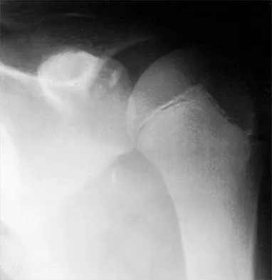
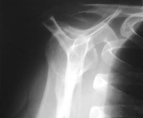
A 12-year-old pitcher has had a 2-month history of pain in his right dominant shoulder after throwing. He reports that the pain has gradually progressed to the point where he cannot throw without pain. He also notes that the pain now awakens him at night if he has been active. Anti-inflammatory drugs have failed to provide relief. Examination reveals no abnormalities except for some localized tenderness over the proximal humerus. Figures 32a and 32b show radiographs of both shoulders. What is the most likely diagnosis?


Explanation

32b The history, examination, and radiographs are pathognomonic for Little League shoulder, a stress syndrome of the proximal humeral physis caused by overuse. Complete fracture rarely occurs, and recovery usually occurs with rest. Night pain is always a serious concern and further work-up is needed if the patient does not respond to activity modification. Occult instability is not a real concern in this patient, although it should be included in the differential diagnosis. Albert MJ, Drvaric DM: Little League shoulder: Case report. Orthopedics 1990;13:779-781.

Question 10

Which of the following ligaments is the primary static restraint against inferior translation of the arm when the shoulder is in 0 degrees of abduction?


Explanation

The superior glenohumeral ligament (SGHL) and coracohumeral ligament serve as primary static restraints against inferior translation of the arm when the shoulder is in 0 degrees of abduction. Of these, the coracohumeral ligament has been shown to have a greater cross-sectional area, greater stiffness, and greater ultimate load than the SGHL. The inferior glenohumeral ligament plays a greater stabilizing role with increasing abduction of the arm. The coracoacromial ligament may help provide superior stability, especially when the rotator cuff is deficient. The coracoclavicular ligaments stabilize the acromioclavicular joint. Boardman ND, Debski RE, Warner JJ, et al: Tensile properties of the superior glenohumeral and coracohumeral ligaments. J Shoulder Elbow Surg 1996;5:249-254.

Question 11

A 16-year-old high school student undergoes a routine preparticipation physical examination at the beginning of the school year. Examination reveals marked laxity of both shoulders but only mild generalized laxity in other joints. The load and shift test allows for anterior humeral translation to the glenoid rim and posterior humeral translation beyond the glenoid rim. The sulcus sign is present. What is the next most appropriate step in management?


Explanation

This patient has shoulder laxity without apprehension. Because there is a wide range of normal laxity in asymptomatic shoulders, the physician should inform the student of these findings, recommend shoulder strengthening exercises, and allow unrestricted sports participation unless symptoms develop. Harryman DT, Sidles JA, Harris SL, Matsen FA III: Laxity of the normal glenohumeral joint: A quantitative in vivo assessment. J Shoulder Elbow Surg 1992;1:66-76. Hawkins RJ, Bokor RJ: Clinical evaluation of shoulder problems, in Rockwood CA Jr, Matsen FA III (eds): The Shoulder. Philadelphia, PA, WB Saunders, 1998, vol 1, p 186. McFarland EG, Campbell G, McDowell J: Posterior shoulder laxity in asymptomatic athletes. Am J Sports Med 1996;24:468-471.

Question 12

A 21-year-old professional baseball player has had painful catching and stiffness in his dominant right elbow for the past year. Examination reveals a flexion contracture of 2 degrees and mild pain with full elbow flexion. Radiographs are shown in Figures 33a and 33b. The most effective management should consist of


Explanation

33b The radiographs show osteochondritis dissecans of the capitellum and a loose body in the anterior compartment. Arthroscopic removal is indicated because symptoms referable to the loose body are present. Baumgarten TE: Osteochondritis dissecans of the capitellum. Sports Med Arthroscopy Rev 1995;3:219-223.

Question 13

A 42-year-old patient has had painful inferior subluxation of the glenohumeral joint following a recent cerebrovascular accident (CVA). Figure 34 shows the AP radiograph of the shoulder. Management should consist of


Explanation

Following a CVA and with the resumption of upright posture, downward subluxation of the glenohumeral joint may occur. Although usually painless, some patients may report pain secondary to stretching of the brachial plexus. This is the result of flaccid paralysis of the deltoid muscle, and it will persist until some motor tone or spasticity returns to the shoulder girdle musculature. Early sling support and range-of-motion exercises to prevent contracture will provide the best relief. Surgical procedures are not indicated. Braun RM, Botte MJ: Treatment of shoulder deformity in acquired spasticity. Clin Orthop 1999;368:54-65.

Question 14

A 50-year-old man who underwent an arthroscopic rotator cuff repair 5 days ago now returns for an early postoperative follow-up because of increasing pain in his shoulder. He reports increasing malaise and has a low-grade fever. Examination reveals no redness or swelling, but he has scant serous drainage from the posterior portal. An emergent Gram stain is positive for gram-positive cocci. The next most appropriate step in management should consist of


Explanation

An infection of the shoulder is considered a surgical emergency unless there are medical reasons that a patient cannot be taken to the operating room. If cultures of wound drainage are in question, then an aspiration should be done emergently, not several days later. The hallmark of infection in any major joint is increasing pain out of proportion to what is expected. Drainage occurring 1 to 2 days after an arthroscopic procedure is not normal, and it should be aggressively treated. Delay in diagnosis can result in sepsis and on a delayed basis, postinfectious arthritis. Both the glenohumeral joint and the subacromial space require debridement and irrigation, followed by antibiotics after both areas are cultured. Mansat P, Cofield RH, Kersten TE, Rowland CM: Complications of rotator cuff repair. Orthop Clin North Am 1997;28:205-213. Settecerri JJ, Pitner MA, Rock MG, Hanssen AD, Cofield RH: Infection after rotator cuff repair. J Shoulder Elbow Surg 1999;8:1-5. Ward WG, Eckardt JJ: Subacromial/subdeltoid bursa abscesses: An overlooked diagnosis. Clin Orthop 1993;288:189-194.

Question 15

A 42-year-old man who is right-hand dominant injured his right shoulder when he fell from a ladder onto his outstretched arm 1 hour ago. Radiographs reveal a two-part greater tuberosity anterior fracture-dislocation. Initial management should consist of


Explanation

Greater tuberosity anterior fractures associated with anterior glenohumeral dislocations respond very well to closed methods in the majority of patients. Closed reduction of the glenohumeral joint often anatomically reduces the greater tuberosity into its cancellous bed, without the need for open fixation or cuff repair. Once closed reduction of the joint is performed, tuberosity displacement and joint articulation should be evaluated radiographically with AP and scapular lateral views as well as an axillary view. The axillary view will not only definitively show the joint articulation but also demonstrate posterior displacement of the greater tuberosity missed on the AP and lateral views. If no or minimal (5 mm) displacement is found, then nonsurgical management consisting of a sling and gentle passive range-of-motion exercises can be instituted. Beaty JH (ed): Orthopaedic Knowledge Update 6. Rosemont, IL, American Academy of Orthopaedic Surgeons, 1999, pp 271-286.

Question 16

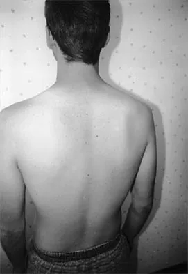
A 19-year-old man who plays college volleyball undergoes a routine preparticipation physical examination. Figure 35 shows a posterior view of his dominant shoulder. An electromyogram shows that this is a chronic injury, and an MRI scan shows no abnormalities. The best course of action should be


Explanation

Isolated palsy of the infraspinatus portion of the suprascapular nerve is common in volleyball players and is seen frequently in the throwing arm of baseball players. The exact cause is not known, but it may be the result of either tethering or traction on the nerve at the spinoglenoid notch. Synovial cysts in the spinoglenoid notch also can be a cause, but the patient's negative MRI findings rule out that entity. Because many isolated nerve palsies of the infraspinatus branch are asymptomatic, initial management should always be nonsurgical. Surprisingly, many athletes with this injury can participate fully in sports. Surgical treatment with decompression at the notch is unpredictable and generally is indicated only if nonsurgical management fails. Ferretti A, Cerullo G, Russo G: Suprascapular neuropathy in volleyball players. J Bone Joint Surg Am 1987;69:260-263.

Question 17

A 59-year-old construction worker who is right-hand dominant has had right shoulder pain for the past 9 months with no history of injury. Nonsurgical management consisting of two cortisone injections, physical therapy for 3 months, and nonsteroidal anti-inflammatory drugs has failed to provide lasting relief. Examination reveals tenderness over the acromioclavicular (AC) joint and over the subacromial bursa. He has positive Neer and Hawkins impingement signs and AC joint pain with adduction of the shoulder. Radiographs are shown in Figures 36a and 36b. An MRI scan reveals an intact rotator cuff. Management should now consist of


Explanation

36b Because the patient has clinical and radiographic signs of AC arthritis and subacromial impingement, the treatment of choice is anterior acromioplasty and distal clavicle excision. Arthroscopic acromioplasty alone would not address the AC arthritis. The rotator cuff is intact; therefore, rotator cuff repair is not indicated. An open Mumford procedure would address the AC arthritis only and not the impingement symptoms. Immobilization might lead to stiffness of the shoulder and is not recommended for treating impingement.

Question 18

What three structures are considered the primary constraints necessary for elbow stability?


Explanation

The three primary constraints necessary for elbow stability in all directions are the ulnar part of the lateral collateral ligament (also called the lateral ulnar collateral ligament), the anterior band of the medial collateral ligament, and the coronoid. The radial head and capsule are secondary constraints to elbow instability. Kasser JR (ed): Orthopaedic Knowledge Update 5. Rosemont, IL, American Academy of Orthopaedic Surgeons, 1996, pp 283-294.

Question 19

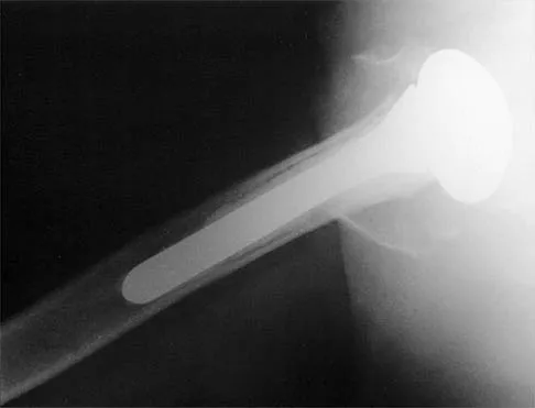
A 68-year-old woman has been progressing slowly after undergoing humeral head replacement for a four-part fracture 3 months ago. She has not regained active elevation, she feels an audible clunk on attempting elevation, and she reports pain and weakness. She used a sling for 2 weeks in the immediate postoperative period. Radiographs are shown in Figure 37a through 37c. Management should consist of


Explanation

37b 37c Immediate repair of the tuberosity and rotator cuff is recommended on identifying the avulsion or nonunion. Revising the humeral component to increase tension and length will overtighten the cuff and increase the chance of tuberosity pull-off. The glenoid is uninvolved and should not be replaced. Attempts to strengthen the rotator cuff will be unsuccessful because the insertions are no longer attached to the humerus when the tuberosities avulse. Brown TD, Bigliani LU: Complications with humeral head replacement. Orthop Clin North Am 2000;31:77-90.

Question 20

What is the most important feature in choosing an outcome instrument to assess shoulder disorders?


Explanation

There has been a recent increase in the use of outcome instruments to document and measure effects of treatment of medical conditions, including shoulder disorders. The most important feature of an instrument is whether it actually measures what it purports to measure; this is defined as its validity. Leggin BG, Iannotti JP: Shoulder outcome measurement, in Iannotti JP, Williams GR (eds): Disorders of the Shoulder: Diagnosis and Management. Philadelphia, PA, Lippincott Williams and Wilkins, 1999, p 1027.

Question 21

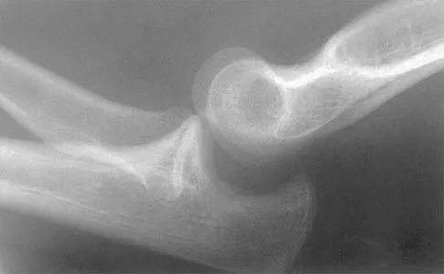
Figure 38 shows the radiograph of a 16-year-old wrestler who injured his elbow when he was thrown to the mat by his opponent. To minimize additional trauma to the medial soft tissues, the elbow should be reduced in


Explanation

The elbow dislocates by a three-dimensional movement of supination and valgus during flexion. Additional trauma during reduction is minimized by recreating the deformity and reducing the elbow in supination. The actual maneuver includes full supination (actually hypersupination) of the elbow in a valgus position. This is followed by pushing the olecranon distally in line with the long axis of the ulna while swinging the elbow into varus, and then relaxing the supination torque. Postreduction stability is enhanced in pronation, except when the soft-tissue disruption is extensive. O'Driscoll SW: Elbow dislocations, in Morrey BF (ed): The Elbow and Its Disorders, ed 2. Philadelphia, PA, WB Saunders, 1993, p 414.

Question 22

In patients older than age 40 years who sustain a first-time anterior dislocation of the shoulder, prolonged morbidity is most commonly associated with


Explanation

In the study done by Pevny and associates, 35% of patients older than age 40 years sustained rotator cuff tears and 8% had axillary nerve palsies. All of the patients with axillary nerve palsy also had rotator cuff tears. Imaging of the rotator cuff is indicated in this age group. The incidence of recurrent instability in patients older than age 40 years is 10% to 15%. Pevny T, Hunter RE, Freeman JR: Primary traumatic anterior shoulder dislocation in patients 40 years of age and older. Arthroscopy 1998;14:289-294. Sonnabend DH: Treatment of primary anterior shoulder dislocation in patients older than 40 years of age: Conservative versus operative. Clin Orthop 1994;304:74-77.

Question 23

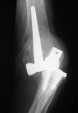
Figure 39 shows the AP radiograph of a 62-year-old man with degenerative osteoarthritis secondary to trauma. History reveals that he underwent total elbow arthroplasty 3 years ago. He continues to report instability and constant pain. A complete work-up, including aspiration and cultures, is negative. Treatment should consist of removal of the components and


Explanation

An unconstrained prosthesis dislocation is a disconcerting problem that is not easily resolved; however, revision to a semiconstrained prosthesis would best achieve both pain relief and stability. Removal of the components and distraction arthroplasty or conversion to a resection arthroplasty are options, but the results would be unpredictable with regards to pain relief, postoperative motion, or elbow stability. Arthrodesis is poorly tolerated. With revision to another unconstrained prosthesis, there is the risk of continued redislocation because of chronic ligamentous insufficiency. Linscheid RL: Resurfacing elbow replacement arthroplasty: Rationale, technique and results, in Morrey BF (ed): The Elbow and Its Disorders, ed 3. Philadelphia, PA, WB Saunders, 2000, pp 602-610.

Question 24

A 67-year-old woman undergoes a revision total shoulder arthroplasty for replacement of a loose glenoid component. Examination in the recovery room reveals absent voluntary deltoid and triceps contraction, weakness of wrist and thumb extension, and absent sensation in the palmar aspect of all fingertips and the radial forearm. The next most appropriate step in management should consist of


Explanation

Neurologic injury after shoulder replacement is relatively uncommon, occurring in 4% of shoulders in one large series. The importance of identifying and protecting the musculocutaneous and axillary nerves cannot be overemphasized; it is especially critical during revision arthroplasty when the normal anatomic relationships have been distorted. The long deltopectoral approach leaving the deltoid attached to the clavicle was found to be significant in the development of postoperative neurologic complications. A correlation was found between surgical time and postoperative neurologic complications, with long surgical times being associated with more neurologic complications. The presumed mechanism of injury is traction on the plexus that occurs during the surgery. A neurologic injury after total shoulder arthroplasty usually does not interfere with the long-term outcome of the arthroplasty itself; it is best managed by protective measures with passive range of motion of the involved extremity. Wirth MA, Rockwood CA Jr: Complications of shoulder arthroplasty. Clin Orthop 1994;307:47-69.

Question 25

Figure 40 shows the radiograph of a 16-year-old wrestler who injured his elbow when he was thrown to the mat by his opponent. Closed reduction is readily accomplished, and the elbow seems stable. Management should now consist of application of a splint for


Explanation

Flexion contractures are the most common complication of elbow dislocations. About 15% of patients lose more than 30 degrees of flexion. The risk of contracture is proportional to the duration of immobilization. Elbows should be moved within the first few days after reduction. The splinting is for comfort and protection only while the pain subsides. Mehlhoff TL, Noble PC, Bennett JB, Tullos HS: Simple dislocation of the elbow in the adult: Results after closed treatment. J Bone Joint Surg Am 1988;70:244-249. Linscheid RL, O'Driscoll SW: Elbow dislocations, in Morrey BF (ed): The Elbow and Its Disorders, ed 2. Philadelphia, PA, WB Saunders, 1993, pp 441-452. O'Driscoll SW, Jupiter JB, King GJ, Hotchkiss RN, Morrey BF: The unstable elbow. Instr Course Lect 2001;50:89-102.

Question 26

During a Latarjet procedure, the coracoid process is transferred to the anterior glenoid. Which nerve is at greatest risk of injury during the coracoid preparation and transfer of the conjoined tendon?


Explanation

The musculocutaneous nerve enters the coracobrachialis 5-8 cm distal to the coracoid process. It is tethered to the conjoined tendon and is at significant risk during retraction and transfer in a Latarjet procedure.

Question 27

An 82-year-old woman sustains a displaced 4-part proximal humerus fracture. She has a documented history of severe osteoarthritis and a massive rotator cuff tear prior to the injury. What is the most appropriate surgical treatment?


Explanation

Reverse total shoulder arthroplasty is the treatment of choice for elderly patients with 4-part proximal humerus fractures and pre-existing rotator cuff arthropathy or massive cuff tears, as it utilizes the deltoid for shoulder elevation.

Question 28

In reverse total shoulder arthroplasty (rTSA), which of the following component positioning strategies is most effective in minimizing the risk of scapular notching?


Explanation

Inferior translation and inferior tilt of the glenosphere, along with increased lateral offset, are crucial strategies to reduce scapular notching in rTSA. This prevents abutment of the humeral tray against the scapular neck during adduction.

Question 29

A 24-year-old rugby player presents with recurrent anterior shoulder instability. A 3D CT scan reveals 27% anterior glenoid bone loss. What is the most appropriate definitive management?


Explanation

In young, high-demand collision athletes with critical glenoid bone loss (typically >20-25%), an arthroscopic Bankart repair has an unacceptably high failure rate. A bony augmentation procedure, such as the Latarjet, is definitively indicated.

Question 30

A 65-year-old man presents with an inability to actively elevate his arm above 60 degrees. Passive elevation is full. An intra-articular injection of local anesthetic completely restores his active elevation to 150 degrees. This clinical finding most likely indicates:


Explanation

True pseudoparalysis implies a mechanical inability to elevate the arm that does not improve with pain relief. Restoration of active motion after a local anesthetic injection confirms pseudoparesis driven by pain inhibition.

Question 31

A 50-year-old man falls on an outstretched arm and experiences acute anterior shoulder pain. Physical examination reveals significantly increased passive external rotation compared to the contralateral side. Which of the following tests is most likely to be positive?


Explanation

Increased passive external rotation following a traumatic event strongly suggests a subscapularis tear. The lift-off test specifically evaluates the integrity and strength of the subscapularis muscle.

Question 32

A 35-year-old man presents to the emergency department after experiencing a grand mal seizure. He holds his right arm internally rotated and is unable to actively or passively externally rotate it. An AP radiograph demonstrates the "lightbulb sign." What is the most appropriate initial management?


Explanation

The clinical presentation (locked internal rotation) and radiographic "lightbulb sign" are classic for a posterior shoulder dislocation. Initial management involves prompt closed reduction under conscious sedation or general anesthesia.

Question 33

A 28-year-old professional volleyball player complains of vague posterior shoulder pain and weakness. Examination reveals isolated atrophy of the infraspinatus with normal supraspinatus bulk and strength. Where is the most likely site of nerve compression?


Explanation

Entrapment of the suprascapular nerve at the spinoglenoid notch affects only the infraspinatus muscle. Entrapment further proximal at the suprascapular notch would affect both the supraspinatus and the infraspinatus.

Question 34

A 55-year-old diabetic woman presents with a 4-month history of severe, diffuse shoulder pain that is progressively worsening, especially at night. Her range of motion is globally decreased but pain is the primary limiting factor. She is currently in which phase of adhesive capsulitis?


Explanation

The freezing phase (lasting 2-9 months) is characterized by severe, progressive pain and a gradual loss of motion. The subsequent frozen phase involves less pain but profound, unyielding stiffness.

Question 35

During a diagnostic shoulder arthroscopy on a 22-year-old pitcher, you identify a bucket-handle tear of the superior labrum. However, probing confirms that the biceps anchor remains firmly attached to the superior glenoid tubercle. This corresponds to which classification of SLAP lesion?


Explanation

A Type III SLAP lesion is defined as a bucket-handle tear of the superior labrum with an intact biceps anchor. Type II, the most common type, involves frank detachment of the biceps anchor from the glenoid.

Question 36

A 30-year-old weightlifter feels a sudden "pop" in his axilla while performing a heavy bench press. He presents with extensive ecchymosis over the anterior axillary fold. He will most likely demonstrate marked weakness in which of the following shoulder motions?


Explanation

The pectoralis major acts primarily as an adductor and internal rotator of the humerus. Ruptures typically occur during eccentric contraction, such as the descent phase of a bench press, resulting in weakness in these specific motions.

Question 37

When evaluating a midshaft clavicle fracture for nonoperative management, which of the following radiographic findings is the most significant risk factor for subsequent nonunion?


Explanation

Significant fracture displacement, specifically initial shortening greater than 2 cm (or 100% displacement), is biomechanically strongly associated with an increased risk of nonunion and poor functional outcomes in nonoperatively treated clavicle fractures.

Question 38

A 25-year-old cyclist falls directly onto his shoulder apex. Radiographs reveal 100% superior displacement of the distal clavicle relative to the acromion. The coracoclavicular distance is increased by 50% compared to the uninjured side. The deltotrapezial fascia remains intact. This represents what type of acromioclavicular (AC) joint injury?


Explanation

A Type III AC joint separation involves complete rupture of both the AC and CC ligaments, resulting in 25-100% superior displacement of the clavicle. Type V involves >100% displacement with disruption of the overlying deltotrapezial fascia.

Question 39

A 19-year-old football player sustains a posterior sternoclavicular dislocation during a pile-up. He presents with shortness of breath and difficulty swallowing. What is the most immediate life-threatening complication that must be ruled out?


Explanation

Posterior sternoclavicular dislocations can compress critical mediastinal structures. Tracheal compression resulting in airway compromise is an immediate, life-threatening emergency requiring prompt reduction.

Question 40

A 13-year-old elite baseball pitcher presents with worsening right shoulder pain occurring strictly during the cocking phase of throwing. AP radiographs reveal widening and irregularity of the proximal humeral physis. What is the underlying pathophysiology?


Explanation

Little League shoulder is a fatigue failure of the proximal humeral physis, representing a Salter-Harris I stress fracture. It is caused by repetitive rotational and distraction forces exerted during the throwing motion.

Question 41

A 32-year-old overhead athlete presents with poorly localized posterior shoulder pain and paresthesias over the lateral deltoid. Angiography demonstrates occlusion of the posterior circumflex humeral artery with the arm in abduction and external rotation. Which nerve is most likely compressed?


Explanation

Quadrilateral space syndrome involves the compression of the axillary nerve and posterior circumflex humeral artery within the quadrilateral space. It is often caused by fibrous bands or hypertrophy of the adjacent teres minor muscle.

Question 42

Despite advancements in implant design, what remains the most common complication and cause for late revision surgery following anatomic total shoulder arthroplasty (aTSA)?


Explanation

Aseptic loosening of the polyethylene glenoid component is the most common reason for late revision in aTSA. The eccentric loading of the component, known as the "rocking horse" phenomenon, significantly contributes to this failure mechanism.

Question 43

A 26-year-old manual laborer complains of painful crepitus and a loud snapping sensation at the superomedial border of his scapula with movement. Following failure of extensive nonoperative management, surgical intervention is planned. Which structure is most commonly targeted for resection?


Explanation

Snapping scapula syndrome is frequently caused by an inflamed scapulothoracic bursa and/or a prominent superomedial scapular angle. Operative treatment typically consists of bursectomy and resection of the superomedial angle.

Question 44

A 21-year-old collegiate tennis player complains of posterior shoulder pain during the late cocking phase of his serve. Arthroscopy reveals undersurface fraying of the posterior supraspinatus and anterior infraspinatus, along with corresponding posterosuperior labral fraying. This triad is characteristic of:


Explanation

Internal impingement occurs in overhead athletes when the arm is placed in extreme abduction and external rotation. This position causes the undersurface of the posterosuperior rotator cuff to mechanically abut against the posterosuperior glenoid labrum.

Question 45

A 40-year-old man presents with sudden, severe right shoulder pain that lasted unremittingly for 2 weeks. As the pain spontaneously subsided, he noticed profound weakness of the shoulder musculature and pronounced winging of the scapula. EMG demonstrates acute denervation. What is the most likely diagnosis?


Explanation

Parsonage-Turner syndrome (acute brachial neuritis) classically presents with acute onset of severe shoulder pain, followed days or weeks later by patchy muscle weakness, atrophy, and painless scapular winging as the pain resolves.

Question 46

Compared to native shoulder anatomy, a traditional Grammont-style reverse total shoulder arthroplasty alters the biomechanics by doing which of the following?


Explanation

The traditional Grammont-style reverse shoulder arthroplasty medializes the center of rotation to the glenoid face and inferiorly displaces the humerus. This design tensions the deltoid and increases its moment arm, allowing it to elevate the arm without a functioning rotator cuff.

Question 47

A 25-year-old rugby player undergoes an open Latarjet procedure for recurrent anterior shoulder instability with 25% glenoid bone loss. Postoperatively, he presents with inability to flex his elbow and decreased sensation over the lateral forearm. Which nerve was most likely injured during the procedure?


Explanation

The musculocutaneous nerve is at risk during the Latarjet procedure when the conjoint tendon is retracted medially. Injury results in weakness of elbow flexion (biceps, brachialis) and sensory loss over the lateral forearm via the lateral antebrachial cutaneous nerve.

Question 48

A 55-year-old manual laborer presents with a massive, irreparable posterosuperior rotator cuff tear. He has intact subscapularis function and no glenohumeral arthritis. He desires to return to heavy labor. Which of the following tendon transfers is most appropriate to restore active external rotation?


Explanation

Latissimus dorsi tendon transfer is indicated for younger, active patients with an irreparable posterosuperior rotator cuff tear, an intact subscapularis, and no arthritis. It helps restore active forward elevation and external rotation.

Question 49

According to the Walch classification, a B2 glenoid is characterized by which of the following?


Explanation

A Walch B2 glenoid is characterized by a biconcave articular surface with asymmetric posterior wear and posterior subluxation of the humeral head. It poses a high risk for glenoid component loosening if not addressed during anatomic total shoulder arthroplasty.

Question 50

A 28-year-old man presents with dull, aching shoulder pain and prominent medial scapular winging that worsens when pushing against a wall. Examination reveals weakness of the serratus anterior. Injury to which nerve is the most likely cause?


Explanation

Medial scapular winging is caused by serratus anterior paralysis due to long thoracic nerve injury. It is classically accentuated by having the patient push forward against a wall.

Question 51

A 32-year-old professional volleyball player presents with poorly localized posterior shoulder pain and paresthesias over the lateral deltoid. An MRI demonstrates isolated atrophy of the teres minor muscle. Which of the following structures is most likely being compressed?


Explanation

Quadrilateral space syndrome involves compression of the axillary nerve and posterior humeral circumflex artery. It typically presents with localized teres minor atrophy, as the anterior branch of the axillary nerve supplying the deltoid may be spared.

Question 52

A 21-year-old collegiate baseball pitcher complains of posterior shoulder pain during the late cocking phase of throwing. Physical exam reveals 15 degrees of internal rotation and 125 degrees of external rotation at 90 degrees of abduction, compared to 60 degrees and 100 degrees respectively on the contralateral side. What is the most appropriate initial management?


Explanation

Glenohumeral internal rotation deficit (GIRD) is common in overhead athletes and is primarily caused by posterior capsular contracture. The initial treatment of choice is a dedicated stretching program, including sleeper stretches, to restore internal rotation.

Question 53

During the surgical approach for a displaced 3-part proximal humerus fracture, preservation of the blood supply to the humeral head is critical. Which artery provides the predominant blood supply to the humeral head?


Explanation

Recent anatomic injection studies have demonstrated that the posterior humeral circumflex artery provides the predominant blood supply to the humeral head. This challenges the historical belief that the anterior humeral circumflex artery was the main supplier.

Question 54

A 45-year-old woman presents with acute, severe shoulder pain that awakens her from sleep. Radiographs show a fluffy, ill-defined calcific deposit in the supraspinatus tendon. During which phase of calcific tendinitis does a patient typically experience the most severe pain?


Explanation

Calcific tendinitis is most painful during the resorptive phase. During this time, the calcific deposit has a toothpaste-like consistency and elicits a vigorous vascular and inflammatory response.

Question 55

A 19-year-old male is brought to the emergency department after a motor vehicle accident. He has severe pain over his medial clavicle, shortness of breath, and dysphagia. Radiographs and a CT scan confirm a posterior sternoclavicular dislocation. What is the most appropriate management?


Explanation

Posterior sternoclavicular dislocations can compress vital mediastinal structures. Due to the high risk of catastrophic vascular injury during manipulation, reduction must be performed in the operating room with cardiothoracic surgery available.

Question 56

Which of the following structures is NOT a component of the rotator interval of the shoulder?


Explanation

The rotator interval is a triangular space bordered by the supraspinatus superiorly, subscapularis inferiorly, and the coracoid process medially. It contains the coracohumeral ligament, superior glenohumeral ligament, joint capsule, and the long head of the biceps tendon, but not the middle glenohumeral ligament.

Question 57

Which of the following is considered an absolute indication for operative fixation of an acute midshaft clavicle fracture?


Explanation

Absolute indications for operative fixation of a clavicle fracture include open fractures, neurovascular compromise, and severe skin tenting that threatens skin integrity. Shortening and displacement are considered relative indications.

Question 58

A 50-year-old woman with diabetes mellitus presents with worsening shoulder stiffness and pain. Arthroscopy reveals hypervascularity and proliferative synovitis without significant capsular fibrosis. This represents which stage of adhesive capsulitis, and what is the typical duration of this stage?


Explanation

Stage 2 of adhesive capsulitis (the "freezing" stage) typically lasts from 3 to 9 months. It is characterized by intense pain, progressive loss of motion, and arthroscopic findings of hypertrophic, hypervascular synovitis with early scar formation.

Question 59

A 30-year-old weightlifter feels a "pop" in his anterior axilla while performing a bench press. Examination reveals an ecchymotic swelling and loss of the anterior axillary fold. MRI confirms a complete rupture of the pectoralis major at its humeral insertion. Which of the following is the most appropriate treatment to restore maximum strength?


Explanation

In young athletic patients seeking to return to high-level activity, surgical repair of a complete pectoralis major rupture at the humeral insertion is indicated. Operative treatment reliably restores peak torque in shoulder adduction and internal rotation.

Question 60

A 42-year-old man presents with sudden onset of severe, unrelenting right shoulder pain lasting for two weeks, followed by profound weakness of his deltoid and supraspinatus. There is no history of trauma. MRI of the shoulder is unremarkable. What is the most likely diagnosis?


Explanation

Parsonage-Turner syndrome (idiopathic brachial neuritis) classically presents with acute, severe shoulder pain that resolves after a few weeks. It is immediately followed by patchy lower motor neuron weakness and atrophy, most commonly affecting the suprascapular and axillary nerve distributions.

Question 61

A 40-year-old man suffers a first-time seizure and sustains a locked posterior shoulder dislocation. CT scan shows an impaction fracture of the anteromedial humeral head involving 35% of the articular surface. Which of the following is the most appropriate surgical treatment?


Explanation

For a reverse Hill-Sachs lesion involving 20% to 40% of the articular surface in a young, active patient, the modified McLaughlin procedure is indicated. Transferring the lesser tuberosity with the attached subscapularis into the defect durably restores joint stability.

Question 62

A 24-year-old overhead athlete presents with deep shoulder pain and clicking. MR arthrogram demonstrates detachment of the superior labrum and the origin of the long head of the biceps tendon from the glenoid. What is the standard arthroscopic management for this specific lesion in this patient?


Explanation

The scenario describes a Type II SLAP tear. In young overhead athletes, the standard treatment to restore function is arthroscopic repair of the superior labrum and biceps anchor back to the superior glenoid using suture anchors.

Question 63

Which of the following radiographic findings is a widely accepted indication for operative fixation of an extra-articular scapular body/neck fracture?


Explanation

Indications for operative treatment of extra-articular scapular fractures include medial/lateral displacement > 20 mm, angular deformity > 45 degrees, and an abnormally low glenopolar angle (typically < 22 degrees).

Question 64

A 35-year-old man presents with posterior shoulder pain and weakness in external rotation. Forward elevation strength is normal. Examination reveals atrophy isolated to the infraspinatus fossa. Where is the most likely location of nerve compression?


Explanation

Compression of the suprascapular nerve at the spinoglenoid notch affects only the distal branch supplying the infraspinatus, causing isolated external rotation weakness. Compression at the suprascapular notch would affect both the supraspinatus and infraspinatus.

Question 65

A 28-year-old professional volleyball player presents with insidious onset of right shoulder pain and weakness. Clinical examination reveals isolated atrophy of the infraspinatus with normal supraspinatus bulk and strength. MRI demonstrates a paralabral cyst in the spinoglenoid notch. What is the most likely location of the labral tear associated with this condition?


Explanation

Spinoglenoid notch cysts compressing the suprascapular nerve typically cause isolated infraspinatus atrophy. They are most commonly associated with posterior-superior labral tears, which allow joint fluid to form a one-way valve mechanism leading to the cyst.

Question 66

In reverse total shoulder arthroplasty (RTSA), which of the following glenosphere design modifications or placements is most effective in decreasing the incidence of inferior scapular notching?


Explanation

Inferior translation (overhang) of the glenosphere and lateralization of the center of rotation help prevent mechanical impingement of the humeral component on the inferior scapular neck. This significantly reduces the risk of scapular notching.

Question 67

A 22-year-old collegiate rugby player presents with recurrent anterior shoulder instability. A 3D CT scan reveals a 25% anterior glenoid bone loss. What is the most appropriate surgical management to restore stability?


Explanation

Anterior glenoid bone loss greater than 20% to 25% is a strong indication for a bony augmentation procedure. The Latarjet procedure (coracoid transfer) successfully restores glenohumeral stability through bone augmentation and the sling effect of the conjoined tendon.

Question 68

A 48-year-old recreational tennis player presents with persistent shoulder pain despite 6 months of conservative management. MRI confirms an isolated Type II SLAP tear. What is the most reliable surgical option for this specific patient?


Explanation

In patients older than 40-45 years with a Type II SLAP tear, primary biceps tenodesis provides more reliable pain relief and functional improvement. It also carries lower complication rates and less postoperative stiffness compared to SLAP repair.

Question 69

A 21-year-old collegiate baseball pitcher complains of posterior shoulder pain during the late cocking phase of throwing. Examination shows a 25-degree loss of internal rotation and a 15-degree gain in external rotation compared to the contralateral side. What is the initial treatment of choice?


Explanation

Glenohumeral internal rotation deficit (GIRD) in overhead athletes is caused by posterior capsular contracture. It is initially managed non-operatively with a targeted physical therapy program emphasizing posterior capsular stretching, such as sleeper stretches.

Question 70

A 32-year-old male bodybuilder presents with an acute tearing sensation in his anterior chest and axilla while performing a heavy bench press. Examination reveals an asymmetric chest wall and a palpable defect in the axillary fold. If surgical repair is planned, which anatomical structure represents the most common site of failure in this injury?


Explanation

Pectoralis major ruptures most commonly occur at the insertion of the sternocostal head onto the humerus. This typically occurs during maximal eccentric loading, such as the descent phase of a bench press.

Question 71

A 65-year-old woman is planning to undergo total shoulder arthroplasty (TSA) for primary osteoarthritis. Preoperative CT imaging reveals a Walch B2 glenoid. What is the recommended strategy to address this specific deformity during primary TSA to prevent early glenoid component loosening?


Explanation

A Walch B2 glenoid is characterized by biconcavity and posterior subluxation of the humeral head. Addressing the pathologic retroversion with a posterior augmented glenoid or asymmetric reaming is critical to prevent eccentric loading and early loosening of the component.

Question 72

A 78-year-old right-hand-dominant woman sustains a 4-part proximal humerus fracture with severe varus collapse and significant comminution of the tuberosities. She lives independently and is medically optimized. What is the most appropriate surgical treatment?


Explanation

In elderly patients with complex, comminuted 4-part proximal humerus fractures, reverse total shoulder arthroplasty (RTSA) provides more predictable functional outcomes and pain relief. It relies less on tuberosity healing compared to ORIF or hemiarthroplasty.

Question 73

A 28-year-old male construction worker falls directly onto his right shoulder. Radiographs demonstrate an acromioclavicular (AC) joint injury with the distal clavicle displaced superiorly by 150% of the normal AC joint distance. What is the most appropriate management?


Explanation

A Type V AC joint separation is characterized by greater than 100% superior displacement of the distal clavicle with detachment of the deltotrapezial fascia. Operative reconstruction of the CC ligaments is indicated to restore normal shoulder mechanics.

Question 74

A 35-year-old man presents with vague posterior shoulder pain and numbness over the lateral aspect of the deltoid. MRI of the shoulder demonstrates isolated atrophy and fatty infiltration of the teres minor muscle. Which of the following structures is most likely compressed?


Explanation

Quadrilateral space syndrome involves compression of the axillary nerve and the posterior circumflex humeral artery. It classically presents with teres minor atrophy, deltoid weakness, and lateral shoulder paresthesias.

Question 75

A 52-year-old woman with a 10-year history of poorly controlled type 1 diabetes presents with severe, progressive shoulder stiffness and pain for 4 months. She has profound loss of active and passive external rotation. Which of the following best describes the underlying pathophysiology of her condition?


Explanation

Adhesive capsulitis (frozen shoulder) is strongly associated with diabetes mellitus. It is characterized by fibroblastic proliferation, capsular fibrosis, and contracture, driven by inflammatory cytokines and altered collagen metabolism.

Question 76

A 72-year-old woman undergoes a reverse total shoulder arthroplasty (RTSA). At 2-year follow-up, radiographs show inferior scapular notching. Which of the following implant positioning strategies most effectively reduces the risk of this complication?


Explanation

Scapular notching in RTSA is caused by mechanical impingement of the humeral cup against the inferior scapular neck. Inferior translation and inferior tilt of the glenosphere, along with lateralization, decrease the risk of notching.

Question 77

A 22-year-old rugby player presents with recurrent anterior shoulder instability. A 3D CT scan reveals 25% anterior glenoid bone loss. What is the most appropriate surgical management?


Explanation

In a collision athlete with significant anterior glenoid bone loss (>20-25%), isolated soft tissue stabilization is associated with high failure rates. The Latarjet procedure provides a triple blocking effect (bone, sling, capsule) and is the gold standard for this scenario.

Question 78

A 78-year-old woman sustains a 4-part proximal humerus fracture with head splitting and osteopenia. She lives independently and is a community ambulator. What is the most reliable surgical option to predictably restore forward elevation?


Explanation

In elderly patients with complex 4-part or head-splitting proximal humerus fractures, RTSA provides more predictable pain relief and functional restoration (especially forward elevation) compared to ORIF or hemiarthroplasty, bypassing the need for tuberosity healing.

Question 79

A 28-year-old elite volleyball player complains of vague posterior shoulder pain and weakness with external rotation. Examination reveals isolated atrophy of the infraspinatus with normal supraspinatus strength. MRI reveals a paralabral cyst. Which nerve is most likely compressed, and at what anatomical location?


Explanation

Compression of the suprascapular nerve at the spinoglenoid notch affects only the infraspinatus muscle, leading to isolated external rotation weakness and atrophy. Compression at the suprascapular notch would affect both the supraspinatus and infraspinatus.

Question 80

A 32-year-old male bodybuilder felt a pop in his anterior chest while performing a heavy bench press. He has bruising over the anterior axillary fold and weakness in internal rotation and adduction. Which segment of the pectoralis major is most commonly injured in this mechanism?


Explanation

Pectoralis major ruptures most commonly occur at the humeral insertion (tendon avulsion) during eccentric contraction, such as bench pressing. The sternal head is tightest in the extended and abducted position, making it most vulnerable to rupture.

Question 81

A 45-year-old woman presents with shoulder pain and difficulty lifting her arm after a lymph node biopsy in the posterior cervical triangle. On examination, the superior angle of the scapula is displaced laterally, and winging worsens with arm abduction. What is the most likely diagnosis?


Explanation

Spinal accessory nerve palsy (often iatrogenic from neck procedures) paralyzes the trapezius, causing lateral scapular winging. Long thoracic nerve palsy paralyzes the serratus anterior, resulting in medial scapular winging.

Question 82

A 60-year-old man with primary glenohumeral osteoarthritis is planned for a total shoulder arthroplasty. A 3D CT scan reveals a Walch B2 glenoid with 20 degrees of retroversion. To prevent early glenoid component loosening, which of the following is the most appropriate management strategy?


Explanation

A Walch B2 glenoid features biconcave wear and significant retroversion. Correcting retroversion to less than 10-15 degrees is critical to prevent eccentric loading and early component failure, achieved via augmented glenoids or structural bone grafting.

Question 83

A 40-year-old man presents with acute, excruciating right shoulder pain that started 2 weeks ago following a viral illness. The severe pain has subsided, but he now has profound weakness in forward elevation and external rotation. MRI of the shoulder is unremarkable. What is the most appropriate next step in diagnosis?


Explanation

The clinical presentation is classic for Parsonage-Turner syndrome (acute brachial neuritis), characterized by severe spontaneous pain followed by patchy weakness and muscle atrophy. EMG/NCS is the diagnostic modality of choice to confirm acute denervation.

Question 84

A 25-year-old manual laborer falls directly onto his shoulder. Radiographs show a Rockwood type III acromioclavicular (AC) joint separation. What is the current consensus regarding the initial management of this injury?


Explanation

Most Rockwood type III AC joint separations are managed nonoperatively initially, yielding good functional outcomes comparable to surgery but with fewer complications. Surgery may be considered later for chronic pain or specific high-demand needs.

Question 85

A 65-year-old man presents with an inability to actively elevate his arm above 45 degrees, despite full passive range of motion. MRI reveals a massive, retracted tear of the supraspinatus and infraspinatus with Goutallier stage 4 fatty infiltration. His acromiohumeral distance is 4 mm. What is the most appropriate surgical treatment?


Explanation

This patient has pseudoparalysis with a massive, irreparable rotator cuff tear, advanced fatty infiltration, and proximal humeral migration. Reverse total shoulder arthroplasty is the treatment of choice to restore stability and active elevation.

Question 86

A 42-year-old man presents with a locked shoulder in internal rotation after a generalized seizure. An axillary lateral radiograph confirms a posterior dislocation with a reverse Hill-Sachs lesion involving 35% of the articular surface. Which of the following is the most appropriate surgical treatment?


Explanation

For posterior shoulder dislocations with a large reverse Hill-Sachs lesion involving 20-40% of the articular surface, structural reconstruction of the defect (osteoarticular allograft) is indicated. Subscapularis transfer is typically reserved for defects <20%.

Question 87

A 72-year-old woman undergoes a reverse total shoulder arthroplasty for cuff tear arthropathy. To minimize the risk of scapular notching, which of the following is the optimal positioning strategy for the glenoid baseplate?


Explanation

Scapular notching occurs when the humeral component impinges on the inferior scapular neck during adduction. Placing the glenoid baseplate with an inferior tilt and inferior translation decreases this impingement risk.

Question 88

A 22-year-old rugby player presents with recurrent anterior shoulder instability. CT evaluation reveals 22% anterior glenoid bone loss and an engaging 'off-track' Hill-Sachs lesion. Which of the following is the most appropriate definitive management?


Explanation

In the setting of significant anterior glenoid bone loss (>20%) and an engaging Hill-Sachs lesion, isolated soft tissue repair has an unacceptably high failure rate. The Latarjet procedure restores the glenoid track and provides a sling effect to prevent engagement.

Question 89

A 28-year-old man presents with persistent shoulder pain and weakness 4 months after a blunt trauma to his right neck and shoulder region. On examination, forward elevation of the arm against resistance exacerbates the prominence of the medial border of the scapula. Which of the following nerves is most likely injured?


Explanation

Prominence of the medial border of the scapula with forward elevation indicates medial winging, which is caused by serratus anterior weakness. The serratus anterior is innervated by the long thoracic nerve.

Question 90

A 55-year-old man presents with anterior shoulder pain and weakness after attempting to catch a falling heavy object. Physical examination reveals increased passive external rotation compared to the contralateral side, and profound weakness on the belly-press test. MRI confirms an isolated full-thickness tendon tear. The involved tendon normally inserts onto which of the following structures?


Explanation

The patient's presentation and belly-press weakness strongly indicate an acute subscapularis tendon tear. The subscapularis tendon anatomically inserts onto the lesser tuberosity of the proximal humerus.

Question 91

A 68-year-old woman sustains a displaced proximal humerus fracture. According to Hertel's criteria for ischemia, which of the following radiographic findings is the strongest predictor of subsequent avascular necrosis of the humeral head?


Explanation

Hertel established that a short metaphyseal head extension (calcar length < 8 mm) and a disrupted medial hinge are excellent predictors of humeral head ischemia. These findings correlate strongly with subsequent avascular necrosis.

Question 92

A 45-year-old manual laborer has a massive, irreparable posterosuperior rotator cuff tear. He exhibits severe weakness in external rotation and elevation but has intact subscapularis function. He is considering a latissimus dorsi tendon transfer. Which of the following is considered an absolute contraindication to this procedure?


Explanation

Latissimus dorsi transfer is used to restore external rotation and active elevation in irreparable posterosuperior tears. However, a deficient subscapularis is a contraindication because the anterior force couple must be intact for the transfer to succeed.

Question 93

A 32-year-old bodybuilder feels a 'pop' and develops severe pain in the anterior axillary fold while performing a heavy bench press. Examination reveals loss of the normal axillary contour. If operative repair is undertaken, the torn tendon should be reattached to which of the following anatomic locations?


Explanation

Pectoralis major ruptures commonly occur during eccentric loading. The sternal head of the pectoralis major tendon anatomically inserts onto the lateral lip of the bicipital groove, which is the surgical target for repair.

Question 94

A 40-year-old man presents with a locked, internally rotated shoulder following a generalized seizure. Radiographs confirm a posterior glenohumeral dislocation with an anteromedial humeral head impaction fracture involving 30% of the articular surface. After closed reduction, what is the most appropriate surgical intervention?


Explanation

Posterior dislocations with a significant reverse Hill-Sachs lesion (20-40% articular surface) are effectively managed with a McLaughlin procedure or its modification. This involves transferring the subscapularis tendon or the lesser tuberosity into the anteromedial defect.

Question 95

A 35-year-old woman presents with acute, severe, unremitting right shoulder pain of 2 weeks' duration, which started spontaneously. The intense pain has recently subsided, but she has now developed profound weakness in shoulder abduction and external rotation. MRI of the shoulder demonstrates no structural tendon pathology. What is the most likely diagnosis?


Explanation

Parsonage-Turner syndrome (neuralgic amyotrophy) classically presents with acute severe shoulder pain lasting days to weeks, followed by patchy muscle weakness and atrophy as the pain subsides. It is a self-limiting brachial neuritis often requiring only supportive care.

Question 96

A 50-year-old woman with type 1 diabetes mellitus presents with insidious onset of progressive shoulder stiffness and pain. Passive range of motion is globally restricted, particularly in external rotation with the arm at the side. Thickening and contracture of which of the following structures is most characteristic of this condition?


Explanation

Adhesive capsulitis typically involves robust fibroblastic proliferation and contracture of the rotator interval and the coracohumeral ligament. Contracture of the coracohumeral ligament is the primary reason for the hallmark loss of passive external rotation with the arm adducted.

Question 97

A 42-year-old mechanic sustains an acute distal biceps tendon rupture and undergoes repair using a two-incision technique. Compared to a single anterior incision approach, the two-incision technique carries a historically higher risk of which of the following postoperative complications?


Explanation

The two-incision technique for distal biceps repair avoids the anterior structures, reducing the risk of lateral antebrachial cutaneous nerve (LABCN) and radial nerve injuries. However, it carries a higher risk of heterotopic ossification and radioulnar synostosis.

Question 98

A 26-year-old elite volleyball player complains of vague posterior shoulder pain and selective weakness in external rotation. Forward elevation and internal rotation strength are 5/5. MRI demonstrates an isolated paralabral ganglion cyst at the spinoglenoid notch. Which of the following physical exam findings is most likely present?


Explanation

A cyst at the spinoglenoid notch selectively compresses the distal branches of the suprascapular nerve, affecting only the motor supply to the infraspinatus. Compression more proximally at the suprascapular notch would affect both the supraspinatus and infraspinatus.

Question 99

A 29-year-old man requires a glenohumeral arthrodesis due to an irreparable total brachial plexus injury. To optimize postoperative function, allowing his hand to reach his mouth and perineum, what is the generally recommended position for fusing the glenohumeral joint?


Explanation

The ideal position for shoulder arthrodesis is approximately 20-30 degrees of abduction, 20-30 degrees of flexion, and 30-40 degrees of internal rotation. This position allows periscapular motion to optimally position the hand for essential daily activities.

Question 100

A 24-year-old cyclist falls directly onto his shoulder. Radiographs demonstrate a complete dislocation of the acromioclavicular (AC) joint with the clavicle displaced superiorly by 200% compared to the contralateral side. The deltotrapezial fascia is completely stripped from the distal clavicle. Which Rockwood classification best describes this injury?


Explanation

A Type V AC joint injury features superior displacement of the distal clavicle by 100% to 300% relative to the acromion, accompanied by severe stripping of the deltotrapezial fascia. This degree of displacement and fascial compromise generally warrants operative intervention.

None

Clinic OS
Medically Verified Content by
Prof. Clinic OS
Consultant Orthopedic & Spine Surgeon
Chapter Index