العربية
Part of the Master Guide

100 High-Yield Orthopedic MCQs: Shoulder & Elbow | Mock Exam 251

Orthopedic Shoulder 2026 MCQs: Board Review Questions & Answers (Part 3)

23 Apr 2026 60 min read 75 Views
Figure for Shoulder 2000 MCQs - Part 3 - Question 51

Key Takeaway

This article provides essential research regarding Orthopedic Shoulder 2026 MCQs: Board Review Questions & Answers (Part 3). Top-rated Orthopedic Shoulder 2026 MCQs bank. Practice with clinical case questions, orthopedic surgery board review, and evidence-based answers updated for 2026.

Orthopedic Shoulder 2026 MCQs: Board Review Questions & Answers (Part 3)

Comprehensive 100-Question Exam


00:00

Start Quiz

Question 1

Flexion and extension of the elbow occur about an axis of rotation that


Explanation

The elbow mimics a true hinge and flexes and extends around an axis that is centered in the centers of the trochlea and capitellum. The medial epicondyle is not perfectly isometrically placed; rather the axis of rotation passes through a point on the anteroinferior aspect of the medial epicondyle. Application of a hinged external fixator is possible because of the fact that there is a single axis of rotation. Morrey BF (ed): The Elbow and Its Disorders, ed 2. Philadelphia, PA, WB Saunders, 1993, pp 53-54. London JT: Kinematics of the elbow. J Bone Joint Surg Am 1981;63:529-535.

Question 2

Figure 27 shows the radiograph of a 26-year-old man who sustained a closed head injury and a closed elbow dislocation 6 weeks ago. Examination reveals 65 degrees to 115 degrees of flexion, and intensive physical therapy has resulted in no improvement. A decision regarding the timing of surgical correction of the contracture should be based on


Explanation

The patient has heterotopic ossification, a more common finding in patients who have sustained head injuries. Treatment will require removal of the heterotopic bone and anterior and posterior capsulectomies. The main concern about timing is the possible recurrence of heterotopic bone. While an extended wait was once thought necessary, this is no longer true. The timing is based on the time since injury and evidence of bone maturation on plain radiographs. A sharp marginal demarcation of the new bone and a trabecular pattern within it are usually present 3 to 6 months after onset, indicating that it is safe to proceed with surgical excision. It is not necessary to wait more than 6 months. Bone scan results are not good indicators because they may remain "hot" for long periods of time. The levels of alkaline phosphatase and serum calcium-phosphorus product do not need to be measured.

Question 3

A 70-year-old man who underwent an uncomplicated large rotator cuff repair 6 months ago is now seeking a second opinion regarding persistent pain and weakness in his shoulder. Examination reveals that his incision is well healed and unreactive. The surgical report suggests that the tendons were secured back to bone with sutures through the greater tuberosity. Figure 28 shows a radiograph that was obtained 1 week ago. What is the most likely diagnosis?


Explanation

Symptoms can persist following a rotator cuff repair for a variety of reasons. In the early postoperative period, infection is the primary concern. Stiffness and loss of motion can occur because of postoperative scarring. Complex regional pain syndrome can occur but is rare, and the diagnosis is not made with a plain radiograph. This radiograph shows a superiorly migrated humeral head that articulates with the acromion, indicating that the repair has failed. While large to massive tears may fail more commonly than once thought, the clinical outcome may be satisfactory in many patients. Mansat P, Cofield RH, Kersten TE, Rowland CM: Complications of rotator cuff repair. Orthop Clin North Am 1997;28:205-213.

Question 4

A 29-year-old man who lifts weights states that he injured his left shoulder while performing a bench press 2 days ago. The following morning he noted ecchymosis and swelling in the left chest wall. Examination reveals ecchymosis and tenderness and deformity in the left anterior chest wall and axillary fold that is accentuated with resisted adduction of the arm. Passive range of motion beyond 90 degrees of forward flexion and 45 degrees of external rotation is extremely painful. Glenohumeral stability is difficult to assess because of severe guarding. Figure 29 shows an MRI scan. Management should consist of


Explanation

Rupture of the pectoralis major tendon most commonly occurs during bench pressing. Wolfe and associates have shown that the most inferiorly located fibers of the sternal head lengthen disproportionately during the final 30 degrees of humeral extension during the bench press. This creates a mechanical disadvantage in the final portion of the eccentric phase of the lift; with forceful flexion of the shoulder these maximally stretched fibers may rupture. In most patients, particularly in young athletes, the treatment of choice is anatomic repair of the ruptured tendon to its insertion in the proximal humerus either with suture anchors or transosseous sutures. Following surgery, most patients experience a near normal return of strength and significant improvement in the cosmetic appearance of the deformity. While more technically challenging, repair of chronic rupture is possible and is indicated in some patients. Wolfe SW, Wickiewicz TL, Cavanaugh JT: Ruptures of the pectoralis major muscle: An anatomic and clinical analysis. Am J Sports Med 1992;20:587-593.

Question 5

What range of motion parameters are required for a patient with posttraumatic elbow stiffness to accomplish all the normal activities of daily living?


Explanation

Activities of daily living such as dressing, eating, and bathing can all be performed with elbow motion through a 100 degrees arc of flexion and extension (30 degrees to 130 degrees) and a 100 degrees arc of forearm rotation (50 degrees pronation, 50 degrees supination). Some patients can accomplish these activities of daily living with 10 degrees less motion at each end point. This is referred to as the functional arc of motion. Kasser JR (ed): Orthopaedic Knowledge Update 5. Rosemont, IL, American Academy of Orthopaedic Surgeons, 1996, pp 283-294.

Question 6

A 24-year-old athlete has a painful right shoulder. Figure 30 shows an intra-articular photograph that was obtained through a posterior portal during arthroscopy; the labrum is indicated by the arrow. Based on these findings, management should consist of


Explanation

The photograph shows a normal variant that is a sublabral hole beneath the anterosuperior labrum. In some instances, the labrum will become confluent with the middle glenohumeral ligament as a stout band. Because this variant is not abnormal, no treatment is necessary. Securing this portion of the labrum to the capsule may tighten the middle glenohumeral ligament complex and restrict external rotation of the arm. Andrews JR, Guerra JJ, Fox GM: Normal and pathologic arthroscopic anatomy of the shoulder, in Andrews JR, Timmerman LA (eds): Diagnostic and Operative Arthroscopy, ed 1. Philadelphia, PA, WB Saunders, 1997, pp 60-76.

Question 7

The use of a screw between the clavicle and the coracoid process to maintain the clavicle and acromioclavicular (AC) joint in a reduced position is a treatment option for AC joint separations. Screw removal is generally recommended after soft-tissue healing. What effect does this rigid coracoclavicular fixation have on shoulder kinematics?


Explanation

This issue has been debated since Inman published his classic study on clavicular rotation in 1944. Subsequently, it has been shown by several authors that the clinical evaluation of patients with either coracoclavicular screws in place or with arthrodesis of the coracoclavicular reveals little to no loss of shoulder motion. This is most likely the result of synchronous motion of the scapula and clavicle in shoulder movements. Flatow EL: The biomechanics of the acromioclavicular, sternoclavicular, and scapulothoracic joints. Instr Course Lect 1993;42:237-245. Kenedy JC, Cameron H: Complete dislocation of the acromioclavicular joint. J Bone Joint Surg Br 1954;36:202-208. Rockwood CA Jr, Williams GR, Young CD: Disorders of the acromioclavicular joint, in Rockwood CA Jr, Matsen FA III (eds): The Shoulder. Philadelphia, PA, WB Saunders, 1998, vol 1, pp 483-553.

Question 8

Figure 31 shows the AP and lateral radiographs of the elbow of a 56-year-old man with chronic polyarticular rheumatoid arthritis. His function continues to be limited by pain with activities of daily living. Examination shows that his total arc of motion is 110 degrees. Nonsurgical management has failed to provide relief. Treatment should now consist of


Explanation

A semiconstrained prosthesis can provide excellent results in carefully selected patients. Because the radiographs show extensive joint destruction with loss of the capitellum and trochlea, a capitellocondylar total elbow (unconstrained) prosthesis is contraindicated. Elbow fusion is poorly accepted, and the radiographs show too much articular destruction for a radial head excision, synovectomy, or interposition arthroplasty to be effective. Ewald FC, Simmons ED Jr, Sullivan JA, et al: Capitellocondylar total elbow replacement in rheumatoid arthritis: Long-term results. J Bone Joint Surg Am 1993;75:498-507.

Question 9

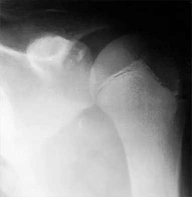
A 12-year-old pitcher has had a 2-month history of pain in his right dominant shoulder after throwing. He reports that the pain has gradually progressed to the point where he cannot throw without pain. He also notes that the pain now awakens him at night if he has been active. Anti-inflammatory drugs have failed to provide relief. Examination reveals no abnormalities except for some localized tenderness over the proximal humerus. Figures 32a and 32b show radiographs of both shoulders. What is the most likely diagnosis?


Explanation

32b The history, examination, and radiographs are pathognomonic for Little League shoulder, a stress syndrome of the proximal humeral physis caused by overuse. Complete fracture rarely occurs, and recovery usually occurs with rest. Night pain is always a serious concern and further work-up is needed if the patient does not respond to activity modification. Occult instability is not a real concern in this patient, although it should be included in the differential diagnosis. Albert MJ, Drvaric DM: Little League shoulder: Case report. Orthopedics 1990;13:779-781.

Question 10

Which of the following ligaments is the primary static restraint against inferior translation of the arm when the shoulder is in 0 degrees of abduction?


Explanation

The superior glenohumeral ligament (SGHL) and coracohumeral ligament serve as primary static restraints against inferior translation of the arm when the shoulder is in 0 degrees of abduction. Of these, the coracohumeral ligament has been shown to have a greater cross-sectional area, greater stiffness, and greater ultimate load than the SGHL. The inferior glenohumeral ligament plays a greater stabilizing role with increasing abduction of the arm. The coracoacromial ligament may help provide superior stability, especially when the rotator cuff is deficient. The coracoclavicular ligaments stabilize the acromioclavicular joint. Boardman ND, Debski RE, Warner JJ, et al: Tensile properties of the superior glenohumeral and coracohumeral ligaments. J Shoulder Elbow Surg 1996;5:249-254.

Question 11

A 16-year-old high school student undergoes a routine preparticipation physical examination at the beginning of the school year. Examination reveals marked laxity of both shoulders but only mild generalized laxity in other joints. The load and shift test allows for anterior humeral translation to the glenoid rim and posterior humeral translation beyond the glenoid rim. The sulcus sign is present. What is the next most appropriate step in management?


Explanation

This patient has shoulder laxity without apprehension. Because there is a wide range of normal laxity in asymptomatic shoulders, the physician should inform the student of these findings, recommend shoulder strengthening exercises, and allow unrestricted sports participation unless symptoms develop. Harryman DT, Sidles JA, Harris SL, Matsen FA III: Laxity of the normal glenohumeral joint: A quantitative in vivo assessment. J Shoulder Elbow Surg 1992;1:66-76. Hawkins RJ, Bokor RJ: Clinical evaluation of shoulder problems, in Rockwood CA Jr, Matsen FA III (eds): The Shoulder. Philadelphia, PA, WB Saunders, 1998, vol 1, p 186. McFarland EG, Campbell G, McDowell J: Posterior shoulder laxity in asymptomatic athletes. Am J Sports Med 1996;24:468-471.

Question 12

A 21-year-old professional baseball player has had painful catching and stiffness in his dominant right elbow for the past year. Examination reveals a flexion contracture of 2 degrees and mild pain with full elbow flexion. Radiographs are shown in Figures 33a and 33b. The most effective management should consist of


Explanation

33b The radiographs show osteochondritis dissecans of the capitellum and a loose body in the anterior compartment. Arthroscopic removal is indicated because symptoms referable to the loose body are present. Baumgarten TE: Osteochondritis dissecans of the capitellum. Sports Med Arthroscopy Rev 1995;3:219-223.

Question 13

A 42-year-old patient has had painful inferior subluxation of the glenohumeral joint following a recent cerebrovascular accident (CVA). Figure 34 shows the AP radiograph of the shoulder. Management should consist of


Explanation

Following a CVA and with the resumption of upright posture, downward subluxation of the glenohumeral joint may occur. Although usually painless, some patients may report pain secondary to stretching of the brachial plexus. This is the result of flaccid paralysis of the deltoid muscle, and it will persist until some motor tone or spasticity returns to the shoulder girdle musculature. Early sling support and range-of-motion exercises to prevent contracture will provide the best relief. Surgical procedures are not indicated. Braun RM, Botte MJ: Treatment of shoulder deformity in acquired spasticity. Clin Orthop 1999;368:54-65.

Question 14

A 50-year-old man who underwent an arthroscopic rotator cuff repair 5 days ago now returns for an early postoperative follow-up because of increasing pain in his shoulder. He reports increasing malaise and has a low-grade fever. Examination reveals no redness or swelling, but he has scant serous drainage from the posterior portal. An emergent Gram stain is positive for gram-positive cocci. The next most appropriate step in management should consist of


Explanation

An infection of the shoulder is considered a surgical emergency unless there are medical reasons that a patient cannot be taken to the operating room. If cultures of wound drainage are in question, then an aspiration should be done emergently, not several days later. The hallmark of infection in any major joint is increasing pain out of proportion to what is expected. Drainage occurring 1 to 2 days after an arthroscopic procedure is not normal, and it should be aggressively treated. Delay in diagnosis can result in sepsis and on a delayed basis, postinfectious arthritis. Both the glenohumeral joint and the subacromial space require debridement and irrigation, followed by antibiotics after both areas are cultured. Mansat P, Cofield RH, Kersten TE, Rowland CM: Complications of rotator cuff repair. Orthop Clin North Am 1997;28:205-213. Settecerri JJ, Pitner MA, Rock MG, Hanssen AD, Cofield RH: Infection after rotator cuff repair. J Shoulder Elbow Surg 1999;8:1-5. Ward WG, Eckardt JJ: Subacromial/subdeltoid bursa abscesses: An overlooked diagnosis. Clin Orthop 1993;288:189-194.

Question 15

A 42-year-old man who is right-hand dominant injured his right shoulder when he fell from a ladder onto his outstretched arm 1 hour ago. Radiographs reveal a two-part greater tuberosity anterior fracture-dislocation. Initial management should consist of


Explanation

Greater tuberosity anterior fractures associated with anterior glenohumeral dislocations respond very well to closed methods in the majority of patients. Closed reduction of the glenohumeral joint often anatomically reduces the greater tuberosity into its cancellous bed, without the need for open fixation or cuff repair. Once closed reduction of the joint is performed, tuberosity displacement and joint articulation should be evaluated radiographically with AP and scapular lateral views as well as an axillary view. The axillary view will not only definitively show the joint articulation but also demonstrate posterior displacement of the greater tuberosity missed on the AP and lateral views. If no or minimal (5 mm) displacement is found, then nonsurgical management consisting of a sling and gentle passive range-of-motion exercises can be instituted. Beaty JH (ed): Orthopaedic Knowledge Update 6. Rosemont, IL, American Academy of Orthopaedic Surgeons, 1999, pp 271-286.

Question 16

A 19-year-old man who plays college volleyball undergoes a routine preparticipation physical examination. Figure 35 shows a posterior view of his dominant shoulder. An electromyogram shows that this is a chronic injury, and an MRI scan shows no abnormalities. The best course of action should be


Explanation

Isolated palsy of the infraspinatus portion of the suprascapular nerve is common in volleyball players and is seen frequently in the throwing arm of baseball players. The exact cause is not known, but it may be the result of either tethering or traction on the nerve at the spinoglenoid notch. Synovial cysts in the spinoglenoid notch also can be a cause, but the patient's negative MRI findings rule out that entity. Because many isolated nerve palsies of the infraspinatus branch are asymptomatic, initial management should always be nonsurgical. Surprisingly, many athletes with this injury can participate fully in sports. Surgical treatment with decompression at the notch is unpredictable and generally is indicated only if nonsurgical management fails. Ferretti A, Cerullo G, Russo G: Suprascapular neuropathy in volleyball players. J Bone Joint Surg Am 1987;69:260-263.

Question 17

A 59-year-old construction worker who is right-hand dominant has had right shoulder pain for the past 9 months with no history of injury. Nonsurgical management consisting of two cortisone injections, physical therapy for 3 months, and nonsteroidal anti-inflammatory drugs has failed to provide lasting relief. Examination reveals tenderness over the acromioclavicular (AC) joint and over the subacromial bursa. He has positive Neer and Hawkins impingement signs and AC joint pain with adduction of the shoulder. Radiographs are shown in Figures 36a and 36b. An MRI scan reveals an intact rotator cuff. Management should now consist of


Explanation

36b Because the patient has clinical and radiographic signs of AC arthritis and subacromial impingement, the treatment of choice is anterior acromioplasty and distal clavicle excision. Arthroscopic acromioplasty alone would not address the AC arthritis. The rotator cuff is intact; therefore, rotator cuff repair is not indicated. An open Mumford procedure would address the AC arthritis only and not the impingement symptoms. Immobilization might lead to stiffness of the shoulder and is not recommended for treating impingement.

Question 18

What three structures are considered the primary constraints necessary for elbow stability?


Explanation

The three primary constraints necessary for elbow stability in all directions are the ulnar part of the lateral collateral ligament (also called the lateral ulnar collateral ligament), the anterior band of the medial collateral ligament, and the coronoid. The radial head and capsule are secondary constraints to elbow instability. Kasser JR (ed): Orthopaedic Knowledge Update 5. Rosemont, IL, American Academy of Orthopaedic Surgeons, 1996, pp 283-294.

Question 19

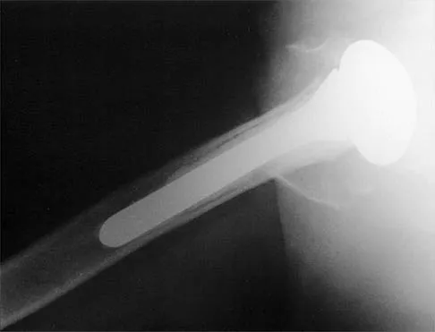
A 68-year-old woman has been progressing slowly after undergoing humeral head replacement for a four-part fracture 3 months ago. She has not regained active elevation, she feels an audible clunk on attempting elevation, and she reports pain and weakness. She used a sling for 2 weeks in the immediate postoperative period. Radiographs are shown in Figure 37a through 37c. Management should consist of


Explanation

37b 37c Immediate repair of the tuberosity and rotator cuff is recommended on identifying the avulsion or nonunion. Revising the humeral component to increase tension and length will overtighten the cuff and increase the chance of tuberosity pull-off. The glenoid is uninvolved and should not be replaced. Attempts to strengthen the rotator cuff will be unsuccessful because the insertions are no longer attached to the humerus when the tuberosities avulse. Brown TD, Bigliani LU: Complications with humeral head replacement. Orthop Clin North Am 2000;31:77-90.

Question 20

What is the most important feature in choosing an outcome instrument to assess shoulder disorders?


Explanation

There has been a recent increase in the use of outcome instruments to document and measure effects of treatment of medical conditions, including shoulder disorders. The most important feature of an instrument is whether it actually measures what it purports to measure; this is defined as its validity. Leggin BG, Iannotti JP: Shoulder outcome measurement, in Iannotti JP, Williams GR (eds): Disorders of the Shoulder: Diagnosis and Management. Philadelphia, PA, Lippincott Williams and Wilkins, 1999, p 1027.

Question 21

Figure 38 shows the radiograph of a 16-year-old wrestler who injured his elbow when he was thrown to the mat by his opponent. To minimize additional trauma to the medial soft tissues, the elbow should be reduced in


Explanation

The elbow dislocates by a three-dimensional movement of supination and valgus during flexion. Additional trauma during reduction is minimized by recreating the deformity and reducing the elbow in supination. The actual maneuver includes full supination (actually hypersupination) of the elbow in a valgus position. This is followed by pushing the olecranon distally in line with the long axis of the ulna while swinging the elbow into varus, and then relaxing the supination torque. Postreduction stability is enhanced in pronation, except when the soft-tissue disruption is extensive. O'Driscoll SW: Elbow dislocations, in Morrey BF (ed): The Elbow and Its Disorders, ed 2. Philadelphia, PA, WB Saunders, 1993, p 414.

Question 22

In patients older than age 40 years who sustain a first-time anterior dislocation of the shoulder, prolonged morbidity is most commonly associated with


Explanation

In the study done by Pevny and associates, 35% of patients older than age 40 years sustained rotator cuff tears and 8% had axillary nerve palsies. All of the patients with axillary nerve palsy also had rotator cuff tears. Imaging of the rotator cuff is indicated in this age group. The incidence of recurrent instability in patients older than age 40 years is 10% to 15%. Pevny T, Hunter RE, Freeman JR: Primary traumatic anterior shoulder dislocation in patients 40 years of age and older. Arthroscopy 1998;14:289-294. Sonnabend DH: Treatment of primary anterior shoulder dislocation in patients older than 40 years of age: Conservative versus operative. Clin Orthop 1994;304:74-77.

Question 23

Figure 39 shows the AP radiograph of a 62-year-old man with degenerative osteoarthritis secondary to trauma. History reveals that he underwent total elbow arthroplasty 3 years ago. He continues to report instability and constant pain. A complete work-up, including aspiration and cultures, is negative. Treatment should consist of removal of the components and


Explanation

An unconstrained prosthesis dislocation is a disconcerting problem that is not easily resolved; however, revision to a semiconstrained prosthesis would best achieve both pain relief and stability. Removal of the components and distraction arthroplasty or conversion to a resection arthroplasty are options, but the results would be unpredictable with regards to pain relief, postoperative motion, or elbow stability. Arthrodesis is poorly tolerated. With revision to another unconstrained prosthesis, there is the risk of continued redislocation because of chronic ligamentous insufficiency. Linscheid RL: Resurfacing elbow replacement arthroplasty: Rationale, technique and results, in Morrey BF (ed): The Elbow and Its Disorders, ed 3. Philadelphia, PA, WB Saunders, 2000, pp 602-610.

Question 24

A 67-year-old woman undergoes a revision total shoulder arthroplasty for replacement of a loose glenoid component. Examination in the recovery room reveals absent voluntary deltoid and triceps contraction, weakness of wrist and thumb extension, and absent sensation in the palmar aspect of all fingertips and the radial forearm. The next most appropriate step in management should consist of


Explanation

Neurologic injury after shoulder replacement is relatively uncommon, occurring in 4% of shoulders in one large series. The importance of identifying and protecting the musculocutaneous and axillary nerves cannot be overemphasized; it is especially critical during revision arthroplasty when the normal anatomic relationships have been distorted. The long deltopectoral approach leaving the deltoid attached to the clavicle was found to be significant in the development of postoperative neurologic complications. A correlation was found between surgical time and postoperative neurologic complications, with long surgical times being associated with more neurologic complications. The presumed mechanism of injury is traction on the plexus that occurs during the surgery. A neurologic injury after total shoulder arthroplasty usually does not interfere with the long-term outcome of the arthroplasty itself; it is best managed by protective measures with passive range of motion of the involved extremity. Wirth MA, Rockwood CA Jr: Complications of shoulder arthroplasty. Clin Orthop 1994;307:47-69.

Question 25

Figure 40 shows the radiograph of a 16-year-old wrestler who injured his elbow when he was thrown to the mat by his opponent. Closed reduction is readily accomplished, and the elbow seems stable. Management should now consist of application of a splint for


Explanation

Flexion contractures are the most common complication of elbow dislocations. About 15% of patients lose more than 30 degrees of flexion. The risk of contracture is proportional to the duration of immobilization. Elbows should be moved within the first few days after reduction. The splinting is for comfort and protection only while the pain subsides. Mehlhoff TL, Noble PC, Bennett JB, Tullos HS: Simple dislocation of the elbow in the adult: Results after closed treatment. J Bone Joint Surg Am 1988;70:244-249. Linscheid RL, O'Driscoll SW: Elbow dislocations, in Morrey BF (ed): The Elbow and Its Disorders, ed 2. Philadelphia, PA, WB Saunders, 1993, pp 441-452. O'Driscoll SW, Jupiter JB, King GJ, Hotchkiss RN, Morrey BF: The unstable elbow. Instr Course Lect 2001;50:89-102.

Question 26

A 72-year-old female undergoes reverse total shoulder arthroplasty (RTSA) for severe rotator cuff tear arthropathy. By what primary biomechanical mechanism does this prosthesis restore active shoulder elevation?


Explanation

RTSA shifts the center of rotation medially and inferiorly. This mechanism increases the deltoid moment arm and tension, allowing the deltoid to effectively recruit more muscle fibers to elevate the arm in the absence of a functioning rotator cuff.

Question 27

During a Latarjet procedure for recurrent anterior shoulder instability, the coracoid process is osteotomized and transferred. To avoid iatrogenic injury to the musculocutaneous nerve during the deep dissection, the surgeon must remember that the nerve typically enters the conjoint tendon at what distance distal to the coracoid tip?


Explanation

The musculocutaneous nerve typically enters the medial aspect of the conjoint tendon between 3 and 8 cm (average 5 cm) distal to the tip of the coracoid. Retraction and dissection in this zone must be performed with caution.

Question 28

A 45-year-old manual laborer presents with a chronic, massive, irreparable tear of the posterosuperior rotator cuff. He demonstrates profound weakness in external rotation (horn blower's sign) but has an intact subscapularis. Which tendon transfer is biomechanically most appropriate to restore external rotation and elevation?


Explanation

The lower trapezius transfer is biomechanically superior for restoring external rotation because its line of pull closely replicates that of the native infraspinatus. Latissimus dorsi is an alternative but its native function is internal rotation, requiring retraining.

Question 29

A 28-year-old professional volleyball player presents with insidious onset of posterior shoulder pain and weakness. MRI reveals isolated atrophy of the teres minor. The entrapped nerve is located in a quadrilateral space bounded superiorly by which anatomic structure?


Explanation

Quadrilateral space syndrome involves compression of the axillary nerve and posterior circumflex humeral artery. The space is bounded superiorly by the teres minor, inferiorly by the teres major, medially by the long head of the triceps, and laterally by the humeral shaft.

Question 30

A 32-year-old weightlifter presents with vague posterior shoulder pain and profound weakness in external rotation, with preserved abduction strength. EMG confirms an isolated nerve entrapment at the spinoglenoid notch. Which finding is expected on MRI?


Explanation

The suprascapular nerve innervates the supraspinatus before passing through the spinoglenoid notch to innervate the infraspinatus. Compression at the spinoglenoid notch isolatedly affects the infraspinatus, presenting as external rotation weakness with preserved abduction.

Question 31

A 65-year-old man with primary glenohumeral osteoarthritis is planned for an anatomic total shoulder arthroplasty. CT imaging demonstrates a biconcave glenoid with severe posterior cartilage wear and posterior subluxation of the humeral head. This represents which Walch classification?


Explanation

Walch Type B2 glenoids are characterized by a biconcave surface with asymmetric posterior cartilage wear and posterior subluxation of the humeral head. This morphology carries a high risk of early glenoid loosening if not properly corrected during arthroplasty.

Question 32

A 68-year-old woman sustains a displaced 4-part proximal humerus fracture. According to Hertel's criteria, which of the following radiographic findings is the strongest predictor of ensuing humeral head ischemia?


Explanation

Hertel's criteria for predicting ischemia of the humeral head include a calcar length of less than 8 mm, disruption of the medial hinge, and an anatomic neck fracture pattern. These factors indicate severe disruption of the vascular supply to the articular segment.

Question 33

A 76-year-old female presents with acute lateral shoulder pain 4 months after undergoing a reverse total shoulder arthroplasty. Radiographs reveal an acromial base stress fracture (Levy Type II). Which surgical factor most significantly increases the risk of this complication?


Explanation

Acromial stress fractures after RTSA are strongly associated with overtensioning of the deltoid, which places excessive load on the acromion. Excessive lateralization or distalization can contribute to this detrimental overtensioning.

Question 34

A 29-year-old competitive weightlifter feels a pop in his anterior chest wall while performing a heavy bench press. Examination reveals loss of the anterior axillary fold and weakness in adduction. Where does the most common pattern of this muscular rupture occur?


Explanation

Pectoralis major ruptures typically occur in weightlifters (e.g., bench press) and most commonly involve the avulsion of the sternal head from its humeral insertion. Surgical repair is indicated for young, active patients to restore full strength.

Question 35

A 24-year-old carpenter presents with a dull ache in his right shoulder and inability to fully elevate his arm. Examination reveals medial winging of the scapula that worsens when he pushes against a wall. Injury to which nerve is the most likely cause?


Explanation

Medial winging of the scapula is caused by paralysis of the serratus anterior muscle, which is innervated by the long thoracic nerve. Lateral winging is typically associated with spinal accessory nerve injury (trapezius paralysis).

Question 36

Which design parameter of a reverse total shoulder arthroplasty most effectively decreases the risk of scapular notching?


Explanation

Inferior placement and overhang of the glenosphere relative to the inferior glenoid rim effectively minimizes scapular notching by avoiding impingement of the humeral cup on the scapular neck during adduction.

Question 37

A 65-year-old man presents with insidious onset of shoulder pain 18 months following an anatomic total shoulder arthroplasty. Inflammatory markers are normal. Aspirate cultures grow Cutibacterium acnes at 10 days. Which of the following best describes this organism?


Explanation

Cutibacterium (formerly Propionibacterium) acnes is a Gram-positive, anaerobic (to aerotolerant) bacillus that is a common cause of indolent prosthetic joint infections in the shoulder.

Question 38

A 28-year-old weightlifter presents with isolated wasting of the infraspinatus muscle and weakness in external rotation. An MRI reveals a paralabral cyst. Where is the most likely location of the cyst and the associated nerve compression?


Explanation

Compression of the suprascapular nerve at the spinoglenoid notch typically causes isolated infraspinatus weakness and atrophy, as the motor branches to the supraspinatus have already departed the nerve proximal to this level.

Question 39

According to Hertel's criteria, which of the following is the strongest predictor of ischemia and subsequent avascular necrosis following a proximal humerus fracture?


Explanation

Hertel identified a short calcar length (metaphyseal extension < 8 mm) and disruption of the medial hinge as the strongest predictors of ischemia and avascular necrosis of the humeral head.

Question 40

A 32-year-old bodybuilder feels a pop in his anterior axilla while bench pressing. Examination reveals loss of the anterior axillary fold and weakness in internal rotation and adduction. Which portion of the pectoralis major is most commonly ruptured and what is its anatomic insertion?


Explanation

The sternal head of the pectoralis major most commonly ruptures. Due to the 180-degree twist of the tendon, the sternal head inserts deep and superior to the clavicular head on the lateral lip of the bicipital groove.

Question 41

A 55-year-old laborer with an irreparable posterosuperior rotator cuff tear and an intact subscapularis undergoes a lower trapezius tendon transfer. The transferred tendon is typically augmented with an allograft and inserted onto which of the following structures?


Explanation

The lower trapezius transfer for an irreparable posterosuperior rotator cuff tear is routed deep to the deltoid and inserted onto the greater tuberosity to restore external rotation and active elevation.

Question 42

In performing a Latarjet procedure for recurrent anterior shoulder instability, the coracoid process is transferred to the anterior glenoid. The dynamic "sling effect" of this procedure is primarily provided by which of the following structures?


Explanation

The conjoint tendon (short head of the biceps and coracobrachialis) provides a dynamic sling effect across the anterior inferior capsule when the arm is abducted and externally rotated, greatly contributing to joint stability.

Question 43

A 35-year-old overhead athlete complains of posterior shoulder pain and numbness over the lateral deltoid. MRI shows isolated teres minor atrophy. Which vascular structure is most likely compressed along with the involved nerve?


Explanation

Quadrilateral space syndrome involves compression of the axillary nerve and the posterior humeral circumflex artery, leading to lateral shoulder numbness and teres minor atrophy.

Question 44

A 22-year-old collegiate baseball pitcher has a Type II SLAP tear confirmed on MRI. Following failure of conservative management, what is the generally recommended initial surgical approach to return him to high-level pitching?


Explanation

In young, high-demand overhead throwing athletes with a Type II SLAP tear, arthroscopic SLAP repair is generally preferred initially to restore the superior labral anchor and optimal kinematics, despite challenging return-to-play rates.

Question 45

A 24-year-old pitcher is diagnosed with Glenohumeral Internal Rotation Deficit (GIRD). He has 130 degrees of external rotation and 30 degrees of internal rotation on the throwing side. Which anatomic change is primarily responsible for this physical exam finding?


Explanation

GIRD is primarily caused by contracture and thickening of the posterior band of the inferior glenohumeral ligament, which limits internal rotation and shifts the humeral head posterosuperiorly in the cocking phase.

Question 46

When converting a patient from an anatomic total shoulder arthroplasty to a reverse total shoulder arthroplasty, what happens to the center of rotation of the glenohumeral joint?


Explanation

A reverse total shoulder arthroplasty moves the center of rotation inferiorly and medially. This increases the deltoid moment arm and tension, allowing the deltoid to effectively recruit more fibers for shoulder elevation.

Question 47

During shoulder arthroscopy, the "comma sign" is a useful anatomic landmark. It is indicative of a tear and retraction of which of the following structures?


Explanation

The comma sign represents a portion of the superior glenohumeral ligament and the coracohumeral ligament that tears and retracts with the superior edge of a torn subscapularis, serving as an important marker for its identification.

Question 48

During a deltopectoral approach to the shoulder, how far distal to the lateral acromion does the axillary nerve typically traverse the deep surface of the deltoid muscle?


Explanation

The axillary nerve generally crosses the deep surface of the deltoid approximately 5 to 7 cm distal to the lateral edge of the acromion. Care must be taken to avoid retractors or dissection that risks injury in this zone.

Question 49

A 30-year-old woman presents with medial scapular winging that is accentuated when she pushes against a wall. Which nerve and corresponding muscle are most likely affected?


Explanation

Medial scapular winging (where the medial border becomes prominent) is caused by paralysis of the serratus anterior, which is innervated by the long thoracic nerve. Trapezius palsy typically causes lateral winging.

Question 50

A 19-year-old football player presents with a closed posterior sternoclavicular joint dislocation following a direct blow. He complains of mild difficulty swallowing. What is the most appropriate next step in management?


Explanation

Posterior sternoclavicular dislocations can compress critical mediastinal structures. A CT scan evaluates the great vessels, and reduction should be performed in the OR with cardiothoracic surgery available due to the risk of catastrophic vascular injury.

Question 51

In a severe acromioclavicular (AC) joint separation, the coracoclavicular (CC) ligaments are disrupted. Which of the following accurately describes the relative anatomy and function of the intact CC ligaments?


Explanation

The conoid ligament is situated medial to the trapezoid ligament and provides the primary restraint against superior translation of the clavicle. The trapezoid provides primary restraint to axial compression.

Question 52

Which of the following systemic conditions has the strongest and most well-documented association with the development of adhesive capsulitis of the shoulder?


Explanation

Diabetes mellitus has a very strong association with adhesive capsulitis. Patients with diabetes often experience a more severe and protracted clinical course that can be resistant to standard conservative management.

Question 53

A 68-year-old man with primary osteoarthritis of the shoulder has a B2 glenoid identified on preoperative CT scan. According to the Walch classification, a B2 glenoid is characterized by:


Explanation

The Walch B2 glenoid in primary glenohumeral osteoarthritis is characterized by posterior subluxation of the humeral head and asymmetric posterior glenoid wear, creating a classic biconcave shape with increased retroversion.

Question 54

A 25-year-old male weightlifter complains of superior shoulder pain localized to the AC joint during bench press. Radiographs reveal subchondral cysts and osteopenia of the distal clavicle. If 6 months of conservative management fails, what is the most appropriate surgical intervention?


Explanation

Distal clavicle osteolysis commonly affects weightlifters. If nonoperative treatment (rest, NSAIDs, injections) fails, arthroscopic or open distal clavicle excision provides reliable symptomatic relief.

Question 55

Which physical examination test is considered most specific for evaluating the integrity of the superior portion of the subscapularis tendon?


Explanation

The Bear-hug test isolates and heavily evaluates the superior portion of the subscapularis tendon. The Belly-press test evaluates the mid/upper portion, while the Lift-off test relies primarily on the inferior portion of the subscapularis.

Question 56

A 72-year-old woman undergoes a reverse total shoulder arthroplasty (rTSA) for cuff tear arthropathy. Postoperatively, she achieves 150 degrees of active forward elevation but complains of profound weakness and inability to actively externally rotate her arm when at her side. Which of the following concurrent procedures would have best addressed this specific postoperative deficit?


Explanation

Loss of active external rotation with the arm at the side is due to an absent or non-functional teres minor (often seen in massive posterosuperior cuff tears). A latissimus dorsi or lower trapezius transfer to the greater tuberosity is indicated to restore active external rotation.

Question 57

A 21-year-old collegiate rugby player sustains a third anterior shoulder dislocation. An MRI reveals a Bankart lesion and 25% anterior glenoid bone loss. If an open Latarjet procedure is performed, the coracoid graft acts primarily through a "sling effect" to stabilize the joint in abduction and external rotation. This sling effect is created by the conjoint tendon interacting with which structure?


Explanation

The dynamic 'sling effect' of the Latarjet procedure is created by the conjoint tendon (short head of biceps and coracobrachialis) tensioning across the lower half of the subscapularis muscle when the arm is abducted and externally rotated.

Question 58

An 82-year-old female presents with a severely comminuted 4-part proximal humerus fracture after a fall. Imaging reveals a disrupted medial hinge, less than 2 mm of metaphyseal extension attached to the articular segment, and severe osteoporosis. A reverse total shoulder arthroplasty (rTSA) is chosen over open reduction internal fixation (ORIF). What biomechanical principle explains the functional success of rTSA in this setting?


Explanation

Reverse TSA medializes and distalizes the joint's center of rotation. This increases the deltoid muscle's moment arm and resting tension, allowing it to substitute for the deficient rotator cuff in elevating the arm.

Question 59

A 28-year-old professional volleyball player presents with isolated weakness of the right shoulder, specifically noting fatigue during the cocking phase of serving. Physical examination reveals isolated atrophy of the infraspinatus fossa with normal supraspinatus bulk and strength. An MRI is most likely to demonstrate an abnormality in which of the following locations?


Explanation

Isolated infraspinatus weakness and atrophy indicate entrapment of the suprascapular nerve at the spinoglenoid notch. This is often secondary to a paralabral cyst associated with a posterior labral tear in overhead athletes.

Question 60


During an anatomic coracoclavicular (CC) ligament reconstruction for a chronic Type V acromioclavicular joint separation, the surgeon drills tunnels in the clavicle to recreate the conoid and trapezoid ligaments. To accurately replicate native anatomy, the conoid tunnel should be placed approximately:


Explanation

The conoid ligament inserts approximately 45 mm medial to the distal end of the clavicle on its posterior aspect. The trapezoid ligament inserts more laterally (approximately 25 mm medial) and anteriorly.

Question 61

A 23-year-old baseball pitcher presents with posterior shoulder pain during the late cocking phase of throwing. He has a positive apprehension sign that is relieved by a posterior-directed force on the proximal humerus, but he complains of deep posterior pain rather than instability. Arthroscopic evaluation is most likely to show which of the following?


Explanation

Internal impingement typically occurs in overhead throwers during abduction and external rotation. It is characterized by the undersurface (articular side) of the posterior rotator cuff abutting the posterosuperior glenoid labrum, causing fraying of both structures.

Question 62

A 32-year-old bodybuilder feels a "pop" in his axilla while performing a heavy bench press. Examination reveals an asymmetric chest wall and weakness in internal rotation and adduction. MRI confirms a complete pectoralis major rupture. Which of the following statements regarding the relevant anatomy and injury pattern is correct?


Explanation

The sternal head of the pectoralis major crosses beneath the clavicular head to insert proximally and deep on the lateral lip of the bicipital groove. This deep, lower portion is under maximum stretch during a bench press and typically tears first.

Question 63

A 45-year-old woman undergoes a posterior triangle lymph node biopsy. Three weeks later, she complains of a dull ache in her shoulder and an inability to elevate her arm above 90 degrees. Examination shows lateral displacement and downward rotation of the scapula. Which nerve was most likely injured, and what is the primary affected muscle?


Explanation

Injury to the spinal accessory nerve during posterior cervical triangle surgery denervates the trapezius. This results in lateral winging and downward rotation of the scapula, distinguishing it from the medial winging seen in long thoracic nerve palsy.

Question 64

A 55-year-old woman with type 1 diabetes presents with insidious onset of progressive shoulder stiffness and pain, currently in the "freezing" stage of idiopathic adhesive capsulitis. Histologic evaluation of her shoulder capsule would most likely show an abundance of which cell type, and contracture of which primary structure in the rotator interval?


Explanation

Adhesive capsulitis is characterized by a fibroblastic proliferation and multilocular cytokine expression (e.g., TGF-beta). The pathognomonic macroscopic finding is a thick, contracted coracohumeral ligament within the rotator interval.

Question 65


A 75-year-old man is 4 years status post a Grammont-style reverse total shoulder arthroplasty. Radiographs show Grade 3 scapular notching. Which intraoperative technical error most significantly increases the risk of this specific complication?


Explanation

Scapular notching is caused by mechanical impingement of the humeral tray against the inferior scapular neck. Superior baseplate positioning fails to provide adequate inferior overhang, significantly increasing the risk of notching.

Question 66

A 64-year-old right-hand-dominant male presents with persistent right shoulder pain and weakness following a fall onto an outstretched hand 3 months ago. Physical examination demonstrates increased passive external rotation compared to the contralateral shoulder, a positive lift-off test, and a positive belly-press test. Which of the following tendons is predominantly affected?


Explanation

The subscapularis is the primary internal rotator of the shoulder. A tear results in increased passive external rotation (due to loss of anterior restraint) and positive lift-off and belly-press tests.

Question 67

An orthopaedic surgeon is planning an anatomic total shoulder arthroplasty for a 68-year-old man with primary osteoarthritis. Preoperative axillary CT imaging demonstrates a biconcave glenoid with severe posterior cartilaginous wear and 22 degrees of retroversion. According to the Walch classification, what type of glenoid morphology is this?


Explanation

The Walch B2 glenoid is defined by an asymmetric, biconcave morphology with posterior wear and increased retroversion. It is highly associated with posterior subluxation of the humeral head in primary osteoarthritis.

Question 68

A 36-year-old man presents to the emergency department after suffering a generalized tonic-clonic seizure. He complains of severe anterior shoulder pain. On examination, his arm is fixed in internal rotation, and he has zero degrees of active or passive external rotation. The AP shoulder radiograph shows a "lightbulb sign." What is the most appropriate initial management?


Explanation

The patient has a posterior shoulder dislocation (fixed internal rotation, 'lightbulb sign' from internal rotation on AP view). Initial management is closed reduction, typically utilizing axial traction with gentle anterior translation and external rotation.

Question 69

A 14-year-old elite Little League pitcher presents with progressive, insidious-onset throwing arm shoulder pain. Radiographs demonstrate widening and irregularity of the proximal humeral physis compared to the contralateral side. What is the standard of care for this condition?


Explanation

Little League shoulder is an epiphysiolysis of the proximal humerus caused by repetitive torsional stress. It is managed nonoperatively with complete cessation of throwing (typically 3 months) until symptoms resolve and radiographs normalize.

Question 70

A 26-year-old man is undergoing arthroscopic stabilization for recurrent anterior shoulder instability. Diagnostic arthroscopy reveals a Bankart lesion and a large, "engaging" Hill-Sachs lesion, with minimal glenoid bone loss (<10%). The surgeon elects to perform a Bankart repair and a Remplissage procedure. Which of the following is the most likely long-term kinematic consequence of adding the Remplissage?


Explanation

The Remplissage procedure involves tenodesis of the infraspinatus and posterior capsule into the Hill-Sachs defect to prevent it from engaging the anterior glenoid. This non-anatomic tethering predictably decreases external rotation, particularly in abduction.

Question 71

A 48-year-old male presents with acute, unprovoked, excruciating right shoulder pain that awakened him from sleep. The severe pain lasts for nearly two weeks and requires narcotic analgesia. As the pain begins to subside, he notes profound weakness in his deltoid and periscapular muscles. An MRI of the shoulder and cervical spine is unremarkable. EMG at 4 weeks shows denervation of the suprascapular and axillary nerves. What is the most likely diagnosis?


Explanation

Parsonage-Turner syndrome (acute brachial neuritis) classically presents with a brief, severe prodrome of acute, unprovoked shoulder pain, followed by patchy weakness and amyotrophy (often C5-C6 nerve distributions) as the pain resolves.

Question 72

A 72-year-old woman undergoes a reverse total shoulder arthroplasty for severe rotator cuff tear arthropathy. To minimize the risk of scapular notching postoperatively, which of the following baseplate and glenosphere configurations is most appropriate?


Explanation

Scapular notching in reverse total shoulder arthroplasty can be minimized by placing the glenoid baseplate inferiorly and using an inferiorly eccentric or overhanging glenosphere. Lateralization and inferior tilt of the baseplate also independently reduce the incidence of inferior scapular impingement.

Question 73

A 35-year-old man presents with persistent shoulder pain following an electric shock injury.

An axillary radiograph reveals a locked posterior shoulder dislocation with an anterior humeral head defect involving 30% of the articular surface. Which of the following is the most appropriate surgical management?


Explanation

Posterior shoulder dislocations frequently present with an anteromedial humeral head defect (reverse Hill-Sachs lesion). For defects involving 20% to 40% of the articular surface, transferring the subscapularis tendon or lesser tuberosity into the defect (McLaughlin or modified McLaughlin procedure) effectively prevents recurrent instability.

Question 74

A 55-year-old manual laborer has a massive, irreparable posterosuperior rotator cuff tear. He is being considered for a latissimus dorsi tendon transfer. Which of the following preoperative clinical or radiographic findings is considered an absolute contraindication for this procedure?


Explanation

An intact and functioning subscapularis is biomechanically essential for a successful latissimus dorsi transfer to maintain anterior-posterior force coupling in the shoulder. A deficient subscapularis or true pseudoparalysis with superior escape are absolute contraindications for this procedure.

Question 75

A 28-year-old professional volleyball player complains of vague posterior shoulder pain and isolated weakness in external rotation. Examination reveals profound wasting of the infraspinatus fossa, while the supraspinatus fossa is well-preserved.

MRI demonstrates a paralabral cyst. Which of the following anatomical locations is the most likely site of nerve compression?


Explanation

Isolated infraspinatus weakness strongly suggests compression of the suprascapular nerve at the spinoglenoid notch. This is frequently caused by a paralabral cyst extending from a posterior SLAP tear, whereas compression at the suprascapular notch would typically affect both the supraspinatus and infraspinatus.

Question 76

A 75-year-old woman sustains a displaced 4-part proximal humerus fracture.

If open reduction and internal fixation is attempted, which of the following initial radiographic findings is the most reliable predictor of subsequent humeral head avascular necrosis?


Explanation

According to Hertel's criteria, a posteromedial metaphyseal head extension (calcar length) of less than 8 mm and disruption of the medial periosteal hinge are the strongest predictors of ischemia. These indicators suggest severe compromise to the ascending branches of the anterior humeral circumflex artery and capsular vessels.

Question 77

During a Latarjet procedure for recurrent anterior shoulder instability, the coracoid process is osteotomized and transferred to the anterior glenoid. Which of the following nerves is at greatest risk of direct injury during the placement of deep medial retractors and mobilization of the coracoid graft?


Explanation

The musculocutaneous nerve typically enters the coracobrachialis muscle 3 to 8 cm distal to the coracoid process tip. It is highly susceptible to neurapraxia or transection from traction or deep retractor placement during the Latarjet procedure.

Question 78

A 68-year-old man presents with chronic right shoulder pain and an inability to actively elevate his arm past 45 degrees. Radiographs reveal superior migration of the humeral head and severe glenohumeral arthritis. Which of the following is the most appropriate definitive surgical management?


Explanation

Reverse total shoulder arthroplasty is indicated for rotator cuff tear arthropathy with pseudoparalysis. It alters the joint's center of rotation, allowing the deltoid to effectively elevate the arm.

Question 79

A 22-year-old collegiate baseball pitcher presents with posterior shoulder pain during the late cocking phase of throwing. Physical examination reveals a positive apprehension test that is relieved by a posterior-directed force on the proximal humerus. What is the most likely diagnosis?


Explanation

Internal impingement occurs in overhead athletes during late cocking, where the articular surface of the rotator cuff gets pinched between the greater tuberosity and the posterosuperior glenoid.

Question 80

A 45-year-old man presents with isolated weakness in shoulder external rotation. Atrophy is noted over the infraspinatus fossa, but supraspinatus strength is normal. An MRI is most likely to show a paralabral cyst in which of the following locations?


Explanation

A cyst at the spinoglenoid notch selectively compresses the suprascapular nerve branch to the infraspinatus, causing isolated external rotation weakness. Compression at the suprascapular notch would affect both the supraspinatus and infraspinatus.

Question 81

A 19-year-old contact athlete experiences recurrent anterior shoulder dislocations. Preoperative imaging reveals a glenoid bone loss of 28%. Which of the following procedures is most appropriate to prevent recurrence?


Explanation

The Latarjet procedure (coracoid transfer) is indicated for recurrent anterior instability with critical glenoid bone loss (>20-25%). Soft tissue stabilization alone carries an unacceptably high recurrence rate in this setting.

Question 82

Which of the following comorbidities is most strongly associated with both the development of adhesive capsulitis and a more refractory course to conservative treatment?


Explanation

Diabetes mellitus is the strongest risk factor for adhesive capsulitis, occurring in up to 20% of diabetic patients. These patients generally have a more protracted course and poorer response to conservative management.

Question 83

During the deltopectoral approach to the shoulder, the cephalic vein is typically identified in the internervous plane between the deltoid and pectoralis major. To minimize the risk of bleeding from its major tributaries, the cephalic vein should ideally be retracted in which direction?


Explanation

The cephalic vein is typically retracted laterally with the deltoid to preserve its major deltoid tributaries. Medial retraction risks avulsing these branches, leading to significant bleeding.

Question 84

A 50-year-old man presents with chronic shoulder pain. Physical examination demonstrates a positive belly-press test and an inability to maintain internal rotation against resistance when the hand is lifted off the lower back. These findings are most indicative of a tear involving which structure?


Explanation

The belly-press and lift-off tests isolate the subscapularis tendon. Weakness or inability to perform these tests strongly correlates with a subscapularis tear.

Question 85

A 32-year-old competitive weightlifter feels a "pop" in his anterior axilla while performing a heavy bench press. Examination shows loss of the normal anterior axillary contour. In an acute complete rupture, which anatomic portion is most commonly avulsed and where is its normal insertion?


Explanation

Pectoralis major ruptures most commonly involve the sternal head avulsing from its humeral insertion. The sternal head twists 180 degrees to insert deep and proximal to the clavicular head on the humerus.

Question 86

A 65-year-old woman with severe glenohumeral osteoarthritis undergoes an anatomic total shoulder arthroplasty. Ten years later, she presents with progressive pain and a loss of active elevation. What is the most common cause of late failure in anatomic total shoulder arthroplasty?


Explanation

Aseptic glenoid component loosening is the most common complication and cause of late failure following anatomic total shoulder arthroplasty. Eccentric wear and rocking horse forces contribute to this failure mode.

Question 87

A 28-year-old man sustains a severe elbow injury. Imaging confirms a terrible triad injury pattern.

What is the recommended sequence of surgical reconstruction to restore elbow stability?


Explanation

The standard surgical algorithm for a terrible triad injury builds stability from deep/anterior to superficial/lateral: coronoid fixation first, followed by radial head repair/replacement, and finally lateral collateral ligament (LCL) repair.

Question 88

A 70-year-old woman sustains a 3-part proximal humerus fracture treated with open reduction and internal fixation using a locking plate. Postoperatively, she develops severe shoulder pain and crepitus with range of motion. What is the most common complication associated with this specific fixation method?


Explanation

Primary or secondary screw cut-out into the articular space is the most frequent complication following locked plating of proximal humerus fractures, particularly in osteoporotic bone.

Question 89

A 40-year-old manual laborer complains of right shoulder ache and a prominent medial scapular border, especially when doing a wall push-up. An injury to which nerve is responsible for this physical finding?


Explanation

Medial winging of the scapula is caused by paralysis of the serratus anterior muscle, which is innervated by the long thoracic nerve. Lateral winging is associated with spinal accessory nerve (trapezius) palsy.

Question 90

A 72-year-old woman undergoes reverse total shoulder arthroplasty (rTSA) for massive, irreparable rotator cuff tear arthropathy. Postoperatively, what surgical technique modification regarding glenoid baseplate and glenosphere positioning has been biomechanically and clinically proven to minimize the risk of inferior scapular notching?


Explanation

Inferior positioning and inferior tilt of the glenosphere in reverse total shoulder arthroplasty limit mechanical impingement of the humeral component against the scapular neck during adduction. This technique effectively minimizes the risk of scapular notching, which is the most common radiographic complication of rTSA.

Question 91

A 65-year-old right-hand-dominant woman sustains the proximal humerus fracture shown in Figure 17 after a fall from a standing height.

According to Hertel's radiographic criteria, which of the following fracture characteristics is the most reliable predictor of humeral head ischemia and subsequent avascular necrosis?


Explanation

Hertel identified a metaphyseal head extension (calcar length attached to the articular segment) of less than 8 mm, disruption of the medial hinge, and a basicervical (anatomic neck) fracture pattern as the most reliable predictors of humeral head ischemia. A short calcar segment indicates severe disruption of the vascular supply from the ascending branch of the anterior humeral circumflex artery and intraosseous vessels.

Question 92

A 25-year-old elite volleyball player presents with vague posterior shoulder pain and decreased spiking power. Physical examination demonstrates isolated weakness in external rotation with the arm at the side. An MRI demonstrates a paralabral cyst isolated to the spinoglenoid notch, as seen in Figure 24.

Based on the anatomic location of this cyst, which of the following clinical findings is most likely PRESERVED?


Explanation

A cyst in the spinoglenoid notch compresses the suprascapular nerve distal to the branches innervating the supraspinatus muscle. Therefore, supraspinatus function (active initiation of shoulder abduction) is preserved, while the infraspinatus suffers isolated denervation leading to external rotation weakness and subsequent atrophy.

None

Clinic OS
Medically Verified Content by
Prof. Clinic OS
Consultant Orthopedic & Spine Surgeon
Chapter Index