ABOS Shoulder MCQs (Set 2): Rotator Cuff, Instability & Fractures | Board Review

Key Takeaway
This high-yield question set (Set 2) for the AAOS/ABOS exams covers critical shoulder pathologies. Topics include diagnosis, management, and surgical considerations for rotator cuff tears, various forms of shoulder instability, and classification of proximal humerus fractures. Prepare effectively for board certification.
ABOS Shoulder MCQs (Set 2): Rotator Cuff, Instability & Fractures | Board Review
Comprehensive 100-Question Exam
00:00
Start Quiz
Question 1
A 37-year-old electrician is diagnosed with a frozen shoulder after sustaining an electrical injury at work 2 weeks ago. Examination reveals that he cannot actively or passively externally rotate or abduct the arm. The glenohumeral joint and scapula move in a 1:1 ratio. Radiographs are shown in Figures 15a and 15b. The best course of action should be
Explanation
Question 2
An 80-year-old man has had increasing shoulder pain for the past 4 months. He reports that it began with soreness and stiffness after chopping some wood. A coronal MRI scan is shown in Figure 16. Initial management should consist of
Explanation
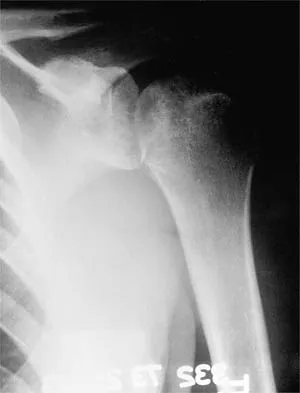
Question 3
Figure 17 shows the radiograph of a 25-year-old professional football player who has superior shoulder pain that prevents him from sports participation. History reveals that he sustained a shoulder injury that was treated with closed reduction and temporary pinning 3 years ago. The best course of action should be
Explanation
Question 4
A 54-year-old man undergoes total shoulder arthroplasty for osteoarthritis. Despite compliance with an early passive range-of-motion exercise program, he does not regain more than 90 degrees of elevation, 10 degrees of external rotation, and has internal rotation to the fifth lumbar vertebra. At 6 months, his motion fails to improve. Radiographs are shown in Figures 18a and 18b. What is the best course of action?
Explanation
Question 5
A 47-year-old patient has had persistent pain and weakness after undergoing a reamed intramedullary nailing for a midshaft humerus fracture 8 months ago. There is no evidence of infection. Radiographs are shown in Figures 19a and 19b. Management should consist of
Explanation
Question 6
An 18-year-old man sustained closed humeral shaft and forearm fractures of his dominant arm in a motor vehicle accident. Neurovascular examination is intact, and his condition is stable. The best course of action for management of the injuries should be
Explanation
Question 7
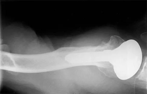
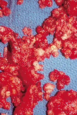
A 32-year-old woman has had pain and a visibly growing mass in the shoulder for 3 years but denies any history of trauma. Examination reveals a swollen, boggy shoulder mass. The AP radiograph and MRI scan are shown in Figures 20a and 20b. Figures 20c through 20e show a portion of the excised mass and the photomicrographs of the biopsy specimen. What is the most likely diagnosis?
Explanation
Question 8
What is the most important factor regarding the risk of recurrent instability in a patient with an acute anterior dislocation of the shoulder?
Explanation
Question 9
A 25-year-old man injured his dominant shoulder after falling on his outstretched arm 4 months ago. Examination reveals that he cannot lift his arm above 90 degrees, and he has pronounced medial scapular winging. Management should consist of
Explanation
Question 10
Treatment of adhesive capsulitis has a high failure rate when the underlying cause is
Explanation
Question 11
Figure 21 shows the AP radiograph of a 41-year-old patient who sustained a closed bicolumnar fracture of the distal humerus that resulted in a painful nonunion. What is the best initial construct for rigid stabilization of this fracture pattern?
Explanation
Question 12
Figure 22 shows the radiographs of a 16-year-old boy who injured his elbow in a fall 1 year ago. Although he has no pain, he reports restricted forearm rotation and elbow flexion. What is the most likely diagnosis?
Explanation
Question 13
A 55-year-old man has had progressive right shoulder pain for the past 2 years. Examination reveals active elevation to 120 degrees, external rotation to 20 degrees, and internal rotation to the sacrum. AP and axillary radiographs are shown in Figures 23a and 23b. Which of the following procedures would result in the most predictable long-term pain relief?
Explanation
Question 14
A 20-year-old professional baseball pitcher has had a 3-year history of increased aching in his shoulder that is associated with pitching, and he is now seeking a second opinion. Nonsurgical management consisting of rest, anti-inflammatory drugs, ice, heat, and cortisone injections has failed to provide relief. A previous work-up that included radiographs and gadolinium-enhanced MRI arthrography was negative. Results of an arteriogram suggest quadrilateral space syndrome. Assuming that this is the correct diagnosis, what nerve needs to be decompressed?
Explanation
Question 15
A right-handed 24-year-old woman underwent an arthroscopic Bankart repair for recurrent anterior dislocations 9 months ago. Despite extensive physical therapy for 8 months, the patient has very limited range of motion (elevation to 130 degrees and external rotation to 10 degrees with the arm at the side). Shoulder radiographs are normal. The next step in management should consist of
Explanation
Question 16
A patient with deficient anteroinferior bone stock undergoes a Latarjet procedure that transfers a portion of the coracoid to the glenoid rim and secures it with two screws. After surgery, the patient reports numbness on the anterolateral forearm. To verify the diagnosis, what muscle should be tested for strength?
Explanation
Question 17
A 34-year-old woman has had painful snapping and popping in the elbow since falling while in-line skating 6 months ago. The popping also occurs when she pushes off with her hands to rise from a seated position. Initial radiographs were normal, and she was told that she had sprained her elbow. Examination reveals few findings except that she is very apprehensive when the forearm is forcefully supinated with the elbow extended or partially flexed. A radiograph taken in that position is shown in Figure 24. Treatment should consist of
Explanation
Question 18
A 49-year-old woman noted pain in her right axilla 1 day after moving heavy furniture. Two weeks later, she now reports persistent numbness and paresthesias along the inner aspect of her upper arm radiating into the ulnar digits. Examination reveals full shoulder motion, tenderness over the first rib, and a decreased radial pulse with the shoulder placed overhead. What is the most likely diagnosis?
Explanation
Question 19
A patient has had a locked posterior dislocation of the shoulder for the past 6 months. After undergoing total shoulder arthroplasty that includes adequate anterior releases and posterior capsulorrhaphy, the patient still exhibits posterior instability intraoperatively. The postoperative rehabilitation regimen should include
Explanation
Question 20
Which of the following factors is associated with failure of arthroscopic excision of the distal clavicle?
Explanation
Question 21
Anterior subluxation in a throwing athlete is most commonly the result of
Explanation
Question 22
What is the most significant prognostic factor in nontraumatic osteonecrosis of the humeral head?
Explanation
Question 23
A 43-year-old former professional hockey player reports severe pain in his chest after being checked from the side in a pick-up hockey game. An MRI scan and plain radiographs are shown in Figures 25a through 25c. What is the most likely diagnosis?
Explanation
Question 24
Which of the following is considered a contraindication to functional bracing for the treatment of humeral shaft fractures?
Explanation
Question 25
A 20-year-old man with fascioscapulohumeral dystrophy has severe scapular winging of both shoulders. He can no longer abduct above 80 degrees, and it affects his activities of daily living. A clinical photograph is shown in Figure 26. Definitive management should consist of
Explanation
Question 26
An 75-year-old female presents with chronic right shoulder pain, limited active elevation to 45 degrees, and a positive hornblower's sign. Radiographs demonstrate superior migration of the humeral head and acetabularization of the acromion.
What is the most appropriate definitive management?
Explanation
Question 27
A 22-year-old rugby player presents with recurrent anterior shoulder instability. CT scan indicates 25% glenoid bone loss and an engaging Hill-Sachs lesion. What is the most appropriate surgical intervention?
Explanation
Question 28
A 30-year-old male bodybuilder reports dull, aching posterior shoulder pain and weakness in external rotation. MRI reveals a paralabral cyst at the spinoglenoid notch. Which muscle is primarily affected?
Explanation
Question 29
A 45-year-old male presents with shoulder pain and inability to externally rotate his arm after a generalized seizure. An axillary radiograph shows a posterior shoulder dislocation with a reverse Hill-Sachs lesion involving 35% of the articular surface.
The dislocation is reduced. What is the most appropriate management?
Explanation
Question 30
A 55-year-old man falls on an outstretched hand and presents with weakness in internal rotation. He has a positive belly-press test and increased passive external rotation compared to the contralateral side. Which structure is most likely injured?
Explanation
Question 31
In reverse total shoulder arthroplasty (RTSA), moving the center of rotation medially and distally relative to the native joint serves to:
Explanation
Question 32
A 22-year-old collegiate rugby player presents with recurrent anterior shoulder instability. A pre-operative computed tomography (CT) scan with 3D reconstruction reveals an anterior inferior glenoid bone defect measuring 26% of the native glenoid width. What is the most appropriate surgical management?
Explanation
Question 33
A 45-year-old male presents with vague posterolateral shoulder pain and weakness. Magnetic resonance imaging (MRI) reveals a large paralabral cyst at the spinoglenoid notch compressing the traversing nerve. Which of the following physical examination findings is most likely present?
Explanation
Question 34
Recent anatomical studies and perfusion analyses (such as those by Hertel) regarding proximal humerus fractures have demonstrated that the primary blood supply to the humeral head is derived from which of the following?
Explanation
Question 35
A 60-year-old man feels a sudden pop in his shoulder while lifting a heavy box. On examination, he has a positive belly-press test and increased passive external rotation compared to the contralateral side. Which structure has most likely been ruptured?
Explanation
Question 36
A 75-year-old female sustains a comminuted 4-part proximal humerus fracture. Radiographs and CT show significant osteopenia, severe tuberosity comminution, and a head-split component. What is the most appropriate definitive surgical treatment?
Explanation
Question 37
Which of the following radiographic criteria is most strongly predictive of humeral head ischemia and avascular necrosis following a proximal humerus fracture?
Explanation
Question 38
A 25-year-old male requires surgery for recurrent anterior shoulder instability. Diagnostic arthroscopy reveals an engaging Hill-Sachs lesion and a 12% anterior glenoid bone defect. Which of the following procedures is most appropriate?
Explanation
Question 39
A 45-year-old construction worker presents with deep anterior shoulder pain. MRI arthrogram reveals an isolated Type II SLAP tear. He has failed 6 months of physical therapy and injections. What is the most appropriate surgical intervention?
Explanation
Question 40
A 35-year-old male presents to the emergency department after a generalized tonic-clonic seizure. His arm is locked in internal rotation and he has severe pain with any attempt at external rotation. Anteroposterior (AP) radiograph shows a 'lightbulb sign'. What is the most likely diagnosis?
Explanation
Question 41
A 40-year-old recreational tennis player has an MRI demonstrating a partial articular supraspinatus tendon avulsion (PASTA) involving 60% of the tendon footprint. He has failed conservative management. What is the recommended surgical management?
Explanation
Question 42
On a coronal T2-weighted MRI of the shoulder in a patient with recurrent instability, the normal U-shaped axillary pouch is disrupted and appears as a 'J-sign'. This finding is pathognomonic for which of the following lesions?
Explanation
Question 43
A 32-year-old male sustains a severe blunt trauma to his shoulder resulting in a scapular body fracture. Which of the following is considered an indication for open reduction and internal fixation of this fracture?
Explanation
Question 44
A 65-year-old man is scheduled for repair of a massive, retracted rotator cuff tear. Which of the following preoperative MRI findings is most strongly associated with a high rate of structural failure following repair?
Explanation
Question 45
A 28-year-old competitive weightlifter experiences a tearing sensation in his anterior chest wall during a heavy bench press. Examination reveals loss of the anterior axillary fold and weakness in internal rotation. In a typical pectoralis major rupture, which head of the muscle tears most commonly?
Explanation
Question 46
An 18-year-old female gymnast presents with bilateral shoulder pain and a feeling of looseness. Examination reveals generalized ligamentous laxity, a prominent bilateral sulcus sign, and apprehension in multiple positions. She has not had any prior treatment. What is the most appropriate initial management?
Explanation
Question 47
A 25-year-old male cyclist falls onto his shoulder and sustains a midshaft clavicle fracture. Which of the following findings is an absolute indication for acute operative intervention?
Explanation
Question 48
During an arthroscopic remplissage for a patient with an engaging Hill-Sachs lesion, which anatomical structures are tenodesed into the humeral head defect?
Explanation
Question 49
In a patient with cuff tear arthropathy, what radiographic feature characterizes Hamada Stage 3 disease?
Explanation
Question 50
How does an Anterior Labroligamentous Periosteal Sleeve Avulsion (ALPSA) lesion differ anatomically from a classic Bankart lesion?
Explanation
Question 51
A 20-year-old male presents to the emergency department following a high-speed motor vehicle collision. He has a posterior sternoclavicular dislocation and complains of progressive shortness of breath and dysphagia. A closed reduction under general anesthesia is attempted but fails. What is the most critical next step in management?
Explanation
Question 52
A 22-year-old competitive rugby player presents with recurrent anterior shoulder instability. A 3D CT scan reveals 26% anterior glenoid bone loss. Which of the following is the most appropriate definitive management?
Explanation
Question 53
A 45-year-old man falls while water skiing and presents with severe shoulder pain and weakness. Physical examination reveals increased passive external rotation compared to the contralateral side, weakness in internal rotation, and a positive belly-press test. Which of the following structures is most likely injured?
Explanation
Question 54
A 68-year-old woman sustains a proximal humerus fracture after a fall from standing height. Which of the following radiographic parameters is the most reliable predictor for the development of avascular necrosis (AVN) of the humeral head?
Explanation
Question 55
A 35-year-old man presents to the emergency department with severe shoulder pain after experiencing a grand mal seizure. On examination, his arm is locked in internal rotation and he is unable to actively or passively externally rotate the shoulder. An axillary lateral radiograph confirms a posterior glenohumeral dislocation with an anteromedial humeral head defect (reverse Hill-Sachs lesion) involving 35% of the articular surface. Which of the following is the most appropriate surgical treatment?
Explanation
Question 56
A 26-year-old professional volleyball player complains of vague posterior shoulder pain and progressive weakness. Physical examination demonstrates isolated weakness in external rotation with the arm at the side, but normal abduction strength. There is visible atrophy of the infraspinatus fossa. Where is the most likely location of nerve compression?
Explanation
Question 57
A 71-year-old man presents with an inability to actively elevate his right arm above 40 degrees. Passive elevation is full. MRI reveals a massive, retracted tear of the supraspinatus and infraspinatus tendons with Goutallier stage 4 fatty infiltration. His deltoid function is intact. What is the most appropriate surgical intervention?
Explanation
Question 58
A 28-year-old cyclist crashes over his handlebars and lands on his shoulder point. Radiographs reveal an acromioclavicular (AC) joint separation with the distal clavicle displaced superiorly by 150% compared to the acromion. Which two ligaments are primarily disrupted in this injury?
Explanation
Question 59
A 31-year-old competitive weightlifter felt a 'pop' in his anterior axilla while performing a heavy bench press. Examination shows loss of the normal anterior axillary contour and weakness in internal rotation and adduction. MRI confirms a complete avulsion of the pectoralis major tendon from its humeral insertion. Surgical repair is most likely to restore strength in which of the following motions?
Explanation
Question 60
A 42-year-old male is brought to the emergency department after a first-time seizure. He holds his right arm locked in internal rotation. Radiographs reveal a posterior shoulder dislocation with an anteromedial humeral head impaction fracture (reverse Hill-Sachs lesion). A subsequent CT scan shows the defect involves 35% of the articular surface. Which of the following is the most appropriate surgical treatment?
Explanation
Question 61
A 72-year-old female presents with chronic severe shoulder pain and an inability to actively elevate her arm above 40 degrees, despite full passive range of motion. Radiographs demonstrate severe glenohumeral osteoarthritis with superior migration of the humeral head articulating with the acromion. Which of the following is the most reliable surgical option?
Explanation
Question 62
A 45-year-old manual laborer presents with deep anterior shoulder pain. Clinical examination reveals a positive O'Brien test and tenderness in the bicipital groove. MRI confirms an isolated type II SLAP tear. After 6 months of failed conservative management, what is the recommended surgical intervention?
Explanation
Question 63
A 22-year-old cyclist sustains a completely displaced midshaft clavicle fracture. Which of the following findings serves as an absolute indication for immediate open reduction and internal fixation?
Explanation
Question 64
A 68-year-old female sustains a 4-part proximal humerus fracture. According to Hertel's criteria, which of the following radiographic features is the most reliable predictor for the development of humeral head avascular necrosis?
Explanation
Question 65
A 70-year-old woman presents with severe shoulder pain and an inability to actively elevate her arm past 45 degrees. Radiographs demonstrate superior migration of the humeral head and glenohumeral osteoarthritis. MRI confirms a massive, retracted, and fatty-infiltrated rotator cuff tear. What is the most appropriate surgical treatment?
Explanation
Question 66
A 22-year-old collegiate rugby player has recurrent anterior shoulder instability. CT scan indicates 25% anterior glenoid bone loss. Which of the following is the most appropriate surgical intervention?
Explanation
Question 67
A 35-year-old male presents with isolated weakness in external rotation of the shoulder following a severe traction injury. Atrophy of the infraspinatus is noted, but the supraspinatus is clinically and radiographically normal. Where is the most likely site of nerve compression or injury?
Explanation
Question 68
A 76-year-old right-hand-dominant woman sustains a highly comminuted 4-part proximal humerus fracture with splitting of the humeral head. The tuberosities are widely displaced. What is the most reliable surgical option to restore active elevation?
Explanation
Question 69
A 32-year-old male bodybuilder feels a sudden 'pop' and tearing sensation in his anterior chest wall while performing a heavy bench press. Examination reveals loss of the anterior axillary fold contour and weakness in internal rotation. MRI confirms a complete distal avulsion of the pectoralis major tendon. What is the recommended management?
Explanation
Question 70
A 40-year-old man presents with persistent shoulder pain and inability to actively lift his hand off his abdomen (positive belly-press test). MRI demonstrates a complete, retracted subscapularis tear with Goutallier stage 4 fatty infiltration. What is the most appropriate tendon transfer for this patient?
Explanation
Question 71
A 65-year-old patient presents with an acute anterior shoulder dislocation after a fall. The joint is successfully reduced. Two weeks later, the patient continues to complain of profound weakness in shoulder abduction and external rotation despite normal radiographs. What is the most likely diagnosis?
Explanation
Question 72
A 24-year-old male falls directly onto his shoulder. Examination shows a prominent distal clavicle, and radiographs confirm a Rockwood Type V acromioclavicular (AC) joint separation with 150% superior displacement. What is the recommended management?
Explanation
Question 73
Which of the following physical examination findings is most specific for identifying a SLAP (Superior Labrum Anterior to Posterior) tear in a throwing athlete?
Explanation
Question 74
A 55-year-old diabetic woman presents with insidious onset of progressive shoulder stiffness and pain over the last 4 months. Passive and active ROM are equally restricted, with external rotation at 0 degrees and forward elevation at 80 degrees. Radiographs are normal. What is the primary pathophysiologic process?
Explanation
Question 75
A 19-year-old male sustains a posterior sternoclavicular dislocation during a rugby match. He presents to the ER with mild dyspnea and dysphagia. What is the most critical next step in management?
Explanation
Question 76
A 30-year-old right-hand-dominant male presents with recurrent anterior shoulder instability. An MRI shows an engaging Hill-Sachs lesion without critical glenoid bone loss. Which of the following procedures specifically addresses the engaging nature of this lesion?
Explanation
Question 77
A 45-year-old man falls on his outstretched hand and presents with a displaced, comminuted fracture of the middle third of the clavicle with 2.5 cm of shortening. What is the most significant long-term consequence of nonoperative management of this specific fracture pattern?
Explanation
Question 78
A 22-year-old competitive rugby player presents with recurrent anterior shoulder instability. Advanced imaging reveals 25% anterior glenoid bone loss and an engaging Hill-Sachs lesion. Which of the following surgical procedures is the most appropriate management to minimize the risk of recurrence?
Explanation
Question 79
A 45-year-old heavy laborer presents with profound external rotation weakness and a massive, irreparable posterosuperior rotator cuff tear. Imaging confirms an intact subscapularis and no evidence of glenohumeral osteoarthritis. Which of the following tendon transfers is most appropriate for this patient?
Explanation
Question 80
A 65-year-old woman sustains a highly displaced 4-part proximal humerus fracture. Which of the following radiographic parameters described by Hertel is the most reliable predictor of humeral head ischemia?
Explanation
Question 81
During an open repair of a massive, retracted subscapularis tendon tear, extensive medial mobilization of the muscle belly is required. Which neural structure is at greatest risk of iatrogenic injury during this mobilization?
Explanation
Question 82
A 75-year-old female presents with a severely comminuted, valgus-impacted 4-part proximal humerus fracture with profound osteopenia. The tuberosities are extensively fragmented. What is the most reliable surgical option to restore active elevation in this patient?
Explanation
Question 83
A 35-year-old male presents with his arm locked in internal rotation following a generalized seizure. A CT scan confirms a posterior shoulder dislocation with an anteromedial humeral head defect (reverse Hill-Sachs lesion) involving 30% of the articular surface. What is the most appropriate management?
Explanation
Question 84
A 25-year-old cyclist sustains a completely displaced midshaft clavicle fracture. Which of the following findings is considered an absolute indication for operative fixation?
Explanation
Question 85
Following a standard arthroscopic rotator cuff repair, by which histological mechanism does the tendon primarily heal to the greater tuberosity?
Explanation
Question 86
A 20-year-old male with recurrent anterior shoulder dislocations is found to have 10% glenoid bone loss and a deep, engaging Hill-Sachs lesion on dynamic arthroscopy. Which of the following is the most appropriate arthroscopic management?
Explanation
Question 87
A 40-year-old male sustains an isolated extra-articular scapular neck fracture in a motor vehicle collision. Which of the following parameters represents an accepted indication for open reduction and internal fixation?
Explanation
Question 88
A 55-year-old female undergoes open reduction and internal fixation of a proximal humerus fracture with a locking plate. Three months postoperatively, she develops new-onset pain, and radiographs demonstrate intra-articular screw penetration. What is the most common mechanical cause for this complication?
Explanation
Question 89
A 45-year-old overhead athlete is diagnosed with a symptomatic Type II SLAP tear that has failed conservative management. Compared to an isolated SLAP repair, performing a primary biceps tenodesis in this age group is typically associated with:
Explanation
Question 90
A 30-year-old elite volleyball player presents with vague posterior shoulder pain. MRI reveals a large paralabral cyst in the spinoglenoid notch. Which of the following physical examination findings is most specific to this anatomic level of nerve compression?
Explanation
Question 91
A 25-year-old male falls directly onto his acromion. Radiographs reveal 150% superior displacement of the distal clavicle relative to the acromion, and an axillary view shows the clavicle displaced posteriorly into the trapezius fascia. What is the Rockwood classification of this acromioclavicular joint injury?
Explanation
Question 92
An 18-year-old football player is tackled directly onto his lateral shoulder and presents with severe pain, shortness of breath, and dysphagia. Examination suggests a posterior sternoclavicular joint dislocation. After securing the airway, what is the gold-standard imaging modality to evaluate this injury?
Explanation
Question 93
A 68-year-old male who underwent an anatomic total shoulder arthroplasty 5 years ago presents with new-onset shoulder pain and clicking. Radiographs reveal superior migration of the humeral head and an asymmetric radiolucent line around the glenoid component, suggestive of the 'rocking horse' phenomenon. What is the primary etiology of this finding?
Explanation
None
