Mastering Revision Total Knee Arthroplasty: Meticulous Removal of Well-Fixed Components
Key Takeaway
Welcome to the OR, fellows. Today, we're tackling a critical aspect of revision total knee arthroplasty: the meticulous removal of well-fixed components. This masterclass will guide you through comprehensive preoperative planning, precise surgical anatomy, and a granular, step-by-step intraoperative execution. We'll emphasize bone stock preservation, critical instrument use, and strategies to avoid and manage potential complications, ensuring optimal outcomes for our patients.
Comprehensive Introduction and Patho-Epidemiology
Fellows and esteemed colleagues, welcome to the operating theater and to this definitive exploration of one of the most intellectually demanding and technically unforgiving procedures in adult reconstruction: Revision Total Knee Arthroplasty (TKA) necessitating the meticulous removal of well-fixed components. As the global population ages and the indications for primary arthroplasty continue to expand into younger, more active demographics, the sheer volume of primary TKAs is projected to skyrocket exponentially over the next two decades. Consequently, the epidemiological burden of revision TKA is rising in parallel, presenting a formidable challenge to the orthopedic community. This surgical endeavor is far removed from the routine replacement of a failed implant; it is an exercise in meticulous surgical artistry, demanding the preservation of precious, often compromised, native bone stock while navigating highly altered and complex anatomical landscapes.
The primary objective during this critical phase of revision surgery is the safe, controlled extrication of well-fixed prosthetic components. In many respects, the execution of this specific phase dictates the biomechanical and clinical success of the entire subsequent revision construct. Careless, impatient, or forceful techniques during extraction can precipitate devastating iatrogenic bone loss, catastrophic periprosthetic fractures, and profound soft tissue compromise. Such intraoperative disasters severely limit our reconstructive options, often forcing reliance on highly constrained, structurally massive implants that carry a higher risk of subsequent failure. Therefore, we must approach the well-fixed component not as an obstacle to be brutally overcome, but as a complex interface requiring precision, patience, and a profound, three-dimensional understanding of both native osteology and proprietary prosthetic geometry.
Understanding the pathogenesis of failure in the presence of well-fixed components is paramount. While aseptic loosening remains a leading cause of revision globally, we are increasingly tasked with revising components that are rigidly integrated into the host bone. In these scenarios, the failure mechanism is typically extrinsic to the bone-implant interface itself. The pathogenesis may involve subtle but clinically significant malrotation of the femoral or tibial components, leading to patellofemoral tracking disorders, anterior knee pain, or mid-flexion instability. Alternatively, the failure may stem from chronic, low-grade periprosthetic joint infection (PJI), where the biofilm resides on a macroscopically stable implant. Furthermore, severe ligamentous instability—whether coronal, sagittal, or multidirectional—may necessitate the removal of well-fixed, unconstrained components to upgrade to a higher degree of prosthetic constraint, such as a varus-valgus constrained (VVC) or rotating hinge knee (RHK) design.
Ultimately, mastering the removal of well-fixed components requires a paradigm shift in the surgeon's mindset. The focus must transition from rapid progression to deliberate, millimeter-by-millimeter deconstruction of the implant-bone or implant-cement interface. This chapter will provide an exhaustive, step-by-step blueprint for this process, detailing the requisite preoperative workup, the nuanced surgical anatomy, the specialized armamentarium required, and the specific surgical techniques necessary to safely liberate these components while optimizing the foundation for the ensuing reconstruction.
Detailed Surgical Anatomy and Biomechanics
Before a scalpel ever touches the skin, the revision arthroplasty surgeon must possess an encyclopedic knowledge of the regional anatomy, specifically how it is distorted by prior surgical intervention, chronic inflammation, and the presence of indwelling hardware. Our primary mandate is the preservation of host tissue; achieving this necessitates an intimate understanding of the anatomical "neighborhood" and the biomechanical consequences of our surgical dissection.
Extensor Mechanism and Anterior Structures
The extensor mechanism is the Achilles heel of revision TKA. The insertion of the patellar tendon onto the tibial tubercle is sacred territory; any iatrogenic avulsion or catastrophic failure at this site represents a monumental complication with notoriously poor salvage outcomes. During exposure, particularly in the stiff knee, the extensor mechanism is subjected to immense tension. The quadriceps tendon, while robust, can be compromised by prior incisions, multiple arthrotomies, or chronic disuse atrophy. The patella itself, especially if previously resurfaced, often presents with critically diminished bone stock. Its vascular supply, primarily derived from the superior and inferior genicular anastomotic ring, is frequently compromised by previous medial parapatellar arthrotomies. Consequently, meticulous handling is required to prevent avascular necrosis or fragmentation during component removal or subsequent preparation.
Neurovascular Proximity and Risk Zones
The neurovascular structures of the popliteal fossa represent the most critical risk zone during revision TKA. The popliteal artery and vein lie directly posterior to the posterior capsule of the knee joint. They are anatomically tethered proximally at the adductor hiatus and distally at the soleal arch, rendering them relatively immobile and highly susceptible to traction or direct penetrating injury. During hyperflexion of the knee, anterior subluxation of the tibia, or aggressive posterior dissection (especially when utilizing osteotomes or reciprocating saws to disrupt the posterior femoral condylar interfaces or the posterior aspect of the tibial tray), these vessels are at extreme risk. The tibial nerve courses intimately with the popliteal vessels, while the common peroneal nerve wraps around the fibular neck laterally. Although the peroneal nerve is less directly threatened during the midline removal phase, excessive traction, aggressive lateral capsular releases, or correction of severe valgus deformities can precipitate devastating neuropraxias.
Ligamentous Stabilizers and Capsular Integrity
The medial collateral ligament (MCL) and lateral collateral ligament (LCL) serve as the primary coronal stabilizers of the knee. Their identification, meticulous protection, and preservation are non-negotiable during exposure and component removal. Iatrogenic injury to the MCL during the removal of a well-fixed tibial tray transforms a routine revision into a complex reconstructive challenge requiring highly constrained implants. The posterior cruciate ligament (PCL), if retained from the primary procedure, or the thickened posterior capsule in a posterior-stabilized (PS) design, must be carefully navigated. The posterior capsule often becomes scarred and contracted, contributing to stiffness; however, it also acts as a vital barrier protecting the popliteal neurovascular bundle. Dissection in this area must be performed with blunt instruments or highly controlled sharp dissection under direct visualization.
Osteology, Bone Stock, and Interface Dynamics
The distal femur, proximal tibia, and patella constitute our working osseous surfaces. In the revision setting, these structures are universally compromised by prior bone resections, stress shielding, potential osteolysis, and the very presence of the well-fixed implants we intend to remove. The surgeon must differentiate between the robust cortical shell, which must be preserved at all costs to provide structural support for revision stems and metaphyseal cones, and the cancellous trabecular bone, which intimately interdigitates with bone cement or porous ingrowth surfaces. The interface dynamics dictate our removal strategy. Cemented components require the disruption of the implant-cement or cement-bone interface, often utilizing flexible osteotomes to meticulously break the mantle. Conversely, cementless, highly porous components demand the precise transection of the bone-implant ingrowth interface, frequently requiring specialized oscillating saws, Gigli saws, or metal-cutting burrs to prevent massive avulsion of the host trabecular bone.
Exhaustive Indications and Contraindications
The decision to revise a well-fixed component is never taken lightly. It requires a definitive diagnosis of the failure mechanism and a clear understanding that the benefits of component removal outweigh the inherent risks of bone loss and morbidity.
| Parameter | Indications for Component Removal | Contraindications / Relative Contraindications |
|---|---|---|
| Infection (PJI) | Definitive acute or chronic periprosthetic joint infection necessitating a one- or two-stage exchange arthroplasty. | Medically unstable patient unfit for major surgery; terminal illness where palliation is preferred; chronic suppressive antibiotics chosen due to unacceptable surgical risk. |
| Instability | Severe coronal (varus/valgus), sagittal (flexion instability), or global instability uncorrectable by isolated poly exchange; requiring upgrade to VVC or hinge. | Mild instability manageable with bracing or physical therapy; isolated bearing wear in a modular system where the tray is perfectly positioned and stable. |
| Malalignment | Symptomatic malrotation of femoral/tibial components causing patellar maltracking, mid-flexion instability, or unexplained chronic pain. | Asymptomatic malalignment discovered incidentally on imaging; minor malrotation where the primary symptom is extrinsic (e.g., lumbar radiculopathy). |
| Aseptic Loosening | Loosening of an adjacent component (e.g., loose femur) where the well-fixed component (e.g., tibia) is incompatible with the required revision system. | Both components are well-fixed, functioning well, and the patient's pain is definitively proven to be from an extra-articular source. |
| Periprosthetic Fracture | Fractures (e.g., Supracondylar femur, proximal tibia) where the fracture line compromises the fixation of the implant or precludes stable internal fixation. | Fractures well distal or proximal to the implant that can be successfully managed with standard osteosynthesis techniques (e.g., locked plating) without disturbing the arthroplasty. |
| Severe Stiffness | Debilitating arthrofibrosis where extensive soft tissue releases and component downsizing/repositioning are required to restore functional range of motion. | Stiffness due to patient non-compliance with rehabilitation; complex regional pain syndrome (CRPS) where further surgery may exacerbate the condition. |
Diagnostic Nuances of Indications
When evaluating a patient for potential revision of a well-fixed component, ruling out periprosthetic joint infection (PJI) is the absolute highest priority. A "well-fixed" but painful total knee must be considered infected until proven otherwise. This requires a rigorous diagnostic algorithm, including serum inflammatory markers (ESR, CRP) and mandatory joint aspiration for synovial fluid cell count, differential, and extended cultures. The emergence of biomarkers such as alpha-defensin and synovial CRP has further refined our diagnostic accuracy. If infection is confirmed, the well-fixed nature of the components becomes irrelevant; they must be completely extricated to eradicate the biofilm, typically as part of a two-stage exchange protocol.
Biomechanical Failures and Malposition
In the absence of infection, biomechanical failure secondary to malpositioning is a frequent culprit. A femoral component placed in internal rotation will obligate the patella to track laterally, leading to chronic anterior knee pain, patellar subluxation, and accelerated polyethylene wear. Similarly, internal rotation of the tibial component externally rotates the tibial tubercle relative to the trochlear groove, increasing the Q-angle and exacerbating patellofemoral kinematics. Even if these components are radiographically and clinically "well-fixed" to the bone, their spatial orientation is fundamentally incompatible with normal knee kinematics. In such cases, the surgeon is obligated to remove the well-fixed implants to correct the rotational profile, a task that requires meticulous preoperative CT imaging to quantify the exact degree of malrotation.
The Dilemma of the Isolated Loose Component
A unique indication arises when one component is catastrophically loose while the opposing component remains rigidly fixed. For instance, a patient may present with massive aseptic loosening of the tibial tray, but the femoral component is perfectly positioned and solidly ingrown. If the original implant system is obsolete, or if the required revision tibial construct (e.g., a highly constrained poly) is incompatible with the existing femoral geometry, the surgeon is forced to remove the well-fixed femoral component. This scenario underscores the critical importance of preoperative planning and implant identification; whenever possible, retaining a well-fixed, optimally positioned component is preferred to minimize surgical morbidity, provided modular compatibility can be assured.
Pre-Operative Planning, Templating, and Patient Positioning
The foundation of a successful revision TKA is laid weeks before the patient enters the operating room. Preoperative planning in revision surgery is not merely a recommendation; it is an absolute necessity. The surgeon must anticipate every anatomical challenge, formulate a primary surgical strategy, and develop robust contingency plans for unexpected intraoperative findings.
Comprehensive Clinical and Radiographic Assessment
The preoperative assessment begins with a meticulous history and physical examination, rigorously differentiating intrinsic joint pathology from extrinsic sources of pain, such as lumbar spondylosis, ipsilateral hip osteoarthritis, or peripheral neuropathy. Radiographic evaluation must be exhaustive. Full-length, weight-bearing anteroposterior (AP) radiographs of the lower extremities are mandatory to assess the overall mechanical axis and identify extra-articular deformities. Standard AP, lateral, and Merchant views of the knee are scrutinized for component sizing, positioning, radiolucent lines, and the extent of osteolysis. The Anderson Orthopaedic Research Institute (AORI) classification system should be utilized to grade bone loss and guide the selection of revision implants (e.g., augments, metaphyseal cones, or diaphyseal stems). Furthermore, a high-resolution CT scan with metal artifact reduction sequence (MARS) is invaluable for quantifying component rotation, assessing the exact volume of osteolytic cysts, and evaluating the integrity of the metaphyseal bone stock.
Implant Identification and Armamentarium Procurement
Identifying the existing implants is a critical step. The surgeon must obtain previous operative notes and implant stickers to determine the manufacturer, specific model, size, and fixation method (cemented vs. cementless) of the components currently in situ. This information dictates the specific extraction tools required and determines whether any components can be safely retained. The surgical armamentarium must be comprehensive. For well-fixed component removal, the standard primary TKA tray is vastly insufficient. The room must be equipped with specialized revision instruments, including an array of flexible and rigid osteotomes, Gigli saws, specialized slap hammers, vice grips, high-speed burrs with metal-cutting attachments, and ultrasonic cement removal devices. Furthermore, a complete revision implant system, including various levels of constraint, augments, metaphyseal fixation options (cones/sleeves), and diaphyseal stems, must be immediately available.
Patient Positioning and Surgical Site Preparation
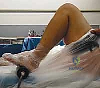
Optimal patient positioning is crucial for facilitating the extensile exposures often required in revision surgery. The patient is positioned supine on a radiolucent operating table to allow for intraoperative fluoroscopy if necessary.
As illustrated above, a rigid foot bump or a specialized leg holder is securely fixed to the table, positioned distal to the foot. This setup is critical; it supports the limb with the knee in varying degrees of flexion, prevents the foot from sliding distally during vigorous manipulation, and allows the surgeon to independently control the flexion-extension arc throughout the procedure. A sterile high-thigh tourniquet is applied to provide a bloodless surgical field, which is absolutely essential for visualizing the subtle interfaces between the implant, cement, and host bone during the meticulous extraction process. The entire lower extremity, from the iliac crest to the toes, is prepped and draped free. This extensive draping ensures that the surgeon has the flexibility to extend the incision proximally into the thigh or distally along the tibial crest should an extensile approach, such as a quadriceps snip or a tibial tubercle osteotomy, become necessary.
Step-by-Step Surgical Approach and Fixation Technique
The surgical execution of removing well-fixed components is a highly orchestrated sequence of events. It requires a delicate balance of controlled force and profound respect for the remaining host bone. The overarching philosophy is to disrupt the fixation interface completely before applying any extractive force.
Extensile Exposure and Joint Debridement
The procedure commences with the selection of the surgical incision. Whenever possible, the most lateral previous anterior incision is utilized to preserve the vascularity of the skin flaps. A standard medial parapatellar arthrotomy is initially performed. However, in the setting of a stiff knee or complex revision, this approach is frequently inadequate and risks avulsion of the patellar tendon. The surgeon must be prepared to seamlessly transition to an extensile exposure. A quadriceps snip—an oblique incision directed superolaterally through the rectus femoris and vastus lateralis tendon—often provides the necessary superior release without altering postoperative rehabilitation. For severe stiffness or when massive tibial exposure is required, a formal Tibial Tubercle Osteotomy (TTO) is performed. Once the joint is exposed, a radical synovectomy is executed. All hypertrophic synovium, scar tissue, and pseudomembranes are meticulously excised to recreate the medial and lateral gutters, mobilize the extensor mechanism, and provide unobstructed visualization of the implant-bone interfaces.
Phase 1: Tibial Polyethylene Extraction
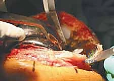
The first critical step in deconstructing the prosthesis is the removal of the modular tibial polyethylene insert. This maneuver is strategically vital; it instantly decompresses the joint space, creating crucial working room to access the femoral and tibial interfaces.
For modular systems, a thin, curved osteotome or a manufacturer-specific extraction tool is driven directly into the interface between the anterior lip of the polyethylene and the metal tibial tray.
By levering the instrument, the locking mechanism is disengaged, and the polyethylene is extracted. In cases of severe wear or if the locking mechanism is jammed, a drill or a specialized screw can be inserted into the polyethylene to provide a handle for extraction. If the implant is an all-polyethylene tibial component or a non-modular monoblock design, a reciprocating saw is used to horizontally transect the polyethylene, removing the articular surface to create the necessary joint space before addressing the fixation interface.
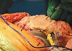
Phase 2: Meticulous Femoral Component Removal
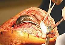
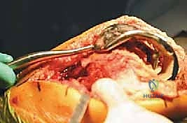
With the joint space opened, attention turns to the femoral component. The goal is to systematically sever the bond between the implant and the bone or cement across all five internal surfaces (anterior flange, anterior chamfer, distal condyles, posterior chamfer, and posterior condyles).
We begin with the most accessible interfaces. Thin, flexible osteotomes are sequentially driven into the anterior flange and distal condylar interfaces. It is imperative that the osteotome remains strictly parallel to the implant surface, hugging the metal to avoid diving into and fracturing the relatively soft metaphyseal bone.
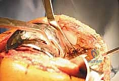
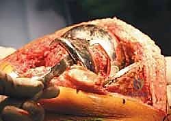
The process is repeated circumferentially. For the posterior condyles and chamfers, which are notoriously difficult to access, curved osteotomes or a Gigli saw passed behind the component can be highly effective. For cementless, porous-coated implants, an oscillating saw with a thin, flexible blade is often required to carefully cut the bony ingrowth directly at the implant surface.
Only after the interface has been completely disrupted circumferentially is extractive force applied. A specialized femoral extraction tool or vice grips are securely attached to the component. A slap hammer is then utilized, directing the force coaxially with the distal femoral geometry.
If the component does not yield with moderate impaction, the surgeon must stop immediately; applying excessive force will result in a catastrophic condylar fracture. The interfaces must be re-evaluated and further disrupted. Once removed, the distal femur is inspected, revealing the remaining bone bed, which must be carefully debrided of residual cement or fibrous tissue using ultrasonic tools or hand curettes.
Phase 3: Tibial Tray and Stem Extrication
The tibial tray presents a unique challenge due to its flat proximal geometry and the presence of a central keel or stem.
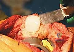
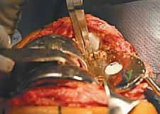
Similar to the femur, the process begins by disrupting the accessible proximal interface. Broad, thin osteotomes are driven horizontally underneath the tibial tray, progressing from anterior to posterior. Extreme caution must be exercised at the posteromedial and posterolateral corners to avoid plunging into the popliteal fossa and injuring the neurovascular structures.
To address the central keel or stem, a specialized technique is often required. If the proximal interface is adequately freed, stacked osteotomes can be used to gently lever the tray upwards, breaking the fixation of the keel. Alternatively, a high-speed burr or a narrow oscillating saw can be used to cut down alongside the keel.
Once the interfaces are thoroughly disrupted, an extraction device is locked onto the anterior aspect of the tray. Upward, coaxial impaction with a slap hammer is applied. As the tray begins to back out, the surgeon must ensure it does not tilt into varus or valgus, which could blow out the fragile medial or lateral cortical walls of the proximal tibia. Following removal, the tibial canal and metaphysis are aggressively debrided of all residual cement and debris to prepare for the revision stem and cones.
Complications, Incidence Rates, and Salvage Management
The removal of well-fixed components is fraught with potential perils. Anticipating these complications and possessing the surgical repertoire to manage them instantly is the hallmark of an expert revision arthroplasty surgeon.
| Complication | Estimated Incidence | Pathophysiology / Risk Factors | Salvage Management and Reconstructive Strategy |
|---|---|---|---|
| Extensor Mechanism Disruption | 1% - 5% | Avulsion of patellar tendon from tibial tubercle due to excessive traction in a stiff knee, or failure to utilize an extensile approach (e.g., quad snip, TTO) when indicated. | Primary repair is rarely successful. Requires major reconstruction using Achilles tendon allograft with bone block, synthetic mesh (e.g., Marlex), or gastrocnemius flap. Post-op requires rigid immobilization in extension for 6-8 weeks. |
| Iatrogenic Bone Loss / Fracture | 10% - 20% | Forceful extraction of components before interfaces are fully disrupted; diving osteotomes into osteoporotic bone; levering against thin cortices. Common at epicondyles and tibial metaphysis. | Immediate intraoperative fixation. Epicondylar fractures may require screw/washer fixation or heavy suture repair of collateral ligaments. Metaphyseal defects dictate the use of highly porous metaphyseal cones/sleeves and long diaphyseal engaging stems to bypass the defect. |
| Neurovascular Injury | < 1% | Direct laceration of popliteal vessels by plunging osteotomes or saws posteriorly; traction injury to peroneal nerve during severe valgus correction or excessive lateral retraction. | Vascular injury requires immediate vascular surgery consultation, tourniquet release, and potential bypass grafting. Neuropraxia requires careful observation; an ankle-foot orthosis (AFO) may be needed for foot drop. |
| Patellar Fracture / Necrosis | 5% - 10% | Aggressive clamping during component removal; excessive bone resection leaving a shell < 10mm; compromised vascularity from multiple previous arthrotomies. | If the remaining bone is viable and >10mm, a biconvex "gull-wing" patelloplasty or a new button can be placed. If fragmented or avascular, a patellectomy with meticulous extensor mechanism repair is the salvage option. |
| Recurrent Infection | 5% - 15% | Failure to completely remove all foreign material (retained cement plugs); inadequate debridement of infected bone; resistant biofilm-producing organisms. | Requires a repeat two-stage exchange arthroplasty. May necessitate long-term suppressive antibiotic therapy or, in extreme, recalcitrant cases, arthrodesis or above-knee amputation (AKA). |
Mitigating Iatrogenic Fractures
The most common major complication during extraction is iatrogenic fracture. The medial and lateral femoral epicondyles, which serve as the origins for the collateral ligaments, are particularly vulnerable when levering a well-fixed femoral component. An avulsion fracture here instantly creates gross coronal instability. The surgeon must abandon extraction, temporarily fix the fracture (often with a screw and washer or heavy non-absorbable sutures), and transition to a highly constrained implant system (VVC or Hinge) with diaphyseal stems to bypass the compromised metaphyseal bone and protect the ligamentous repair.
Management of Massive Bone Defects
Even with flawless technique, the removal of a well-fixed, porous-coated implant will inevitably result in some degree of cavitary or segmental bone loss. The surgeon must be adept at classifying this bone loss (e.g., AORI classification) and applying the appropriate reconstructive techniques. While impaction bone grafting was historically popular, modern revision arthroplasty relies heavily on highly porous titanium metaphyseal cones or sleeves. These devices provide immediate structural support, fill massive metaphyseal voids, and allow for biologic fixation, transferring stress away from the compromised joint line and into the robust diaphyseal bone via fluted, tapered stems.
Phased Post-Operative Rehabilitation Protocols
The surgical procedure is only half the battle; the postoperative rehabilitation dictates the ultimate functional outcome. The protocol following a revision TKA must be highly individualized, taking into account the quality of the remaining bone, the method of implant fixation, and the integrity of the extensor mechanism.
Phase 1: Immediate Postoperative Period (Weeks 0-2)
The primary goals in the immediate postoperative phase are wound healing, edema control, prevention of deep vein thrombosis (DVT), and the initiation of early, safe mobilization. Weight-bearing status is entirely dependent on intraoperative factors. If fully cemented components or fully engaging diaphyseal stems with metaphyseal cones were utilized in good quality bone, weight-bearing as tolerated (WBAT) is generally permitted. However, if structural allografts were used, or if an intraoperative fracture occurred, partial weight-bearing (PWB) or strict non-weight-bearing (NWB) may be mandated for 6 to 8 weeks. Continuous Passive Motion (CPM) machines are rarely used today;
