Part of the Master Guide

Orthopedic Prometric MCQs - Chapter 3 Part 1

Orthopedic Prometric MCQs - Chapter 3 Part 46

27 Apr 2026 44 min read 21 Views
Orthopedic Prometric MCQs - Chapter 3 Part 46

Orthopedic Prometric MCQs - Chapter 3 Part 46

Comprehensive 100-Question Exam


00:00

Start Quiz

Question 1

When using the tibial external rotation test on a patient, increased external rotation at 30° and 90° of knee flexion is indicative of:


Explanation

The tibial external rotation test is performed at 30° and 90° of knee flexion. The degree of foot external rotation with regard to the femur is evaluated. Increased external rotation at 30° is consistent with an isolated posterolateral corner injury. Increased external rotation at 30° and 90° is consistent with a combined posterolateral and posterior cruciate ligament injury.

Question 2

The recommended treatment for an acute combined anterior cruciate ligament and complete posterolateral corner disruption in a young athlete is:


Explanation

In cases of combined cruciate ligament and posterolateral corner injuries, most surgeons recommend addressing both injuries. In one study, the most common cause of anterior cruciate ligament failure was unrecognized and untreated concomitant posterolateral corner injuries.

Question 3

The ideal timing for repair of an acute posterolateral corner knee injury is:


Explanation

Surgical repair of posterolateral corner injuries is recommended within the first several weeks because dissection can be difficult and can result in the need for a reconstruction with longer delays. Results of chronic posterolateral corner injury repairs are inferior to those for acute posterolateral corner injuries.

Question 4

Which of the following exercises must be delayed for up to 3 months after posterolateral corner repair or reconstruction of the knee:


Explanation

Postoperative rehabilitation for posterolateral corner repair or reconstruction involves early protected or nonweight bearing, early range of motion exercises, and quadriceps exercises. Avoidance of hamstring exercises for up to 12 weeks is recommended to decrease external rotational torque and posterior subluxation forces at the knee joint.

Question 5

For patients who sustain a knee dislocation, the role of clinical history, physical examination, and magnetic resonance imaging (MRI) is:


Explanation

In a study of 17 knee dislocations, the accuracy of clinical examination ranged from 53% to 82% correct compared to an accuracy of 85% to 100% with MRI. The limitations of clinical examination were mainly due to associated injuries.

Question 6

After high velocity knee dislocations, there is serious injury to the popliteal vessels in approximately what percentage of patients:


Explanation

After reviewing several series from 1963 to 1992, investigators found serious injury to the popliteal vessels in approximately 30% of cases and peroneal nerve injuries in 25% of cases. The incidence of arterial and nerve injury with lower velocity mechanisms (some athletic injuries) is lower.

Question 7

After high velocity knee dislocations, there is serious injury to the peroneal nerve in approximately what percentage of patients:


Explanation

After reviewing several series from 1963 to 1992, investigators found serious injury to the popliteal vessels in approximately 30% of cases and peroneal nerve injuries in 25% of cases. The incidence of arterial and nerve injury with lower velocity mechanisms (some athletic injuries) is lower.

Question 8

The strongest bundle in the posterior cruciate ligament is the:


Explanation

The posterior cruciate ligament is made up of two bundles (anterolateral and posteromedial) that are named according to their origin on the femur and insertion on the tibia. The anterolateral bundle is the larger and stronger of the two bundles. The anterolateral bundle is tight in flexion, and the posteromedial bundle is tight in extension.

Question 9

In the posterior cruciate ligament the anterolateral bundle is tight in __ and the posteromedial bundle is tight in ____:


Explanation

The posterior cruciate ligament is made up of two bundles (anterolateral and posteromedial) that are named according to their origin on the femur and insertion on the tibia. The anterolateral bundle is the larger and stronger of the two bundles. The anterolateral bundle is tight in flexion, and the posteromedial bundle is tight in extension.

Question 10

When applying valgus stress, over which arc of motion is the anterior band of the anterior oblique component of the ulnar collateral ligament of the elbow under tension:


Explanation

Biomechanical studies demonstrate that the anterior band of the oblique component of the ulnar collateral ligament of the elbow is at greatest tension from full extension to 85° of elbow flexion.

Question 11

When applying valgus stress, over which arc of motion is the posterior band of the anterior oblique component of the ulnar collateral ligament of the elbow under tension:


Explanation

Biomechanical studies demonstrate that the posterior band of the oblique component of the ulnar collateral ligament of the elbow is at greatest tension from 55° to full elbow flexion.

Question 12

Which of the following structures is the main stabilizer of the elbow to valgus stress:


Explanation

The anterior oblique component of the ulnar collateral ligament is the most important stabilizer of the elbow to valgus stress. The most important secondary stabilizer is the radiohumeral articulation. The transverse oblique component of the ulnar collateral ligament imparts little stability to the elbow.

Question 13

Disruption of which of the following ligaments represents the primary lesion in posterolateral rotatory instability of the elbow:


Explanation

Of Driscoll and associates demonstrated that the radial ulnohumeral ligament must be disrupted to produce posterolateral rotator instability of the elbow.

Question 14

Elbow injury usually occurs during which phase of throwing:


Explanation

Peak valgus stresses on the elbow occur during the acceleration phase of throwing making it the phase during which the elbow is most vulnerable to injury.

Question 15

Which of the following structures is the most important dynamic stabilizer of the elbow to valgus stresses during throwing:


Explanation

The flexor-pronator muscle mass on the medial side of the elbow dynamically resists valgus stresses during throwing. Compromise or fatigue of this muscle group with activity may be a predecessor to injury to the ligamentous stabilizing structures.

Question 16

The following is a hip magnetic resonance image (MRI) of a 28-year-old male triathlete who has noticed progressive activityrelated left hip pain. Recommended treatment includes:
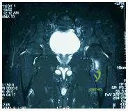
Orthopedic Prometric Exam Chapter 3 Image


Explanation

The MRI depicts a compression sided incomplete femoral neck fracture. Compression sided fractures of the femoral neck are treated with nonweight bearing and close observation. In the advent of fracture line extension, these fractures must be urgently percutaneously pinned. Complete stress fractures and incomplete tension sided fractures of the femoral neck must be urgently percutaneously pinned.

Question 17

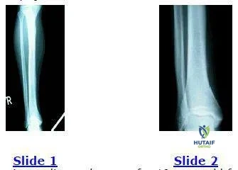
The following radiographs are of a 19-year-old female collegiate distance runner who complained of pain in her right distal tibia. She reports having shin splints 2 years earlier that affected her right proximal tibia. She has been unable to run secondary to symptoms for 3 weeks. She reports being amenorrheic for approximately the last 3 years. Which of the following should be included in her initial treatment regimen:

Orthopedic Prometric Exam Chapter 3 Image


Explanation

This individual has a distal tibial stress fracture as evidenced by the early periosteal reaction shown on radiography. Radiographs also show a healed proximal tibial stress fracture. Amenorrhea is a risk factor for stress fractures and should be addressed with hormone replacement therapy. The other possible answers are inappropriate for initial treatment.

Question 18

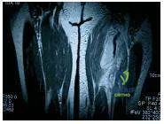
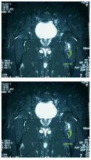
The following figure is the magnetic resonance image (MRI) of a 40-year- old avid female water-skier who felt a pop in her left hip as she was pulled over the front of her ski. Recommended treatment includes:


Explanation

The MRI shows a complete avulsion of the hamstring tendons off the ischial tuberosity. In active individuals, operative repair is recommended for complete avulsions. Nonoperative treatment of complete hamstring avulsion injury yields a low rate of return to sport at preinjury activity level.

Question 19

During which phase of throwing is the flexor-pronator muscle mass most electrically active:

Orthopedic Prometric Exam Chapter 3 Image


Explanation

Peak valgus stresses on the elbow occur during the acceleration phase of throwing making it the phase during which the elbow is most vulnerable to injury. The flexor-pronator muscle mass peaks in activity during the acceleration phase to dynamically stabilize the elbow.

Question 20

Which of the following is a risk factor for anterior cruciate ligament (AC L) injury in noncontact athletes:


Explanation

A high coefficient of friction at the shoe-surface interface is a risk factor for AC L injury in noncontact athletes. Insufficient evidence exists to definitively implicate the other possible answers as risk factors.

Question 21

Which of the following structures constitute the primary static stabilizers of the posterolateral corner (PLC) of the knee?


Explanation

The primary static stabilizers of the PLC are the lateral collateral ligament (LCL), the popliteus tendon, and the popliteofibular ligament (PFL). These structures primarily resist varus stress and external rotation of the tibia.

Question 22

During the tibial external rotation (dial) test, a patient exhibits 15 degrees of increased external rotation at 30 degrees of knee flexion compared to the contralateral side, but normal external rotation at 90 degrees. This finding is most indicative of an isolated injury to which of the following?


Explanation

Increased external rotation at 30 degrees of flexion only indicates an isolated posterolateral corner (PLC) injury. If increased rotation is present at both 30 and 90 degrees, a combined PLC and PCL injury is suspected.

Question 23

A 22-year-old athlete undergoes anterior cruciate ligament (ACL) reconstruction using a bone-patellar tendon-bone (BPTB) autograft. Which of the following is the most frequently reported complication associated with this specific graft choice?


Explanation

Anterior knee pain (or donor site morbidity) is the most common complication following BPTB autograft ACL reconstruction. Patellar fracture and tendon rupture are severe but rare complications.

Question 24

Which of the following clinical tests is most specific for diagnosing a posterolateral corner (PLC) deficiency in the knee?


Explanation

The reverse pivot shift test evaluates PLC instability. The knee is brought from flexion to extension with a valgus and external rotation force; a palpable clunk occurs as the posteriorly subluxated lateral tibial plateau reduces.

Question 25

A 30-year-old male presents after a high-energy dashboard injury resulting in a knee dislocation that spontaneously reduced. His pedal pulses are palpable. What is the most appropriate initial screening tool to evaluate for an occult popliteal artery injury?


Explanation

Ankle-brachial index (ABI) is the most appropriate initial screening tool for vascular injury following knee dislocation. An ABI of less than 0.9 mandates further investigation with CT angiography or prompt surgical exploration.

Question 26

The posterior cruciate ligament (PCL) consists of two main functional bundles. Which of the following statements regarding the anterolateral (AL) bundle is correct?


Explanation

The PCL is composed of a larger anterolateral (AL) bundle and a smaller posteromedial (PM) bundle. The AL bundle is tight in flexion and loose in extension, whereas the PM bundle is tight in extension.

Question 27

A 25-year-old soccer player sustains an acute combined Grade III femoral-sided medial collateral ligament (MCL) tear and an anterior cruciate ligament (ACL) rupture. What is the most widely accepted treatment strategy for this injury pattern?


Explanation

The standard treatment for a combined ACL tear and a femoral-sided Grade III MCL tear is ACL reconstruction with conservative management of the MCL in a hinged brace, as the MCL has excellent healing potential.

Question 28

A 28-year-old skier presents with a swollen knee after a twisting fall. Radiographs demonstrate a small avulsion fracture of the lateral tibial plateau.

This radiographic finding (Segond fracture) is virtually pathognomonic for an injury to which of the following structures?


Explanation

A Segond fracture is an avulsion of the anterolateral capsule and anterolateral ligament (ALL) from the proximal lateral tibia. It is considered highly specific (pathognomonic) for an underlying anterior cruciate ligament (ACL) tear.

Question 29

A 10-year-old boy falls from his bicycle and sustains a Meyers and McKeever Type III tibial eminence fracture. What is the most appropriate definitive management?


Explanation

Type III tibial eminence (spine) fractures are completely displaced. The standard of care is anatomic reduction and internal fixation (using sutures or screws) via an arthroscopic or open approach to restore ACL tension and joint congruity.

Question 30

Which of the following arteries provides the primary blood supply to the anterior cruciate ligament (ACL)?


Explanation

The middle genicular artery, a branch of the popliteal artery, pierces the posterior capsule to provide the primary blood supply to the cruciate ligaments and the synovial fold.

Question 31

During the evaluation of a patient with a suspected knee ligament injury, the quadriceps active test is performed. The knee is flexed to 90 degrees, and the patient is asked to slide their foot forward against resistance. An anterior shift of the tibia is noted. This finding indicates a deficiency of the:


Explanation

The quadriceps active test assesses for a PCL tear. In a PCL-deficient knee flexed to 90 degrees, the tibia sags posteriorly. Contraction of the quadriceps pulls the tibia anteriorly into its reduced anatomical position.

Question 32

A 24-year-old male sustains a traumatic knee dislocation resulting in a combined ACL, PCL, and posterolateral corner (PLC) injury. He is noted to have foot drop on the affected side. Injury to which of the following nerves is most likely?


Explanation

The common peroneal nerve is the most frequently injured nerve in knee dislocations, particularly those involving disruption of the posterolateral corner and lateral side structures. It typically presents as a foot drop.

Question 33

In the posteromedial corner of the knee, which structure serves as the primary restraint to valgus stress and internal rotation when the knee is near full extension?


Explanation

The posterior oblique ligament (POL) is a primary stabilizer of the posteromedial corner. It heavily contributes to the restraint of valgus stress and internal tibial rotation, particularly when the knee is in full or near-full extension.

Question 34

A clinical examination of a knee shows increased external rotation of the tibia at 30 degrees of flexion that normalizes at 90 degrees.

To reconstruct the primary deficient structures in this specific injury pattern, grafts should be routed to replicate the functions of which anatomical structures?


Explanation

The described examination indicates an isolated PLC injury. Anatomical reconstruction of the PLC aims to replicate the functions of the lateral collateral ligament, popliteus tendon, and popliteofibular ligament.

Question 35

A 35-year-old man presents with chronic medial knee pain months after a valgus injury. Radiographs reveal a linear calcification adjacent to the medial femoral condyle. What is the eponymous name for this radiographic sign?


Explanation

A Pellegrini-Stieda lesion is a calcification at the medial femoral condyle at the origin of the medial collateral ligament (MCL). It typically occurs secondary to a previous chronic MCL injury.

Question 36

While femoral-sided Grade III MCL tears are typically managed nonoperatively when combined with an ACL tear, a tibial-sided Grade III MCL avulsion requires surgical repair. Why is operative intervention specifically indicated for this variant?


Explanation

In a tibial-sided Grade III MCL tear, the distal end of the superficial MCL can become entrapped outside the pes anserinus tendons, preventing healing. This creates a Stener-like lesion of the knee that requires surgical repair.

Question 37

According to the Schenck classification of knee dislocations (KD), a patient who sustains tears to both the anterior cruciate ligament (ACL) and the posterior cruciate ligament (PCL), with intact collateral ligaments, is classified as:


Explanation

In the Schenck classification, KD II refers to tears of both the ACL and PCL, with intact collateral ligaments. KD III involves both cruciates plus one collateral, and KD IV involves all four major ligaments.

Question 38

A patient presents with a knee injury. The dial test shows >10 degrees of external rotation asymmetry at 30 degrees of knee flexion, but symmetric rotation at 90 degrees. Which structure is most likely injured?


Explanation

Isolated asymmetry at 30 degrees of flexion indicates an isolated posterolateral corner (PLC) injury. If asymmetry is present at both 30 and 90 degrees, it suggests a combined PLC and PCL injury.

Question 39

In evaluating a patient with a suspected posterolateral corner injury, the reverse pivot shift test is performed. During the test, a clunk is felt as the knee is brought from flexion into extension. This clunk represents:


Explanation

In a PLC injury, the lateral tibial plateau subluxates posteriorly during knee flexion. As the knee is extended during the reverse pivot shift test, the iliotibial band becomes an extensor and reduces the plateau at roughly 20 to 30 degrees of flexion.

Question 40

A 35-year-old male presents with chronic posterolateral knee instability and varus malalignment following an old injury. He is scheduled for a posterolateral corner (PLC) reconstruction. What is the most appropriate initial surgical management?


Explanation

In patients with chronic PLC instability and varus mechanical alignment, a high tibial osteotomy (HTO) should be performed first or concurrently. Performing a soft tissue reconstruction without correcting the bony varus alignment will predictably lead to graft stretching and failure.

Question 41

Which of the following nerves is most frequently injured in the setting of a severe posterolateral corner (PLC) injury or knee dislocation?


Explanation

The common peroneal nerve is uniquely tethered around the fibular neck and is highly susceptible to traction injury during a varus and hyperextension mechanism typical of PLC injuries. It occurs in up to 15-30% of knee dislocations.

Question 42

A patient suffers a high-velocity knee dislocation. Following closed reduction, the Ankle-Brachial Index (ABI) is measured at 0.85. What is the next best step in management?


Explanation

An ABI less than 0.9 after a knee dislocation is highly suspicious for a vascular injury. CT angiography or arterial duplex ultrasonography is indicated to precisely evaluate the popliteal artery and plan potential surgical intervention.

Question 43

A 25-year-old athlete sustains a valgus blow to the knee. MRI reveals a complete rupture of the superficial and deep medial collateral ligament (MCL) with the distal end of the superficial MCL displaced superficial to the pes anserinus. What is the recommended treatment?


Explanation

A Stener-like lesion of the knee occurs when the distal MCL avulses and flips superficial to the pes anserinus. Unlike typical isolated MCL tears that heal well non-operatively, this displaced lesion prevents native healing and requires surgical repair.

Question 44

When reconstructing the anterior cruciate ligament (ACL), the surgeon must identify the lateral intercondylar ridge (resident's ridge). What is the anatomical relationship of the native ACL footprint to this ridge?


Explanation

The lateral intercondylar ridge (resident's ridge) marks the anterior boundary of the ACL femoral footprint when the knee is in 90 degrees of flexion. No native ACL fibers attach anterior to this ridge, making it a critical landmark for femoral tunnel placement.

Question 45

What is the primary restraint to posterior tibial translation at 90 degrees of knee flexion?


Explanation

The anterolateral (AL) bundle of the Posterior Cruciate Ligament (PCL) is the primary restraint to posterior tibial translation at 90 degrees of flexion. It is the larger and stronger bundle and becomes tight in flexion.

Question 46

A patient presents with a chronic grade III isolated PCL injury treated non-operatively 15 years ago. Which compartments of the knee are at the highest risk for developing secondary osteoarthritis?


Explanation

Chronic PCL deficiency leads to altered knee kinematics, specifically increasing contact pressures in the medial compartment and the patellofemoral joint. This predictably leads to early degenerative changes in these specific areas over time.

Question 47

During a physical examination for a suspected knee ligament injury, the examiner actively flexes the patient's quadriceps while the knee is held at 90 degrees of flexion. The tibia is observed to translate anteriorly. This finding is pathognomonic for an injury to which structure?


Explanation

The quadriceps active test is highly specific for a complete posterior cruciate ligament (PCL) tear. In a PCL-deficient knee resting at 90 degrees of flexion, the tibia sags posteriorly; active quadriceps contraction pulls the tibia anteriorly into its normal reduced position.

Question 48

Which of the following anatomical structures forms the primary static restraint to varus opening at 0 and 30 degrees of knee flexion?


Explanation

The fibular collateral ligament (FCL/LCL) is the primary static restraint to varus gapping of the knee at both 0 and 30 degrees of flexion. The popliteus and popliteofibular ligament act as the primary restraints to external rotation.

Question 49

A 22-year-old football player sustains a Schenck KD-III knee dislocation. To minimize the risk of compartment syndrome from fluid extravasation during arthroscopic multiligamentous reconstruction, what is the generally recommended optimal timing for surgery if no vascular injury is present?


Explanation

Allowing 2 to 3 weeks before arthroscopic reconstruction allows the capsular tears to seal, significantly reducing the risk of fluid extravasation and subsequent compartment syndrome. It also allows the patient to regain some range of motion, decreasing the risk of postoperative arthrofibrosis.

Question 50

The meniscofemoral ligaments of the knee are intimately associated with the posterior cruciate ligament (PCL). Which of the following correctly describes their anatomical relationship?


Explanation

The ligament of Humphrey runs anterior to the PCL, whereas the ligament of Wrisberg runs posterior to the PCL. They attach the posterior horn of the lateral meniscus to the medial femoral condyle.

Question 51

A 45-year-old female presents with chronic medial knee pain. Radiographs reveal a calcification near the medial femoral epicondyle.

What is the eponymous name for this radiographic finding?


Explanation

A Pellegrini-Stieda lesion is a calcification at the medial femoral epicondyle, which typically results from a chronic, healed avulsion or injury to the proximal Medial Collateral Ligament (MCL).

Question 52

During a posterolateral corner (PLC) reconstruction using an anatomical technique (e.g., LaPrade), three main structures are reconstructed. Which of the following is NOT one of the three primarily reconstructed structures?


Explanation

Anatomic PLC reconstruction aims to recreate the three major static restraints: the fibular collateral ligament (LCL), the popliteus tendon, and the popliteofibular ligament. The iliotibial band is a dynamic stabilizer and is not routinely reconstructed in this procedure.

Question 53

An isolated posterolateral corner injury is diagnosed. Which physical exam finding would most accurately differentiate an isolated PLC injury from a combined PLC and PCL injury?


Explanation

The posterior drawer test at 90 degrees evaluates the integrity of the PCL. An isolated PLC injury will have a normal posterior drawer at 90 degrees, whereas a combined PLC/PCL injury will demonstrate increased posterior translation.

Question 54

A patient sustains an avulsion fracture of the anterolateral proximal tibia.

This finding is considered pathognomonic for a tear of which major knee ligament?


Explanation

A Segond fracture is an avulsion of the anterolateral capsular complex from the proximal tibia. It is highly associated (pathognomonic) with an underlying Anterior Cruciate Ligament (ACL) tear.

Question 55

The superficial medial collateral ligament (sMCL) has distinct femoral and tibial attachments. Where is its primary distal (tibial) attachment located?


Explanation

The superficial MCL attaches 4 to 5 cm distal to the joint line on the anteromedial surface of the tibia, deep to the pes anserinus tendons. The deep MCL attaches much closer to the articular margins of the joint line.

Question 56

A patient is evaluated for a knee dislocation. Which specific knee dislocation classification implies a tear of the anterior cruciate ligament, posterior cruciate ligament, and medial collateral ligament, while the posterolateral corner remains intact?


Explanation

In the Schenck classification, KD-III refers to injuries involving both cruciates and one collateral ligament. KD-III M indicates the Medial collateral ligament is torn, while KD-III L indicates the Lateral (PLC) structures are torn.

Question 57

A 30-year-old male undergoes a single-bundle anterior cruciate ligament (ACL) reconstruction. To optimally control both anterior translation and rotational instability, where is the most appropriate location for the femoral tunnel within the native footprint?


Explanation

Modern ACL reconstruction emphasizes anatomical placement central within the native footprint. Vertical placement (12 o'clock) fails to control rotational instability, and placement anterior to the resident's ridge leads to graft impingement against the PCL or notch.

Question 58

A 25-year-old male is brought to the emergency department after a high-speed motorcycle accident. Clinical examination reveals a grossly deformed knee that is reduced under sedation. Post-reduction Ankle-Brachial Index (ABI) is 0.85. What is the most appropriate next step in management?


Explanation

An ABI of < 0.9 after a knee dislocation is highly suspicious for a vascular injury and mandates further imaging with CT angiography. Immediate surgical exploration without imaging is generally reserved for hard signs of vascular ischemia, such as absent pulses, an expanding hematoma, or active pulsatile bleeding.

Question 59

Which of the following structures form the primary static stabilizing complex of the posterolateral corner (PLC) of the knee?


Explanation

The primary static stabilizers of the PLC are the lateral collateral ligament (fibular collateral ligament), the popliteus tendon, and the popliteofibular ligament. These structures work together synergistically to resist varus, external rotation, and posterior translation of the tibia.

Question 60

During physical examination of a patient with a suspected multiligamentous knee injury, the dial test reveals 15 degrees of increased external tibial rotation compared to the contralateral side at 30 degrees of knee flexion. At 90 degrees of flexion, the side-to-side difference in external rotation is 2 degrees. What is the most likely injury pattern?


Explanation

An isolated PLC injury characteristically results in increased external rotation at 30 degrees of flexion with a normal (symmetric) dial test at 90 degrees. A combined PCL and PLC injury will demonstrate increased external rotation at both 30 and 90 degrees of knee flexion.

Question 61

A patient sustains a high-energy knee dislocation. On examination, there is a transverse skin furrow (dimple sign) over the medial joint line, and the knee cannot be closed-reduced. What structure is most likely interposing and preventing reduction?


Explanation

The 'dimple sign' or 'pucker sign' occurs in an irreducible posterolateral knee dislocation when the medial joint capsule and medial collateral ligament buttonhole through the joint. An open reduction via a medial approach is urgently required to extricate these structures and achieve reduction.

Question 62

The posterior cruciate ligament (PCL) consists of two main functional bundles. Which statement best describes the biomechanical properties of the anterolateral (AL) bundle?


Explanation

The PCL is composed of the larger, stronger anterolateral (AL) bundle and the smaller posteromedial (PM) bundle. The AL bundle is tight in knee flexion and lax in extension, whereas the PM bundle is tight in extension and lax in flexion.

Question 63

Review the radiograph demonstrating a small cortical avulsion fracture from the medial aspect of the proximal tibia (Reverse Segond fracture).

This radiographic finding is virtually pathognomonic for which of the following combined injuries?


Explanation

A reverse Segond fracture is an avulsion of the deep capsular component of the medial collateral ligament. It is highly associated with posterior cruciate ligament (PCL) tears and peripheral tears of the medial meniscus.

Question 64

During surgical reconstruction of the posterolateral corner (PLC) using a fibular-based technique, a surgeon must be extremely careful to protect the common peroneal nerve. Where is this nerve most vulnerable during the approach?


Explanation

The common peroneal nerve runs posterior to the biceps femoris tendon and wraps around the fibular neck. It must be identified and protected during a lateral approach or fibular-based PLC reconstruction to avoid iatrogenic foot drop.

Question 65

According to the Schenck classification of knee dislocations, a patient with an MRI confirming complete tears of the ACL, PCL, and the posterolateral corner (PLC), with an intact medial collateral ligament (MCL), is classified as:


Explanation

The Schenck classification describes multiligamentous knee injuries based on the involved structures. KD III-L involves the ACL, PCL, and the lateral structures (PLC/LCL), whereas KD III-M involves the ACL, PCL, and medial structures.

Question 66

A 28-year-old male presents with recurrent instability 2 years after an anterior cruciate ligament (ACL) reconstruction using a bone-patellar tendon-bone autograft. Clinical examination reveals an intact ACL graft on Lachman testing but significant varus thrust during gait and a positive reverse pivot shift. Failure to address which of the following at the index procedure most likely caused this outcome?


Explanation

Unrecognized or untreated posterolateral corner (PLC) deficiency results in pathologically increased varus and external rotation forces. This abnormally increased stress on an ACL graft frequently leads to chronic graft elongation and ultimate failure.

Question 67

A 22-year-old collegiate football player sustains an acute combined grade III anterior cruciate ligament (ACL) tear and grade III medial collateral ligament (MCL) tear. Assuming non-operative treatment of the MCL is chosen, what is the most widely accepted management strategy?


Explanation

The standard management for combined ACL and MCL injuries often involves bracing the knee to allow the MCL to heal, followed by delayed ACL reconstruction once full range of motion is restored. This staged approach significantly minimizes the high risk of postoperative arthrofibrosis.

Question 68

The posteromedial corner of the knee provides primary restraint against valgus forces in full extension and anteromedial rotatory instability. Which of the following structures is the most crucial static component of this complex?


Explanation

The posterior oblique ligament (POL) is the primary stabilizer of the posteromedial corner, heavily restricting valgus in full extension and internal tibial rotation. It intimately blends with the semimembranosus tendon and posteromedial capsule.

Question 69

Which type of knee dislocation is most strongly associated with a stretch or complete rupture injury to the common peroneal nerve?


Explanation

Posterolateral knee dislocations, and multiligament injuries involving the posterolateral corner (KD III-L), have the highest rate of common peroneal nerve injury. This is due to severe traction across the nerve as it wraps around the fibular neck.

Question 70

During normal knee kinematics, the popliteus muscle plays a vital role in initiating knee flexion from a fully extended position. What is its primary biomechanical action during this phase?


Explanation

To 'unlock' the fully extended knee during closed-chain kinematics (when the foot is planted), the popliteus muscle acts to externally rotate the femur on the fixed tibia. In an open-chain state, it internally rotates the tibia on the femur.

Question 71

An isolated rupture of the posterolateral corner (PLC) is suspected in a 25-year-old athlete. During the Dial test, what are the expected physical examination findings that differentiate an isolated PLC injury from a combined PLC and posterior cruciate ligament (PCL) injury?


Explanation

An isolated PLC injury exhibits excessive external rotation at 30 degrees of knee flexion but reduces to normal at 90 degrees due to an intact PCL. If external rotation is increased at both 30 and 90 degrees, it indicates a combined PLC and PCL injury.

Question 72

Which of the following anatomic structures is considered the primary restraint to varus opening at 30 degrees of knee flexion?


Explanation

The fibular collateral ligament (LCL) is the primary restraint to varus stress at 5 and 25-30 degrees of knee flexion. The popliteus and popliteofibular ligament act as primary restraints to external rotation.

Question 73

A 25-year-old athlete sustains an acute traumatic knee dislocation. In the emergency department, the Ankle-Brachial Index (ABI) is measured at 0.8. A CTA confirms a popliteal artery intimal tear. The vascular surgeon repairs the artery via a posterior approach. What is the most appropriate next orthopedic step?


Explanation

Following a vascular repair in the setting of a knee dislocation, a spanning external fixator is recommended to provide absolute skeletal stability and protect the delicate vascular anastomosis.

Question 74

What is the primary anatomical and biomechanical rationale for utilizing a tibial inlay technique over a standard transtibial technique during posterior cruciate ligament (PCL) reconstruction?


Explanation

The tibial inlay technique bypasses the acute angle (killer turn) at the posterior tibial tunnel opening, which can lead to graft attenuation and failure seen in transtibial PCL reconstructions.

Question 75

Which of the following physical examination tests is most specific for evaluating posterolateral corner (PLC) rotatory instability?


Explanation

The reverse pivot shift test specifically evaluates for PLC injury. A positive test occurs when a posteriorly subluxated lateral tibial plateau reduces as the knee is flexed past 20 to 30 degrees.

Question 76

A 35-year-old patient presents with a chronic posterolateral corner (PLC) deficiency and a noticeable varus thrust during gait. Radiographs show medial compartment narrowing and significant mechanical varus alignment. What is the most appropriate initial surgical management?


Explanation

In chronic PLC deficiency associated with varus malalignment, a proximal tibial valgus-producing osteotomy must be performed (either staged before or concurrently) to prevent the failure of the soft-tissue PLC reconstruction.

Question 77

A 28-year-old football player presents with acute knee pain and lateral swelling after a direct blow to the anteromedial tibia. A radiograph is obtained.

Based on the classical significance of the "arcuate sign" shown, what associated structural injury is almost certainly present?


Explanation

The arcuate sign is an avulsion fracture of the fibular styloid. It is highly pathognomonic for an injury to the posterolateral corner (PLC) structures.

Question 78

During a single-bundle posterior cruciate ligament (PCL) reconstruction, which specific bundle is typically reconstructed, and at what angle of knee flexion should the graft be tensioned?


Explanation

The anterolateral bundle is the larger and stronger component of the PCL. It is tightest in flexion and is standardly reconstructed and tensioned at 90 degrees of knee flexion.

Question 79

A patient is diagnosed with a grade III MCL tear combined with an ACL rupture. MRI reveals the distal MCL has avulsed from its tibial insertion and retracted superficial to the pes anserinus (Stener-like lesion of the knee). What is the recommended management strategy?


Explanation

While most combined ACL/MCL injuries are treated with bracing for the MCL and delayed ACL reconstruction, a distal MCL avulsion trapped superficial to the pes anserinus cannot heal nonoperatively and requires acute surgical repair.

Question 80

Which neurological structure is at highest risk during an acute posterolateral corner injury, and what specific clinical finding dictates the poorest prognosis for conservative recovery?


Explanation

The common peroneal nerve is injured in up to 30% of severe PLC injuries. A complete palsy documented immediately at the time of injury has a much lower rate of spontaneous recovery compared to partial or delayed-onset lesions.

Question 81

A 22-year-old female skier presents after a twisting knee injury. Radiographs reveal the finding shown.

This classical fracture (Segond fracture) represents an avulsion of which structure and strongly correlates with which associated injury?


Explanation

The Segond fracture is a cortical avulsion off the lateral tibial plateau involving the anterolateral capsule and anterolateral ligament (ALL). It is highly pathognomonic for an anterior cruciate ligament (ACL) tear.

Question 82

When performing an anatomical posterolateral corner (PLC) reconstruction, at what knee position should the fibular collateral ligament (FCL) graft be properly tensioned?


Explanation

During PLC reconstruction, the FCL graft is typically tensioned at 20 degrees of knee flexion while a valgus force is applied. In contrast, the popliteus graft is tensioned at 60 degrees of flexion in neutral rotation.

Question 83

The popliteofibular ligament (PFL) plays a crucial role in posterolateral knee stability. Which of the following best describes its primary anatomical origin and insertion?


Explanation

The popliteofibular ligament (PFL) branches off the popliteus tendon at the musculotendinous junction and inserts firmly into the posteromedial aspect of the fibular styloid, acting as a major restraint to external rotation.

Question 84

A 40-year-old male presents with a suspected posterior cruciate ligament (PCL) injury. On examination, a positive posterior sag sign is noted. Which radiographic view is most accurate for quantifying the exact degree of posterior tibial translation?


Explanation

Bilateral kneeling lateral radiographs provide a consistent, gravity and body-weight directed posterior force to the proximal tibia. This allows for highly accurate quantification of posterior tibial translation compared to the uninjured side.

Question 85

A 50-year-old patient presents with acute posteromedial knee pain after deep flexion. MRI demonstrates the lesion shown.

If left untreated, a complete posterior horn medial meniscal root tear is biomechanically equivalent to which of the following?


Explanation

A meniscal root tear disrupts the crucial circumferential hoop stresses of the meniscus, rendering it functionally incompetent. Biomechanically, it is completely equivalent to a total meniscectomy and leads to rapid articular cartilage degeneration.

Question 86

During an anatomical posterolateral corner (PLC) reconstruction, tunnels must be placed accurately in the lateral femoral condyle. What is the spatial relationship of the normal fibular collateral ligament (FCL) femoral attachment relative to the popliteus tendon attachment?


Explanation

On the lateral femoral condyle, the popliteus tendon insertion is located at the anterior end of the popliteal sulcus. The origin of the FCL is located 18.5 mm proximal and posterior to the popliteus insertion.

None

Dr. Mohammed Hutaif
Medically Verified Content by
Prof. Dr. Mohammed Hutaif
Consultant Orthopedic & Spine Surgeon
Chapter Index