Part of the Master Guide

Orthopedic Prometric MCQs - Chapter 3 Part 1

Orthopedic Prometric MCQs - Chapter 3 Part 16

25 Apr 2026 42 min read 19 Views
Orthopedic Prometric MCQs - Chapter 3 Part 16

Orthopedic Prometric MCQs - Chapter 3 Part 16

Comprehensive 100-Question Exam


00:00

Start Quiz

Question 1

The best way to diagnose dysplasia epiphysealis hemimelica, in addition to history and physical, is:


Explanation

Dysplasia epiphysealis hemimelica, or Trevor disease, is an epiphyseal osteochondroma. The characteristic location, continuity with the epiphysis and alteration in growth of the epiphysis, is the most specific confirmation of the disease. Histology is less specific, as are bone scan and ultrasound.

Question 2

Which of the following commonly used statements regarding the pelvis in patients with bladder exstrophy is true:


Explanation

In patients with bladder exstrophy, computerized modeling shows that the forces across the hip are increased by approximately 30%. However, limp is not significant and the pelvis is not more prone to fracture. Most patients can be made continent through surgery.

Question 3

A 1-year-old child requires evaluation of limb length inequality. The surgeon notices a disproportional increase in length, as well as width, of the ipsilateral upper and lower extremity. No other physical abnormalities are evident. Recommended treatment includes which of the following:


Explanation

Idiopathic hemihypertrophy is associated with an increased incidence of intra-abdominal tumors such as Wilmsâ tumor and hepatoblastoma. Periodic screening by ultrasound throughout childhood is recommended.

Question 4

Which of the following statements describes the growth plate biomechanics of the distal femur:


Explanation

Whereas the the proximal tibial physis is protected by the collateral ligaments and tibial tubercle epiphysis, the distal femoral physis is vulnerable to injury because it is not protected by any ligaments.

Question 5

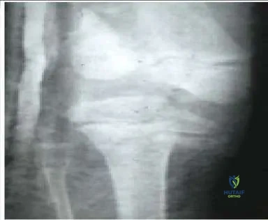
A 200-lb 13-year-old boy sustained a fracture while playing football. His radiographs are presented (Slide 1). Which of the following treatments should be attempted first:

Orthopedic Prometric Exam Chapter 3 Image


Explanation

The radiographs show a Salter II fracture with a large metaphyseal fragment. A high likelihood exists for successful closed reduction, and the metaphyseal fragment should allow rigid fixation. This patient received closed reduction and was internally fixed with three percutaneous 7.3-mm screws (Slide 2). He did not require a cast.

Question 6

An 8-year-old boy was injured by a lawnmower. The resulting proximal tibial injury presented in the radiograph (Slide) is classified as a:

Orthopedic Prometric Exam Chapter 3 Image


Explanation

A Salter IV injury to the proximal tibia is apparent in the patientâ s radiograph. The fracture traverses the epiphysis, physis, and metaphysis medial to the tibial spine. No injury to the lateral portion of the plateau is present. Incidentally, the distal femoral injury is also classified as Salter IV.

Question 7

A 12-year-old boy twisted his knee while riding a bicycle. Based on his radiographs (Slide), which of the following is the most appropriate diagnosis:
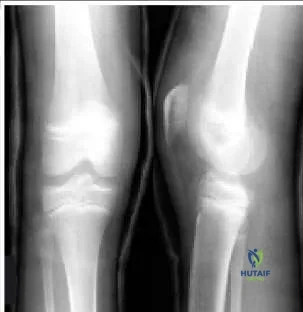
Orthopedic Prometric Exam Chapter 3 Image


Explanation

This patient has a tibial spine avulsion, classified as type III by McKeever and Meyers. Treatment options include closed immobilization or arthroscopic or open reduction and internal fixation.

Question 8

What is the most common cause of intoeing in children with bilateral cerebral palsy:


Explanation

The most common cause of intoeing in children with bilateral spasticity is internal hip rotation. For children with hemiplegia, the most common cause of intoeing is tibial torsion. In some patients, several causes may coexist to cause the condition.

Question 9

Risk factors for superior mesenteric artery syndrome after adolescent idiopathic scoliosis surgery include all of the following except:


Explanation

Superior mesenteric artery syndrome occurs more often in patients with decreased BMI, larger and stiffer thoracic curves, lumbar apical translation of Lenke B or C , and two staged procedures.

Question 10

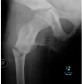
An 8-year-old girl fractures her right femur when starting a sprint. Based on her radiograph (Slide), which of the following is the most likely diagnosis:


Explanation

This patient has signs of a pre-existing fibrous dysplasia lesion, including a central expansion of the subtrochanteric region with a ground-glass (not lucent) appearance. She was diagnosed with fibrous dysplasia.

Question 11

An 8-year old girl with fibrous dysplasia fractures her femur (Slide). Which of the following procedures is the best treatment option:


Explanation

This lesion requires mechanical support because it is vulnerable to malunion and further deformity. Bone graft would be rapidly reabsorbed and has no lasting value in this patient. A screw with a long side plate that extends well into normal bone is the best treatment option.

Question 12

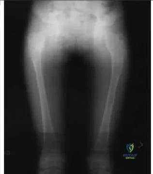
The patient presented in the radiograph (Slide) has a slight, painless limp. Which of the following is the most likely diagnosis:

Orthopedic Prometric Exam Chapter 3 Image


Explanation

This patient has multiple â ground-glassâ lesions and one cystic lesion, as well as bowing in the subtrochanteric region. The patient was diagnosed with fibrous dysplasia.

Question 13

A 14-year-old boy sustains an avulsion of the tibial tubercle while playing basketball. The fracture extends vertically into the joint, crossing the proximal tibial articular surface. He is at greatest risk for which of the following complications:

Orthopedic Prometric Exam Chapter 3 Image


Explanation

Fracture of the tibial tubercle has been reported to lead to compartment syndrome, presumably due to bleeding from the geniculate vessels and the exposed bone. This would not be expected from such a proximal fracture, but orthopedic surgeons should be aware of the possible association.

Question 14

An infant presents with idiopathic scoliosis. Which of the following factors indicates observation as the treatment of choice:


Explanation

Rib-vertebral angle difference >20° rib-vertebra phase 2, and curves >45° indicate a likely progression. The majority of infantile curves are left thoracic, so this is not a factor. A patient younger than 1 year of age has a higher chance of spontaneous resolution than an older patient. Therefore, absent other risk factors, observation is the initial recommend treatment.

Question 15

Which of the following factors predicts a lesser degree of impairment of pulmonary function in patients with adolescent idiopathic scoliosis:


Explanation

An increased Cobb angle, increased cephalad apex of the curve, decreased thoracic kyphosis, and pectus excavatum in addition to the scoliosis are factors that predict a greater degree of pulmonary impairment. A lower number of vertebrae in the curve signals less impairment.

Question 16

All except which of the following neurologic/muscular disorders can present undiagnosed in a patient with scoliosis:


Explanation

Scoliosis can be caused by any neurologic disorder that affects trunk balance. Scoliosis is seen in most patients with Friedreichâ s ataxia, a disorder in which patients present with an ataxic gait in preadolescence or adolescence. The curve becomes significant at about the same time as the ataxia. Scoliosis can also be seen in 10% of patients with C harcot-Marie- Tooth disease. This condition is characterized by cavus feet, intrinsic atrophy, and occasional hip dysplasia; no significant pain or clumsiness is present. Scoliosis is seen in many patients with syringomyelia. The syrinx and scoliosis both develop silently with no noticeable weakness until both the syrinx and the curve are significant. Spinal cord tumor may present with scoliosis in an ambulatory patient. Duchenne muscular dystrophy leads to scoliosis in most patients, but the patients become nonambulatory several years before the curve develops.

Question 17

All except which of the following structural disorders often causes scoliosis and presents undiagnosed in patients:


Explanation

Although achondroplasia causes kyphosis, it is not associated with scoliosis to a significant degree. Marfan syndrome, Ehlers- Danlos syndrome, osteogenesis imperfecta, and Loeys-Dietz syndrome (a defect in TGF-beta receptor protein) are frequently associated with scoliosis.

Question 18

Which of the following levels of evidence should be assigned to a prospective, randomized therapeutic study with 80% follow-up:


Explanation

A study may be level I as long as it has at least 80% follow-up.

Question 19

A prospective comparative study should be assigned which level of evidence:


Explanation

A comparative study is one in which patients treated one way are compared with patients treated in another manner at the same institution. As long as the study is prospective, it can be assigned level II.

Question 20

A case-control study should be assigned which level of evidence:


Explanation

A case-control study is considered level III evidence, as is a retrospective comparison study or a meta-analysis in which the lowest level of primary study is level III.

Question 21

A 3-year-old boy presents with short-limbed dwarfism, frontal bossing, and midface hypoplasia. Radiographs show narrowing of the interpedicular distances in the lumbar spine. Which of the following gene mutations is most likely responsible for this condition?


Explanation

Achondroplasia is the most common form of short-limbed dwarfism and is caused by an activating mutation in the FGFR3 gene. This mutation inhibits chondrocyte proliferation in the proliferative zone of the physis.

Question 22

A 10-year-old child presents with a waddling gait, joint pain, and short stature. Radiographs show delayed, irregular ossification of multiple epiphyses and a "double-layered" patella laterally. A mutation in which of the following is most commonly implicated?


Explanation

Multiple epiphyseal dysplasia (MED) is often caused by a mutation in the COMP gene. A double-layered patella on a lateral knee radiograph is a highly characteristic, pathognomonic finding for this condition.

Question 23

A newborn is evaluated for multiple congenital joint dislocations involving the knees, hips, and elbows. Physical examination reveals spatulate thumbs, a depressed nasal bridge, and a prominent forehead. What is the most critical initial imaging study to obtain in this patient?


Explanation

The patient has Larsen syndrome, which presents with multiple joint dislocations and characteristic spatulate thumbs. Cervical spine kyphosis and instability are common and can lead to severe neurologic compromise, making cervical radiographs critical.

Question 24

A newborn infant is noted to have short limbs, rigid clubfeet, cystic swelling of the ear pinnae (cauliflower ears), and proximally placed, abducted thumbs (hitchhiker thumbs). This condition is caused by a defect in which of the following?


Explanation

Diastrophic dysplasia is an autosomal recessive disorder caused by a mutation in the SLC26A2 (DTDST) gene, leading to defective sulfate transport. Classic findings include hitchhiker thumbs, cauliflower ears, and rigid clubfeet.

Question 25

A 2-year-old child presents with an asymptomatic, painless mass over the mid-clavicle. There is no history of trauma. Radiographs reveal a defect in the middle third of the right clavicle with smooth, rounded bone ends. What is the most appropriate management?


Explanation

Congenital pseudarthrosis of the clavicle almost always occurs on the right side and presents as a painless mass. Surgery is indicated if the patient is symptomatic, has skin compromise, or for cosmetic concerns, typically delayed until 3 to 5 years of age.

Question 26

A 4-year-old boy presents with progressive, asymmetric limb overgrowth, plantar cerebriform connective tissue nevi, and epidermal nevi. A mutation in which of the following genes is classically associated with this syndrome?


Explanation

The presentation is classic for Proteus syndrome, characterized by cerebriform connective tissue nevi, epidermal nevi, and asymmetric overgrowth. It is caused by a somatic mosaic mutation in the AKT1 gene.

Question 27

A 6-year-old child presents with severe disproportionate short-trunk dwarfism, corneal clouding, and normal intelligence. Radiographs show universal platyspondyly and hypoplasia of the odontoid. The patient is deficient in which of the following enzymes?


Explanation

Morquio syndrome (MPS IV) is characterized by normal intelligence, severe skeletal dysplasia, and odontoid hypoplasia predisposing to atlantoaxial instability. It is caused by a deficiency in the enzyme galactosamine-6-sulfatase.

Question 28

A 5-year-old boy has short-trunk dwarfism, coxa vara, and myopia. Radiographs reveal delayed ossification of the femoral heads and platyspondyly. Which of the following genetic mutations is responsible for this condition?


Explanation

Spondyloepiphyseal dysplasia (SED) congenita is caused by mutations in the COL2A1 gene encoding type II collagen. Clinical features include short-trunk dwarfism, coxa vara, myopia, and atlantoaxial instability.

Question 29

A 3-year-old child with multiple café-au-lait spots and axillary freckling presents with anterolateral bowing of the tibia. Radiographs show medullary canal narrowing and early pseudarthrosis. Which of the following is true regarding surgical management?


Explanation

Congenital pseudarthrosis of the tibia is highly associated with NF1 and is notoriously difficult to treat. Standard surgical management involves aggressive resection of the hamartomatous tissue, intramedullary rodding, and robust bone grafting.

Question 30

A 10-year-old girl is noted to have a large head with delayed closure of the fontanelles, dental anomalies, and the ability to appose her shoulders anteriorly. Radiographs reveal hypoplastic clavicles. Which of the following genes is affected in this autosomal dominant condition?


Explanation

Cleidocranial dysplasia is caused by a mutation in the RUNX2 (CBFA1) gene, which is essential for osteoblast differentiation. Patients typically have absent or hypoplastic clavicles, delayed fontanelle closure, and supernumerary teeth.

Question 31

A newborn is diagnosed with osteogenesis imperfecta. According to the Sillence classification, which type is characterized by normal or near-normal stature and blue sclerae throughout life, making it the most common and mildest form?


Explanation

Sillence Type I is the most common and mildest form of OI, characterized by blue sclerae, recurrent fractures that decrease after puberty, and near-normal stature. Type II is perinatally lethal, and Type III is the most severe non-lethal form.

Question 32

A newborn presents with a deep constriction ring around the left lower leg and a distal clubfoot deformity. The toes exhibit acrosyndactyly. Which of the following is the most appropriate management for the deep constriction ring if it is causing distal vascular compromise?


Explanation

Constriction bands in amniotic band syndrome that threaten limb viability require surgical excision. This is performed using staged Z-plasties (releasing 50% of the circumference at a time) to prevent vascular compromise to the distal limb.

Question 33

A 1-year-old girl is evaluated for a short right leg. Examination reveals an absent 5th ray, anteromedial bowing of the tibia, and a dimple over the anterior shin. Radiographs confirm complete absence of the fibula. Which of the following knee ligament anomalies is most commonly associated?


Explanation

Fibular hemimelia is the most common congenital long bone deficiency. It is frequently associated with anteromedial bowing of the tibia, absence of lateral foot rays, and absent or hypoplastic anterior/posterior cruciate ligaments.

Question 34

A 5-year-old girl presents with an enlarged left lower extremity, varicose veins, and a port-wine stain extending from her thigh to her foot. No significant arteriovenous fistulas are noted. Which of the following is the most appropriate initial management for her projected 4 cm leg length discrepancy?


Explanation

Klippel-Trenaunay syndrome involves capillary malformations, venous varicosities, and limb hypertrophy without high-flow AV fistulas. Management of the resulting limb length discrepancy typically involves appropriately timed epiphysiodesis of the hypertrophied (affected) limb.

Question 35

A newborn has multiple rigid joint contractures, featureless limbs lacking normal skin creases, and internal rotation of the shoulders. Sensation is intact, and intellect appears normal. Muscle biopsy is likely to show which of the following?


Explanation

Amyoplasia is the most common form of arthrogryposis multiplex congenita. It is characterized by severe joint contractures and the replacement of skeletal muscle with dense fibrous tissue and fat.

Question 36

A child is born with a severely shortened right thigh held in flexion, abduction, and external rotation. Radiographs show absence of the proximal femur. According to the Aitken classification, a Type C Proximal Focal Femoral Deficiency (PFFD) is characterized by which of the following?


Explanation

In Aitken Type C PFFD, the acetabulum is severely dysplastic, the femoral head is absent, and there is no connection between the shaft and the pelvis. Type D is the most severe, with no acetabulum or femoral head.

Question 37

A 2-year-old boy presents with short stature, frontal bossing, and rhizomelic shortening of the limbs. Genetic testing reveals a mutation in the FGFR3 gene. Which of the following is true regarding the pathophysiology and inheritance of this condition?


Explanation

Achondroplasia is caused by a gain-of-function mutation in FGFR3, which abnormally inhibits chondrocyte proliferation at the physis. It is an autosomal dominant condition strongly associated with advanced paternal age.

Question 38

A newborn presents with micromelic shortening, severe rigid clubfeet, "hitchhiker" thumbs, and cauliflower ears. What is the most likely underlying genetic defect?


Explanation

Diastrophic dysplasia is an autosomal recessive disorder caused by a defect in the SLC26A2 (DTDST) gene, an essential sulfate transporter. Clinical hallmarks include hitchhiker thumbs, severe clubfeet, and auricular cysts.

Question 39

A 7-year-old girl is evaluated for a broad forehead, delayed tooth eruption, and the ability to appose her shoulders anteriorly. Radiographs show hypoplastic clavicles. What is the primary molecular defect associated with this syndrome?


Explanation

Cleidocranial dysplasia is an autosomal dominant condition caused by mutations in the RUNX2 (CBFA1) gene. This gene encodes a master transcription factor essential for normal osteoblast differentiation.

Question 40

A 1-year-old child presents with a shortened right lower extremity. Radiographs reveal a severe proximal focal femoral deficiency (Aitken Class D) with absent hip joint.

Which of the following conditions is most frequently associated with this specific congenital anomaly?


Explanation

Proximal focal femoral deficiency (PFFD) has a very high association (up to 70-80%) with fibular hemimelia. Both anomalies are considered part of a spectrum of postaxial longitudinal deficiencies of the lower extremity.

Question 41

A 4-year-old child presents with an enlarged left lower extremity, cutaneous port-wine stains, and prominent varicose veins. There is a 3 cm leg length discrepancy (left longer than right). What is the most appropriate initial management for the limb length inequality?


Explanation

Klippel-Trenaunay syndrome involves overgrowth of the affected limb. Initial management of the limb length discrepancy is conservative with a shoe lift and observation until skeletal maturity approaches or the discrepancy warrants epiphysiodesis.

Question 42

An infant is born with shortening of the leg, an anteromedial bow of the tibia, absent lateral rays of the foot, and a severe equinovalgus foot deformity. What is the most likely diagnosis?


Explanation

Fibular hemimelia is the most common longitudinal deficiency of the long bones. It classically presents with anteromedial bowing of the tibia, absent lateral rays of the foot, and an equinovalgus foot deformity.

Question 43

A 3-year-old child with anterolateral bowing of the tibia develops a fracture that fails to heal despite casting.

Which of the following systemic conditions is most strongly associated with this bony pathology?


Explanation

Congenital pseudarthrosis of the tibia (CPT) with anterolateral bowing is highly associated with Neurofibromatosis type 1 (NF1). Up to 50% of patients with this specific tibial deformity have underlying NF1.

Question 44

A 12-year-old boy presents with multiple painless bony bumps around his knees and ankles. Radiographs demonstrate multiple osteochondromas. Which gene mutation is responsible, and what is its normal cellular function?


Explanation

Multiple Hereditary Exostoses is caused by mutations in the EXT1 or EXT2 genes, which encode glycosyltransferases involved in the synthesis of heparan sulfate. Loss of this function alters signaling pathways in the growth plate, leading to osteochondroma formation.

Question 45

A neonate sustains a clavicle fracture during birth. Sclerae are distinctly blue, and radiographs show osteopenia but no severe long bone bowing. By age 2, the child has had minimal fractures and is ambulatory. This presentation best fits which Sillence type of Osteogenesis Imperfecta?


Explanation

Sillence Type I OI is the most common and mildest form, characterized by distinctly blue sclerae, mild-to-moderate bone fragility, normal teeth, and minimal or no long bone deformity. Type II is perinatally lethal, while III is severe and progressively deforming.

Question 46

A 6-year-old boy presents with short stature, normal intelligence, knock-knees, and a short trunk. Radiographs reveal platyspondyly with central anterior beaking of the vertebral bodies. He is at greatest risk for which of the following potentially life-threatening orthopedic complications?


Explanation

Morquio syndrome (MPS IV) is characterized by normal intelligence, platyspondyly with central anterior beaking, and severe atlantoaxial instability due to odontoid hypoplasia. These patients require careful cervical spine evaluation before any anesthesia or sports participation.

Question 47

A child with short-trunk dwarfism, a barrel chest, and coxa vara is diagnosed with Spondyloepiphyseal dysplasia congenita (SEDC). Which of the following genetic defects is characteristic of this disorder?


Explanation

SED congenita is caused by mutations in the COL2A1 gene, resulting in defective type II collagen. This primarily affects hyaline cartilage, leading to short-trunk dwarfism, severe coxa vara, and an increased risk of retinal detachment.

Question 48

A 9-year-old child presents with a waddling gait and bilateral knee pain. Radiographs reveal delayed, irregular ossification of the femoral heads and other epiphyses, but the spine appears completely normal.

Which of the following is true regarding this condition?


Explanation

Multiple Epiphyseal Dysplasia (MED) is characterized by irregular epiphyses with a normal spine (differentiating it from SED). The most common genetic cause is an autosomal dominant mutation in the COMP (Cartilage Oligomeric Matrix Protein) gene.

Question 49

A 3-year-old girl is evaluated for asymmetric overgrowth of her right leg. She has a history of an abdominal wall defect at birth, neonatal hypoglycemia, and macroglossia. She should be routinely screened with ultrasound for which of the following?


Explanation

Beckwith-Wiedemann syndrome is an overgrowth disorder characterized by hemihypertrophy, macroglossia, and omphalocele. Patients have a significantly increased risk of embryonal tumors, particularly Wilms tumor and hepatoblastoma, necessitating routine ultrasound screening.

Question 50

A 5-year-old boy presents with progressive, disproportionate overgrowth of the right lower extremity, macrodactyly, and cerebriform connective tissue nevi on his foot. The mutation associated with this syndrome is most likely located in which gene?


Explanation

Proteus syndrome is a rare, sporadic overgrowth disorder caused by a somatic mosaic mutation in the AKT1 gene. It is clinically characterized by progressive, disproportionate overgrowth of limbs, bone abnormalities, and characteristic cerebriform connective tissue nevi.

Question 51

A 4-year-old child presents with short-limb dwarfism but was of normal length at birth. Radiographs show small, irregular epiphyses and fragmented metaphyses. The face and head are normal. Genetic testing shows a defect in the COMP gene. What is the diagnosis?


Explanation

Pseudoachondroplasia presents with normal birth length, subsequent short-limb dwarfism, normal facies, and marked epiphyseal/metaphyseal irregularities. Like MED, it is caused by mutations in the COMP gene, but the clinical phenotype includes significant metaphyseal involvement and dwarfism.

Question 52

An infant is born with ring-like constrictions on multiple digits of both hands, and a severe clubfoot on the right side.

Which of the following is true regarding this syndrome?


Explanation

Congenital constriction band syndrome (amniotic band syndrome) is a sporadic, non-genetic condition resulting from in utero amnion rupture and entanglement of fetal parts. The associated clubfeet are often rigid, resistant to casting, and require surgical release.

Question 53

A 2-year-old child presents with a shortened lower extremity, anteromedial bowing of the tibia, and an absent lateral ray of the foot. What is the most common associated anomaly of the knee in this condition?


Explanation

Fibular hemimelia is the most common long bone deficiency. It is frequently associated with anteromedial tibial bowing, absent lateral rays, ball-and-socket ankle, and knee instability due to an absent anterior cruciate ligament (ACL).

Question 54

A newborn is evaluated for a markedly shortened thigh and flexed, abducted, and externally rotated hip. Radiographs reveal absence of the proximal femur but a normal acetabulum. Which of the following conditions is most likely to be present ipsilaterally?


Explanation

Proximal focal femoral deficiency (PFFD) is commonly associated with ipsilateral fibular hemimelia in up to 50-70% of cases. The acetabulum can range from normal to severely dysplastic depending on the Aitken classification.

Question 55

A mother brings her 3-month-old infant to the clinic because of a painless lump over the right collarbone. There is no history of birth trauma. Radiographs show a distinct lucency in the middle third of the right clavicle with smooth, rounded bone ends. What is the most appropriate management?


Explanation

Congenital pseudarthrosis of the clavicle typically occurs on the right side and presents as a painless mass. Management is primarily observation; surgical repair with bone grafting is indicated if the patient is symptomatic or has significant cosmetic deformity, usually delayed until 3-5 years of age.

Question 56

A 7-year-old boy presents with delayed tooth eruption, frontal bossing, and the ability to approximate his shoulders in the midline anteriorly. A mutation in which of the following genes is responsible for this condition?


Explanation

The child has cleidocranial dysplasia, characterized by hypoplastic or absent clavicles, delayed cranial suture closure, and dental anomalies. It is an autosomal dominant condition caused by a mutation in the RUNX2 (CBFA1) gene, which regulates osteoblast differentiation.

Question 57

A 4-year-old child with achondroplasia presents with progressively declining motor milestones, hyperreflexia, and sleep apnea. What is the most critical next step in evaluation?


Explanation

Infants and young children with achondroplasia are at risk for foramen magnum stenosis. This can cause cervicomedullary compression leading to sleep apnea, hyperreflexia, and sudden death, necessitating urgent MRI evaluation for potential decompression.

Question 58

A 5-year-old child presents with disproportionate short stature, a waddling gait, and ligamentous laxity. The facial features are completely normal. Radiographs reveal delayed ossification of the epiphyses and irregular metaphyses. A mutation in which gene is most likely responsible?


Explanation

Pseudoachondroplasia is caused by a mutation in the Cartilage Oligomeric Matrix Protein (COMP) gene. Unlike achondroplasia, patients have normal facial features and head circumference, but present with severe dwarfism, joint laxity, and early-onset osteoarthritis.

Question 59

A 12-year-old boy presents with bilateral knee pain and a waddling gait. Radiographs show delayed, irregular ossification of the epiphyses and a "double-layer" patella on the lateral view. Which of the following best describes this condition?


Explanation

Multiple Epiphyseal Dysplasia (MED) can be caused by mutations in the COMP gene or type IX collagen. A "double-layer" patella is a classic pathognomonic radiographic sign, and spinal involvement is typically absent or very mild.

Question 60

A 6-year-old child with severe short-trunk dwarfism, a barrel chest, and cleft palate is diagnosed with spondyloepiphyseal dysplasia congenita. Which of the following orthopedic complications requires meticulous monitoring in this patient?


Explanation

SED congenita is a type II collagenopathy (COL2A1 mutation). Patients have a high incidence of odontoid hypoplasia leading to atlantoaxial instability, which requires careful monitoring to prevent catastrophic neurologic injury.

Question 61

A neonate presents with short-limbed dwarfism, rigid clubfeet, swelling of the pinnae ("cauliflower ears"), and abducted thumbs. What is the most common spinal deformity associated with this condition?


Explanation

Diastrophic dysplasia (SLC26A2 mutation) presents with "hitchhiker" thumbs, cauliflower ears, and rigid clubfeet. The most common and challenging spinal deformity is progressive, severe rigid kyphoscoliosis, which often requires surgical intervention.

Question 62

A neonate is born with internally rotated shoulders, extended elbows, flexed wrists, dislocated hips, and rigid equinovarus foot deformities bilaterally. Sensation and intelligence are normal. Muscle biopsy would most likely show:


Explanation

The clinical picture is classic for Amyoplasia, the most common form of Arthrogryposis Multiplex Congenita. The primary pathology is a failure of muscle development due to anterior horn cell deficiency, leading to fibrofatty replacement of muscle and rigid joint contractures.

Question 63

A newborn presents with bilateral knee dislocations, bilateral hip dislocations, and clubfeet. The face exhibits a depressed nasal bridge and prominent forehead. What critical radiographic evaluation must be performed before any manipulative casting or surgery?


Explanation

Larsen syndrome (FLNB mutation) is characterized by multiple joint dislocations and distinct facial features. Cervical kyphosis and instability are extremely common and can be lethal if unrecognized, making cervical spine radiographs mandatory before any manipulation.

Question 64

A newborn has an amputation of the right hand at the wrist and a deep, circumferential constriction band around the left lower leg. The left foot is swollen and cyanotic. What is the appropriate management for the left leg?


Explanation

Amniotic band syndrome (Streeter dysplasia) can cause severe vascular compromise due to deep constriction rings. If the distal extremity is neurovascularly compromised, emergent surgical release of the band with Z-plasty is required to salvage the limb.

Question 65

A neonate presents with a markedly shortened lower leg, varus foot positioning, and an absent hallux. Radiographs reveal complete absence of the tibia but an intact fibula (Jones Type 1a). Which of the following inheritance patterns is associated with this condition when part of Werner syndrome?


Explanation

While isolated tibial hemimelia is often sporadic, it can occur in Werner syndrome (tibial hemimelia-polydactyly syndrome), which has an autosomal recessive inheritance pattern. These patients typically present with absent tibiae and preaxial polydactyly.

Question 66

A 4-year-old child with multiple café-au-lait spots and axillary freckling presents with progressive anterolateral bowing of the tibia. Radiographs show a narrowing of the tibial diaphysis with a cystic lesion. Which of the following treatments has the highest success rate if a pseudarthrosis develops?


Explanation

Congenital pseudarthrosis of the tibia is highly associated with Neurofibromatosis Type 1. The standard surgical treatment with the highest union rate involves complete excision of the hamartomatous tissue, intramedullary rodding, and robust autogenous bone grafting.

Question 67


Which of the following statements regarding the pelvic anatomy in patients with classic bladder exstrophy is correct?


Explanation

In classic bladder exstrophy, the anterior pelvic ring is open. The bony pelvis is characterized by external rotation of the posterior ilium, external rotation of the pubic rami, and profound retroversion of the acetabulum, leading to an out-toeing gait.

Question 68

A 12-year-old boy presents with recurrent ankle sprains and a painful, rigid flatfoot.

CT confirms a calcaneonavicular coalition. What is the most common radiographic sign of this condition seen on a lateral plain film?


Explanation

The anteater nose sign is seen on the lateral radiograph and represents an elongated anterior process of the calcaneus, indicative of a calcaneonavicular coalition. The C-sign is typically associated with talocalcaneal coalitions.

Question 69

A 13-year-old obese boy presents with thigh pain and obligatory external rotation with hip flexion. He is diagnosed with a slipped capital femoral epiphysis (SCFE). Which of the following is an absolute indication for prophylactic pinning of the contralateral asymptomatic hip?


Explanation

Prophylactic pinning of the contralateral hip in SCFE is strongly recommended for patients with an underlying endocrine disorder (e.g., hypothyroidism, renal osteodystrophy) due to the very high risk of bilateral involvement. Age less than 10 or open triradiate cartilage are also considered risk factors for bilateral slips.

Question 70

A 3-year-old child presents with progressive bilateral genu varum. Radiographs demonstrate changes consistent with Langenskiöld stage III Blount's disease.

The primary pathophysiology of infantile Blount's disease involves which of the following?


Explanation

Infantile tibia vara (Blount's disease) is characterized by a growth disturbance of the posteromedial aspect of the proximal tibial physis, leading to progressive varus, flexion, and internal rotation deformities. This is largely secondary to excessive mechanical compressive forces (Heuter-Volkmann principle).

Question 71

A 6-year-old boy is diagnosed with Legg-Calvé-Perthes disease. Which of the following radiographic findings is recognized as a 'head-at-risk' sign indicating a poorer prognosis and risk for hinge abduction?


Explanation

Catterall's 'head-at-risk' signs include the Gage sign (a V-shaped radiolucency in the lateral portion of the epiphysis and adjacent metaphysis), lateral calcification, lateral subluxation, horizontal physis, and metaphyseal cysts. These imply a poorer prognosis and risk of deformity.

Question 72

A 2-week-old infant is being evaluated for developmental dysplasia of the hip (DDH). In which of the following clinical scenarios is the use of a Pavlik harness strictly contraindicated?


Explanation

The Pavlik harness relies on active hip motion and normal muscle tone to achieve and maintain reduction. It is contraindicated in rigid teratologic dislocations, such as those associated with arthrogryposis or spina bifida, which typically require surgical intervention.

Question 73

A newborn is noted to have a shortened leg, anterior bowing of the tibia, and an absent lateral ray of the foot.

Radiographs confirm fibular hemimelia. Which of the following knee conditions is most commonly associated with this anomaly?


Explanation

Fibular hemimelia is frequently associated with knee instability, most notably an absent or hypoplastic anterior cruciate ligament (ACL). Other associations include ball-and-socket ankle joint and tarsal coalitions.

Question 74

A 7-year-old boy with spastic diplegic cerebral palsy has progressive hip pain. AP pelvis radiograph demonstrates a migration percentage of 55%. What is the most appropriate definitive management?


Explanation

In a child with cerebral palsy, a migration percentage >50% generally represents a true dislocation or severe subluxation that will not respond to soft tissue release alone. Bony reconstruction with a varus derotational osteotomy (VDRO) and concurrent pelvic osteotomy (e.g., Dega or San Diego) is required.

Question 75

A 12-year-old boy with Duchenne muscular dystrophy has developed a 50-degree scoliotic curve. His forced vital capacity (FVC) is currently 40% of predicted. What is the recommended management?


Explanation

Scoliosis in Duchenne muscular dystrophy is highly progressive and does not respond to bracing, which can further restrict pulmonary function. Early posterior spinal fusion to the pelvis is indicated when the curve exceeds 20-30 degrees and before pulmonary function severely declines.

Question 76

A 5-year-old child with blue sclerae, dentinogenesis imperfecta, and multiple prior fractures is started on pamidronate therapy. What is the primary mechanism of action of this medication?


Explanation

Pamidronate is a bisphosphonate used to treat Osteogenesis Imperfecta. It acts by inducing osteoclast apoptosis and inhibiting osteoclast-mediated bone resorption, thereby increasing bone mineral density and decreasing fracture rates.

Question 77

During the initial phases of the Ponseti method for correcting clubfoot (talipes equinovarus), the foot is externally rotated/abducted. To avoid creating a midfoot breach, counter-pressure must be correctly applied to which specific anatomical structure?


Explanation

In the Ponseti technique, the deformity is corrected by abducting the supinated foot while applying counter-pressure to the lateral aspect of the head of the talus. Pressing on the calcaneocuboid joint or fifth metatarsal risks blocking the calcaneus from abducting, leading to a midfoot break.

Question 78

A 9-year-old girl is diagnosed with Klippel-Feil syndrome based on a short neck, low posterior hairline, and limited cervical range of motion. Screening of which of the following organ systems is mandatory due to a high association with this syndrome?


Explanation

Klippel-Feil syndrome is associated with several systemic anomalies, most notably genitourinary abnormalities (seen in over 30% of patients), such as unilateral renal agenesis. Congenital heart defects and hearing loss are also highly prevalent and require screening.

Question 79

A 3-year-old child presents with progressive bowing of the lower extremities, short stature, and a waddling gait.

Laboratory studies show a normal serum calcium, markedly low serum phosphate, elevated alkaline phosphatase, and normal PTH. What is the most likely diagnosis?


Explanation

X-linked hypophosphatemic rickets results from a mutation in the PHEX gene, leading to excessive FGF23 and renal phosphate wasting. Classic lab findings include low phosphate, normal calcium, normal PTH, and normal vitamin D levels, distinguishing it from nutritional rickets.

Question 80

Congenital pseudarthrosis of the tibia (CPT) presents with anterolateral bowing of the tibia and is notoriously difficult to treat. It is most strongly associated with which of the following systemic conditions?


Explanation

Approximately 50% of patients with congenital pseudarthrosis of the tibia (CPT) have Neurofibromatosis type 1 (NF1). The pathology involves a thickened periosteum containing hamartomatous fibrous tissue that impedes normal bone healing.

Question 81

A 2-month-old boy is brought to the clinic for congenital muscular torticollis. His head is tilted to the right and rotated to the left. A firm mass is palpated in the right sternocleidomastoid muscle. Which of the following screening evaluations is highly recommended?


Explanation

There is a known association between congenital muscular torticollis and developmental dysplasia of the hip (DDH), occurring in up to 20% of cases. Screening with a clinical hip exam and an ultrasound of the hips is standard practice.

Question 82

A 5-year-old child with achondroplasia presents with newly developed central sleep apnea, hyperreflexia, and clonus.

What is the most likely anatomical etiology of these neurological symptoms?


Explanation

Children with achondroplasia have abnormal endochondral ossification, which can lead to a narrowed foramen magnum. This stenosis can compress the cervicomedullary junction, resulting in central sleep apnea, hyperreflexia, sudden death, or high cervical myelopathy.

Question 83

A 6-year-old boy sustains a severely displaced extension-type supracondylar humerus fracture. On examination, he has a completely absent radial pulse, but his hand is warm, pink, and has a capillary refill of less than 2 seconds. What is the most appropriate next step in management?


Explanation

A pulseless but well-perfused (pink and warm) hand following a supracondylar humerus fracture is usually due to brachial artery kinking or spasm. The immediate treatment of choice is urgent closed reduction and percutaneous pinning, after which perfusion is reassessed.

Question 84

Sprengel deformity is characterized by a high-riding, dysplastic scapula. It is often associated with an omovertebral bone or fibrous band. The omovertebral connection typically attaches from the cervical spine to which part of the scapula?


Explanation

In Sprengel deformity, the omovertebral bone or fibrous connection typically runs from the spinous processes or lamina of the lower cervical vertebrae to the superomedial angle of the elevated scapula. Surgical resection of this connection (e.g., Woodward procedure) is often required for correction.

Question 85

According to the Aitken classification for Proximal Focal Femoral Deficiency (PFFD), Class A is defined by which of the following characteristics?


Explanation

Aitken Class A PFFD is characterized by the presence of a femoral head and a functional acetabulum. The femoral head is connected to the shaft by a cartilaginous model that eventually ossifies, though there is often a severe subtrochanteric varus deformity and limb shortening.

Question 86

A 10-year-old girl has a projected limb length discrepancy of 3.5 cm at skeletal maturity due to overgrowth following a previous femur fracture. What is the most appropriate treatment option?


Explanation

For projected limb length discrepancies between 2 and 5 cm at skeletal maturity, the treatment of choice is typically a timed epiphysiodesis of the longer limb. Discrepancies >5 cm generally require lengthening procedures, while those <2 cm can be managed non-operatively.

None

Dr. Mohammed Hutaif
Medically Verified Content by
Prof. Dr. Mohammed Hutaif
Consultant Orthopedic & Spine Surgeon
Chapter Index