العربية
Part of the Master Guide

Updated Orthopedic Review | Dr Hutaif General Orthopedi -...

Orthopedic Upper Extrem Review | Dr Hutaif General Orth -...

23 Apr 2026 56 min read 124 Views
Conquer Arthroscopic Bankart Repair: Upper Extremity MCQs Online

Key Takeaway

In this comprehensive guide, we discuss everything you need to know about ORTHOPEDIC MCQS ONLINE 014 UPPER EXTREMITY. An arthroscopic Bankart repair is a minimally invasive surgical procedure performed to treat shoulder instability, specifically a Bankart lesion. This injury involves the detachment of the labrum from the anterior glenoid rim after a shoulder dislocation. The procedure reattaches the torn labrum and tightens the joint capsule, aiming to restore shoulder stability and prevent future dislocations through small incisions.

Orthopedic Upper Extrem Review | Dr Hutaif General Orth -...

Comprehensive 100-Question Exam


00:00

Start Quiz

Question 1

A 25-year-old male sustains a midshaft clavicle fracture following a motorcycle accident.

According to current AAOS guidelines, which of the following is considered an ABSOLUTE indication for open reduction and internal fixation?


Explanation

Absolute indications for operative fixation of clavicle fractures include open fractures, skin tenting that threatens skin integrity, and progressive neurologic deficit. Shortening >2cm, severe comminution (Z-type), and floating shoulder are considered relative indications.

Question 2

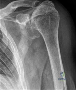
A 65-year-old female presents with a 3-part proximal humerus fracture.

An axillary nerve injury is suspected. Which of the following muscles is primarily evaluated to assess the motor function of this nerve?


Explanation

The axillary nerve innervates the deltoid and teres minor muscles. Teres major is innervated by the lower subscapular nerve, subscapularis by upper and lower subscapular nerves, and infra/supraspinatus by the suprascapular nerve.

Question 3

A 30-year-old male falls and sustains a closed humeral shaft fracture at the distal third (Holstein-Lewis type). He is unable to extend his wrist or digits at presentation. He is placed in a coaptation splint. At 12 weeks of follow-up, radiographs show bridging callus, but there is no clinical sign of nerve recovery. What is the next best step in management?


Explanation

Initial management for closed humeral shaft fractures with primary radial nerve palsy is observation. If there is no clinical evidence of recovery by 12 weeks (3 months), an EMG/NCS should be ordered to evaluate for subclinical reinnervation or severe denervation, which would dictate the need for surgical exploration.

Question 4

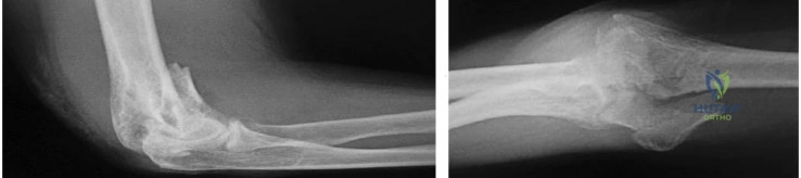
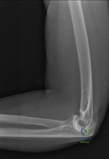
A 40-year-old male presents with a Terrible Triad injury of the elbow.

Surgical management is planned. What is the generally accepted sequence of repair to restore elbow stability?


Explanation

The standard surgical algorithm for a terrible triad injury progresses from deep to superficial and medial to lateral: 1) Coronoid fixation to restore the anterior buttress, 2) Radial head fixation or replacement to restore the lateral column, and 3) Lateral ulnar collateral ligament (LUCL/LCL complex) repair. The MCL is only addressed if the elbow remains unstable after these steps.

Question 5

A 22-year-old male undergoes open reduction and internal fixation of a Galeazzi fracture-dislocation. Intraoperatively, following anatomic fixation of the radius, the distal radioulnar joint (DRUJ) remains irreducible in neutral and pronation, and blocks supination. What is the most likely interposed structure preventing DRUJ reduction?


Explanation

In Galeazzi fracture-dislocations, if the DRUJ is irreducible following anatomic radius fixation, soft tissue interposition must be suspected. The most common interposed structure is the extensor carpi ulnaris (ECU) tendon, requiring open exploration and extraction from the joint.

Question 6

In a patient with a volar Barton's fracture, the volar lunate facet fragment escapes fixation and displaces volarly. Which of the following carpal ligaments is at greatest risk of disruption, leading to volar subluxation of the lunate?


Explanation

The short radiolunate ligament originates on the volar lunate facet of the distal radius and inserts on the volar aspect of the lunate. It acts as a primary stabilizer of the lunate. Loss of this volar marginal fragment (the 'critical corner') leads to volar lunate subluxation and severe carpal instability.

Question 7

A 28-year-old male presents with a scaphoid waist fracture nonunion.

MRI demonstrates avascular necrosis (AVN) of the proximal pole with a structural humpback deformity. Which of the following is the most appropriate graft option to achieve union and correct the deformity?


Explanation

A free vascularized medial femoral condyle (MFC) bone graft is highly effective for scaphoid nonunions complicated by proximal pole AVN and structural collapse (humpback deformity), as it provides both robust vascularity and a strong corticocancellous strut. The 1,2 ICSRA graft provides less reliable vascularity in advanced AVN and less structural support.

Question 8

A 32-year-old male sustains a perilunate dislocation. The 'Space of Poirier' represents an area of capsular weakness allowing the lunate to dislocate volarly. This space is located between which of the following capsuloligamentous structures?


Explanation

The Space of Poirier is a naturally occurring V-shaped area of weakness in the volar wrist capsule, located between the radioscaphocapitate (RSC) ligament and the long radiolunate (LRL) ligament. It opens up during wrist hyperextension, allowing volar dislocation of the lunate in stage IV perilunate injuries.

Question 9

A 35-year-old female with severe carpal tunnel syndrome exhibits thenar atrophy.

Which of the following intrinsic muscles of the hand is innervated by the recurrent motor branch of the median nerve?


Explanation

The recurrent motor branch of the median nerve innervates the 'OAF' muscles: Opponens pollicis, Abductor pollicis brevis, and the superficial head of the Flexor pollicis brevis. The deep head of FPB, adductor pollicis, palmaris brevis, and interossei are innervated by the ulnar nerve.

Question 10

A 45-year-old male is undergoing an anterior submuscular transposition of the ulnar nerve for recalcitrant cubital tunnel syndrome. During the procedure, the nerve is placed deep to which of the following muscular structures?


Explanation

In a submuscular transposition, the ulnar nerve is moved anteriorly and placed deep to the flexor-pronator mass (which includes the pronator teres, flexor carpi radialis, palmaris longus, flexor digitorum superficialis, and flexor carpi ulnaris origins). It is typically laid against the brachialis and flexor digitorum profundus.

Question 11

During repair of a Zone II flexor tendon injury, understanding the blood supply is critical. The vinculum breve to the flexor digitorum superficialis (FDS) tendon inserts at which anatomic location?


Explanation

The vincula are critical for tendon nutrition. The vinculum breve of the FDS inserts on the distal aspect of the proximal phalanx, blending with the volar plate of the PIP joint. The vinculum longum of the FDS is located more proximally.

Question 12

A 45-year-old male presents with a chronic mallet deformity of the ring finger and a secondary swan neck deformity. What is the primary pathoanatomic mechanism causing the PIP joint hyperextension (swan neck) in this setting?


Explanation

In a chronic mallet finger, the terminal extensor tendon is ruptured, leading to a loss of extension at the DIP joint. The extensor mechanism subsequently retracts proximally, which pulls the lateral bands dorsally and proximally. This translates into an exaggerated extension force across the PIP joint, resulting in a secondary swan neck deformity.

Question 13

In a complete tear of the ulnar collateral ligament (UCL) of the thumb (Skier's thumb), a Stener lesion may occur. This lesion is characterized by the proximal stump of the torn UCL displacing superficial to which of the following structures?


Explanation

A Stener lesion occurs when the distal attachment of the thumb UCL avulses and gets flipped proximally and superficial to the adductor aponeurosis. The aponeurosis interposes between the torn ligament ends, preventing spontaneous healing and thus necessitating surgical repair.

Question 14

A 28-year-old volleyball player presents with insidious onset of shoulder pain and weakness.

Examination reveals weakness in external rotation but normal strength in abduction. Entrapment of the suprascapular nerve is suspected. At what anatomical site is the compression most likely occurring?


Explanation

The suprascapular nerve innervates the supraspinatus (shoulder abduction) and infraspinatus (external rotation). Compression at the spinoglenoid notch, often due to a paralabral cyst from a posterior labral tear, affects only the branch to the infraspinatus, sparing the supraspinatus. Compression at the suprascapular notch would affect both.

Question 15

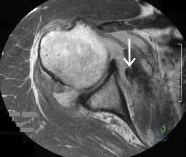
A 24-year-old athlete sustains recurrent anterior shoulder dislocations. An MRI reveals a Hill-Sachs lesion. Advanced imaging and 3D modeling demonstrate that the lesion 'engages' the anterior glenoid rim when the arm is in abduction and external rotation. According to the glenoid track concept, how is this lesion classified?


Explanation

In the glenoid track concept, if a Hill-Sachs lesion extends medially beyond the glenoid track, it will drop over the anterior rim of the glenoid during abduction and external rotation (engaging). This is termed an 'off-track' lesion and typically requires a remplissage procedure or bone block in addition to a Bankart repair.

Question 16

A patient sustains a Type III acromioclavicular (AC) joint separation. The coracoclavicular (CC) ligaments are ruptured. Which CC ligament originates from the base of the coracoid and inserts onto the conoid tubercle of the clavicle?


Explanation

The coracoclavicular (CC) ligament complex consists of the conoid and trapezoid ligaments. The conoid ligament is the more medial and posterior of the two, originating at the base of the coracoid and inserting on the conoid tubercle. The trapezoid ligament is more lateral and anterior.

Question 17

During arthroscopic evaluation of the shoulder, a Type II SLAP tear is identified. The 'peel-back' mechanism is tested to assess the dynamic instability of the biceps anchor. This mechanism is maximally provoked in which shoulder position?


Explanation

The 'peel-back' mechanism occurs when the arm is placed in abduction and external rotation (ABER position). In this position, the vector of the biceps tendon shifts posteriorly and medially, creating a torsional force that 'peels back' the superior labrum off the glenoid rim.

Question 18

A 30-year-old manual laborer presents with progressive dorsal wrist pain.

Radiographs show sclerosis and collapse of the lunate, consistent with Kienbock's disease. Based on the Lichtman classification, what specific radiographic finding differentiates Stage IIIA from Stage IIIB?


Explanation

Lichtman Stage III Kienbock's disease is characterized by lunate collapse. Stage IIIA has lunate collapse but maintains normal carpal alignment (normal scaphoid). Stage IIIB is defined by lunate collapse with fixed scaphoid flexion (rotatory subluxation of the scaphoid), which decreases carpal height.

Question 19

A 42-year-old tennis player undergoes surgical debridement for refractory lateral epicondylitis. Histologic examination of the excised tissue from the extensor carpi radialis brevis (ECRB) origin will most typically demonstrate which of the following?


Explanation

Lateral epicondylitis (tennis elbow) is fundamentally a tendinosis, not an acute tendinitis. Histologic examination classically shows angiofibroblastic hyperplasia, characterized by disorganized collagen, fibroblasts, and poorly formed blood vessels, with an absence of acute inflammatory cells.

Question 20

A 45-year-old bodybuilder feels a pop in his anterior elbow during a heavy deadlift. Clinical exam shows a positive Hook test. A single-incision anterior approach is planned to repair the avulsed distal biceps tendon. During this approach, which nerve is at greatest risk of iatrogenic injury?


Explanation

The lateral antebrachial cutaneous nerve (LABCN) runs superficially in the lateral aspect of the antecubital fossa and is the most commonly injured nerve during the anterior single-incision approach to the distal biceps. The posterior interosseous nerve (PIN) is at higher risk during a two-incision approach or with deep, overzealous lateral retraction.

Question 21

A 45-year-old male feels a sudden 'pop' in his right elbow while attempting to lift a heavy box. On examination, he has a positive hook test and a visible proximal retraction of the biceps muscle belly. A single anterior incision approach is planned for distal biceps tendon repair. Which of the following nerves is at the greatest risk of iatrogenic injury during this specific surgical approach?


Explanation

The lateral antebrachial cutaneous nerve (LABC), which is the terminal sensory branch of the musculocutaneous nerve, exits lateral to the biceps tendon and is highly susceptible to neuropraxia or transection during the single-incision anterior approach to the distal biceps. The posterior interosseous nerve (PIN) is more at risk when exposing the radial tuberosity, particularly if retractors are placed too far laterally or distally, and is the classic nerve injured in a two-incision approach if the muscle splitting is incorrect, but the most frequently injured nerve overall in the single anterior incision is the LABC.

Question 22

A 24-year-old man falls onto an outstretched hand and presents with anatomic snuffbox tenderness. Radiographs reveal a non-displaced fracture through the proximal third of the scaphoid. The high risk of avascular necrosis in this fracture pattern is directly related to its blood supply. Which of the following arteries provides the primary blood supply to the proximal pole of the scaphoid?


Explanation

The dorsal carpal branch of the radial artery provides 70-80% of the blood supply to the scaphoid. It enters the bone at the dorsal ridge (distal to the waist) and flows in a retrograde fashion to supply the proximal pole. Because of this retrograde intraosseous blood supply, fractures at the waist or proximal third sever the blood flow to the proximal pole, leading to a high rate of avascular necrosis and nonunion. The volar carpal branch supplies only the distal 20-30% of the bone.

Question 23

A 55-year-old male with a history of a chronic untreated scapholunate ligament tear presents with increasing wrist pain. Radiographs reveal degenerative narrowing specifically at the articulation between the radial styloid and the scaphoid, with preservation of the rest of the radioscaphoid and midcarpal joints. What is the correct stage of Scapholunate Advanced Collapse (SLAC) in this patient?


Explanation

The SLAC wrist sequence follows a predictable pattern of degenerative changes based on altered biomechanics. Stage I: Osteoarthritis localized to the radial styloid and the distal scaphoid. Stage II: Involvement of the entire radioscaphoid joint. Stage III: Progression to involve the capitolunate joint. Note that the radiolunate joint is classically spared in SLAC wrist due to the concentric articulation of the lunate in the lunate fossa, which is maintained despite the scaphoid rotation.

Question 24

A 30-year-old manual laborer presents with chronic, aching dorsal wrist pain. MRI demonstrates avascular necrosis of the lunate. Plain radiographs show lunate sclerosis but normal architecture without collapse. Radiographic evaluation reveals an ulnar variance of -3 mm. Which of the following is the most appropriate initial surgical intervention if conservative management fails?


Explanation

The patient has Lichtman Stage II Kienbock's disease (lunate sclerosis without collapse) with ulnar minus (negative) variance. The goal of joint-leveling procedures is to decrease the compressive forces across the radiolunate joint. In a patient with negative ulnar variance, a radial shortening osteotomy (or alternatively, an ulnar lengthening osteotomy) is the procedure of choice. Proximal row carpectomy or four-corner fusion are salvage procedures reserved for advanced stages (Stage IIIb or IV) with carpal collapse and arthritis.

Question 25

A 50-year-old diabetic woman presents with triggering and locking of her long finger that has been refractory to two corticosteroid injections. She undergoes surgical release. During the procedure, the surgeon must identify and release the specific structure responsible for the pathology while preserving the critical mechanical pulleys. What is the correct proximal-to-distal sequence of the flexor tendon pulleys in the digit?


Explanation

The flexor tendon sheath contains five annular (A) pulleys and three cruciate (C) pulleys. The correct proximal-to-distal sequence is A1 (at the MCP joint), A2 (proximal phalanx), C1, A3 (PIP joint), C2, A4 (middle phalanx), C3, and A5 (DIP joint). The A2 and A4 pulleys are mechanically critical to prevent bowstringing and must be preserved. Trigger finger is caused by a stenosing tenosynovitis at the A1 pulley, which is the structure divided during release.

Question 26

A 25-year-old male sustains a deep glass laceration over the volar aspect of the proximal phalanx of his index finger. Exploration reveals that both the flexor digitorum superficialis (FDS) and flexor digitorum profundus (FDP) tendons are completely severed. In which flexor tendon zone did this injury occur?


Explanation

Flexor tendon Zone II extends from the proximal edge of the A1 pulley to the insertion of the FDS on the middle phalanx. Historically known as 'no man's land', lacerations in this zone frequently involve both the FDS and FDP tendons. Because the tendons are tightly enclosed within the fibro-osseous sheath in this zone, repairs are technically demanding and prone to restrictive adhesions. Zone I is distal to the FDS insertion (only FDP is present).

Question 27

A 22-year-old rugby player presents after a match unable to actively flex the DIP joint of his ring finger after grabbing an opponent's jersey. Radiographs reveal a large bony avulsion fragment from the volar base of the distal phalanx that is situated intra-articularly at the DIP joint. According to the Leddy-Packer classification of flexor digitorum profundus (FDP) avulsions, what type of injury is this?


Explanation

Jersey fingers are classified by Leddy and Packer based on the level of tendon retraction. Type I: Tendon retracts into the palm; requires repair within 7-10 days due to compromised blood supply. Type II: Tendon retracts to the level of the PIP joint, held by the intact vinculum longum. Type III: A large bony fragment is avulsed and catches at the A4 pulley, preventing proximal retraction; the fragment remains intra-articular at the DIP joint. Type IV involves both a bony avulsion and an independent avulsion of the tendon off the bony fragment.

Question 28

A 35-year-old skier falls while gripping his ski pole and presents with weakness in thumb pinch and pain over the ulnar aspect of the thumb MCP joint. MRI reveals a complete rupture of the ulnar collateral ligament (UCL). The distal end of the torn ligament is displaced, preventing spontaneous healing. Which structure is the torn UCL typically displaced superficial to in this classic lesion?


Explanation

The patient has a Stener lesion, a common complication of a complete thumb UCL rupture (Skier's/Gamekeeper's thumb). In a Stener lesion, the distal end of the completely torn UCL flips back and becomes trapped superficial to the adductor pollicis aponeurosis. Because the aponeurosis interposes between the torn ligament and its insertion on the proximal phalanx, anatomic healing cannot occur without surgical intervention.

Question 29

A 40-year-old female presents with aching pain in her anterior forearm and numbness in the radial 3.5 digits. On examination, she has a negative Tinel's sign at the wrist. She demonstrates numbness over the thenar eminence. Furthermore, she has weakness in thumb interphalangeal joint flexion and index finger DIP joint flexion. What is the most likely primary site of nerve compression?


Explanation

This patient has Pronator Syndrome, a proximal median neuropathy. The key findings differentiating this from Carpal Tunnel Syndrome (CTS) are the numbness over the thenar eminence (supplied by the palmar cutaneous branch of the median nerve, which branches proximal to the transverse carpal ligament) and motor weakness in the Anterior Interosseous Nerve (AIN) distribution (FPL and FDP to the index finger). The most common site of compression in Pronator Syndrome is between the two heads of the pronator teres muscle. The Ligament of Struthers is a much rarer cause of proximal median nerve compression that would also affect the pronator teres muscle itself.

Question 30

A competitive cyclist presents with numbness in his small finger and the ulnar half of his ring finger on the volar aspect, along with weakness of finger abduction and adduction. Sensation over the dorso-ulnar aspect of the hand is completely intact. Based on these findings, compression of the ulnar nerve is most likely occurring in which zone of Guyon's canal?


Explanation

Guyon's canal is divided into three zones. Zone 1 is proximal to the bifurcation of the ulnar nerve into superficial and deep branches; compression here causes both motor (intrinsic muscle weakness) and volar sensory deficits (numbness in volar small/ring fingers). The dorsal ulnar cutaneous nerve branches 5-8 cm proximal to the wrist, so its territory (dorso-ulnar hand) is spared in Guyon's canal compression, distinguishing it from cubital tunnel syndrome. Zone 2 compression affects only the deep motor branch. Zone 3 compression affects only the superficial sensory branch.

Question 31

A 45-year-old mechanic complains of lateral elbow pain radiating down the dorsal forearm. The pain is exacerbated by repetitive pronation and supination. On examination, maximal point tenderness is localized approximately 4 to 5 cm distal to the lateral epicondyle in the extensor muscle mass. Resisted active extension of the middle finger (with the elbow extended) reproduces the pain. There is no demonstrable motor weakness or finger drop. What is the most likely diagnosis?


Explanation

Radial tunnel syndrome represents a painful compression of the radial nerve (typically the posterior interosseous nerve branch) in the proximal forearm without classic motor weakness. It is often confused with lateral epicondylitis; however, the point of maximal tenderness in radial tunnel syndrome is 4-5 cm distal to the lateral epicondyle over the supinator/mobile wad, whereas in lateral epicondylitis it is directly over or immediately adjacent to the epicondyle. PIN syndrome typically presents with motor weakness (inability to extend digits/thumb) rather than just pain. Wartenberg's syndrome is compression of the superficial sensory branch of the radial nerve in the distal forearm.

Question 32

A 28-year-old gymnast presents with ulnar-sided wrist pain and clicking after a fall. Examination reveals a positive fovea sign. Arthroscopy confirms a traumatic avulsion of the Triangular Fibrocartilage Complex (TFCC) from its insertion at the fovea of the ulnar head. According to the Palmer classification, what type of TFCC tear is this?


Explanation

The Palmer classification divides TFCC tears into traumatic (Class 1) and degenerative (Class 2). Class 1A is a central perforation (avascular zone, typically debrided). Class 1B is an ulnar avulsion (from the ulnar styloid or fovea, highly vascular, amenable to repair). Class 1C is a distal avulsion involving the ulnocarpal ligaments. Class 1D is a radial avulsion (from the sigmoid notch of the radius). Class 2 tears involve degenerative wear.

Question 33

A 50-year-old male is undergoing in-situ decompression of the ulnar nerve for severe cubital tunnel syndrome. The surgeon meticulously releases the nerve along its entire course around the elbow to prevent postoperative tethering. Which of the following is the most common anatomical site of ulnar nerve compression in this region?


Explanation

The most common site of ulnar nerve compression at the elbow is at the cubital tunnel retinaculum, also known as Osborne's ligament or fascia. This thick band extends from the medial epicondyle to the olecranon and bridges the two heads of the flexor carpi ulnaris (FCU). Other potential sites of compression include the Arcade of Struthers (proximal), medial intermuscular septum, the deep flexor-pronator aponeurosis, and occasionally an anomalous muscle like the anconeus epitrochlearis, but Osborne's ligament is the most frequently implicated structure.

Question 34

A 60-year-old female presents with pain at the base of her thumb. She has a positive grind test. Radiographs demonstrate joint space narrowing, subchondral sclerosis, and small osteophytes (< 2 mm) at the trapeziometacarpal joint. The scaphotrapezial joint appears completely normal. According to the Eaton-Littler classification of thumb CMC arthritis, what is her stage?


Explanation

The Eaton-Littler classification for thumb CMC arthritis: Stage I: Normal joint contours, possible widening from effusion/laxity. Stage II: Mild joint space narrowing, sclerosis, and osteophytes < 2 mm. Stage III: Significant joint destruction, cystic changes, and osteophytes > 2 mm, but the scaphotrapezial (ST) joint is normal. Stage IV: Involvement of both the trapeziometacarpal and the scaphotrapezial (pantrapezial) joints.

Question 35

A 32-year-old construction worker falls from a scaffolding. Lateral wrist radiographs demonstrate the lunate completely displaced volar to the radius and tipped forward like a 'spilled teacup', while the capitate remains aligned with the longitudinal axis of the radius. According to the Mayfield classification of carpal instability, what stage of injury has occurred?


Explanation

Mayfield described a progressive sequence of perilunate instability based on the path of force around the lunate. Stage I: Scapholunate dissociation. Stage II: Perilunate dislocation (capitate dislocates dorsally while lunate remains in the radial fossa). Stage III: Lunotriquetral disruption. Stage IV: Lunate dislocation (the lunate is squeezed out volarly into the carpal tunnel, appearing as a 'spilled teacup' on the lateral radiograph, and the capitate drops back to articulate with the radius).

Question 36

A macrosomic newborn is noted to have a flaccid right upper extremity immediately after a difficult vaginal delivery involving shoulder dystocia. On examination, the infant has an absent grasp reflex and an ipsilateral ptosis and miosis. However, shoulder abduction and elbow flexion are spontaneous and intact. Which nerve roots are predominantly injured in this pattern of brachial plexus birth palsy?


Explanation

The patient's presentation is classic for Klumpke's palsy, which is a rare lower brachial plexus injury involving roots C8 and T1. It results in absent hand and wrist function (absent grasp reflex) but preserved proximal muscle function (shoulder abduction/elbow flexion, supplied by C5/C6). The presence of Horner's syndrome (ptosis, miosis, anhidrosis) indicates avulsion of the T1 root, which carries sympathetic fibers to the superior cervical ganglion. Erb's palsy (C5, C6) presents with the classic 'waiter's tip' posture and intact hand grasp.

Question 37

A 40-year-old male presents with sudden, severe, unprovoked pain in his right shoulder that awoke him from sleep. The pain lasted for four days and required opioids. As the pain subsided, he noticed profound weakness in shoulder abduction and external rotation. He denies any trauma. MRI of the shoulder is unremarkable. EMG obtained 4 weeks later demonstrates denervation potentials in the supraspinatus and infraspinatus muscles. What is the most likely diagnosis?


Explanation

Parsonage-Turner syndrome (idiopathic brachial neuritis) classically presents with an acute onset of severe, unremitting shoulder or arm pain followed days to weeks later by patchy muscle weakness and atrophy as the pain resolves. It often affects the suprascapular nerve, long thoracic nerve, or anterior interosseous nerve. The lack of trauma, unremarkable MRI, and classic sequence of severe pain followed by flaccid paresis point away from mechanical tears or structural entrapments. Entrapment at the spinoglenoid notch would typically spare the supraspinatus.

Question 38

A 35-year-old diabetic patient presents to the emergency department with a swollen, throbbing index finger 3 days after sustaining a puncture wound. The physician suspects acute purulent flexor tenosynovitis. Which of the following is NOT one of Kanavel's cardinal signs for this condition?


Explanation

Kanavel's four cardinal signs of acute flexor tenosynovitis are: 1) Fusiform (sausage-like) swelling of the digit. 2) The digit is held in a posture of slight FLEXION (not extension) to minimize tension on the inflamed sheath. 3) Tenderness along the entire course of the flexor tendon sheath. 4) Disproportionate, severe pain with passive extension of the digit.

Question 39

A 45-year-old male with a 10-year history of an untreated scaphoid waist fracture presents with progressive wrist pain and stiffness. Radiographs demonstrate advanced carpal collapse. In Scaphoid Nonunion Advanced Collapse (SNAC) wrist, the earliest arthritic changes typically manifest at which of the following articulations?


Explanation

In a SNAC wrist, the scaphoid is ununited. The proximal pole remains attached to the lunate via the intact scapholunate ligament and maintains a concentric articulation with the scaphoid fossa of the radius. The distal pole flexes and rotates, creating an incongruent articulation with the radial styloid. Therefore, Stage I of SNAC wrist arthritis begins at the articulation between the radial styloid and the distal pole of the scaphoid. Stage II involves the scaphocapitate joint, and Stage III involves the capitolunate joint.

Question 40

A 55-year-old male sustains a displaced transverse olecranon fracture after a ground-level fall. The surgeon decides to fix the fracture using tension band wiring. Biomechanically, the primary purpose of tension band fixation in this scenario is to convert which type of force at the dorsal cortex into what type of force at the articular surface?


Explanation

The biomechanical principle of a tension band is to counteract distractive (tensile) forces on the convex (tension) side of a bone and convert them into compressive forces on the concave (articular) side during joint movement. In an olecranon fracture, the pull of the triceps creates tensile forces on the dorsal cortex. The figure-of-eight wire placed on the dorsal surface absorbs these tensile forces, compressing the articular surface of the greater sigmoid notch together during elbow flexion.

Question 41

A 45-year-old male sustains a 'terrible triad' injury of the elbow after a fall on an outstretched hand. Which of the following represents the most widely accepted sequence of surgical reconstruction to restore elbow stability?


Explanation

The standard surgical protocol for a terrible triad of the elbow (elbow dislocation, radial head fracture, coronoid fracture) typically proceeds from deep to superficial: 1) Coronoid fixation or anterior capsule repair, 2) Radial head fixation or arthroplasty, and 3) Lateral ulnar collateral ligament (LUCL) repair. If the elbow remains unstable after these steps, the MCL may be repaired or an external fixator applied.

Question 42

A 55-year-old manual laborer presents with advanced Scapholunate Advanced Collapse (SLAC) of the wrist. Radiographs reveal diffuse radiocarpal and midcarpal arthritis. Which of the following carpal articulations is classically SPARED in a SLAC wrist, allowing for motion-preserving salvage procedures such as a four-corner fusion?


Explanation

In SLAC wrist, the radiolunate joint is classically spared. This is due to the spherical congruence of the radiolunate articulation, which does not experience the same sheer forces and translation that lead to articular wear at the radioscaphoid and capitolunate joints. This anatomic feature allows for proximal row carpectomy or scaphoid excision with four-corner fusion to be viable options.

Question 43

A 24-year-old male sustains a C5-C6 brachial plexus avulsion injury (Erb's palsy). Six months post-injury, he has no elbow flexion but normal hand function. An Oberlin transfer is planned. Which of the following describes the classic Oberlin I transfer?


Explanation

The classic Oberlin transfer (Oberlin I) involves taking a redundant fascicle from the ulnar nerve (typically one supplying the FCU) and transferring it to the motor branch of the biceps (part of the musculocutaneous nerve) to restore elbow flexion in upper trunk brachial plexus injuries.

Question 44

A 19-year-old male presents with a scaphoid waist fracture. Which of the following best describes the predominant vascular supply to the scaphoid that dictates its risk of nonunion?


Explanation

The major blood supply to the scaphoid comes from the dorsal carpal branch of the radial artery, which enters the bone at the dorsal ridge (distal to the waist) and provides retrograde blood flow to the proximal 70-80% of the scaphoid. This retrograde supply is the primary reason proximal pole fractures are at a high risk for avascular necrosis and nonunion.

Question 45

According to the Mayfield classification of perilunate instability, what is the anatomic sequence of ligamentous disruption around the lunate as the severity of injury progresses from Stage I to Stage IV?


Explanation

The Mayfield progression of perilunate instability occurs in a specific sequence around the lunate: Stage I (Scapholunate ligament disruption), Stage II (Capitolunate joint disruption/dislocation), Stage III (Lunotriquetral ligament disruption), and Stage IV (Lunate dislocation, failing the dorsal radiocarpal ligament and extruding the lunate volarly).

Question 46

A 32-year-old female sustains an Essex-Lopresti injury. She undergoes radial head excision without prosthetic replacement. Which of the following is the most likely late complication?


Explanation

An Essex-Lopresti injury consists of a radial head fracture, tear of the interosseous membrane, and disruption of the DRUJ. If the radial head is excised without being replaced, the radius will migrate proximally due to the loss of the stabilizing interosseous membrane and radial head. This leads to positive ulnar variance and severe ulnar impaction syndrome.

Question 47

A patient presents with intrinsic muscle weakness in the hand but normal sensation over the volar small finger and dorsal ulnar aspect of the hand. Electromyography confirms compression of the ulnar nerve in Guyon's canal. This presentation is most consistent with compression at which zone?


Explanation

Guyon's canal is divided into 3 zones. Zone 1 is proximal to the nerve bifurcation (motor and sensory deficits). Zone 2 encompasses the deep motor branch after the bifurcation, typically at the hook of the hamate (motor deficits only). Zone 3 encompasses the superficial sensory branch (sensory deficits only).

Question 48

A 40-year-old male develops a boutonniere deformity of the index finger following a crush injury. What is the underlying pathoanatomy responsible for this specific deformity?


Explanation

A boutonniere deformity is characterized by PIP joint flexion and DIP joint hyperextension. It is caused by rupture or attenuation of the central slip of the extensor mechanism, which allows the lateral bands to subluxate volarly to the axis of rotation of the PIP joint. They then act as flexors of the PIP while continuing to extend the DIP joint.

Question 49

A 65-year-old female presents with severe pain at the base of the thumb. Radiographs demonstrate Eaton-Littler Stage IV carpometacarpal (CMC) arthritis. Which radiographic finding distinguishes Stage IV from Stage III?


Explanation

The Eaton-Littler classification of thumb CMC arthritis: Stage I (subtle widening), Stage II (slight narrowing, osteophytes <2mm), Stage III (significant narrowing, osteophytes >2mm, subluxation), and Stage IV (pantrapezial arthritis, which includes involvement of the scaphotrapezotrapezoidal [STT] joint).

Question 50

A 6-year-old boy sustains a supracondylar humerus fracture. Radiographs show posterolateral displacement of the distal fragment. Which nerve is at the highest risk of injury in this specific displacement pattern?


Explanation

In a supracondylar fracture with posterolateral displacement of the distal fragment, the proximal fragment is directed anteromedially, placing the median nerve (specifically the AIN) and brachial artery at highest risk. Conversely, posteromedial displacement of the distal fragment directs the proximal fragment anterolaterally, risking the radial nerve.

Question 51

A 5-year-old girl is treated nonoperatively for a seemingly minimally displaced pediatric lateral condyle fracture. One year later, she is noted to have a nonunion. What is the most common long-term deformity and associated neurologic complication if left untreated?


Explanation

Nonunion of a lateral condyle fracture in a child typically leads to a progressive cubitus valgus deformity as the medial condyle continues to grow while the lateral side does not. This valgus deformity stretches the ulnar nerve over time, leading to a tardy ulnar nerve palsy.

Question 52

During surgical decompression of the ulnar nerve for cubital tunnel syndrome, the surgeon must divide the roof of the cubital tunnel. What structure forms the roof of this tunnel?


Explanation

The roof of the cubital tunnel is formed by Osborne's ligament (the aponeurosis between the humeral and ulnar heads of the flexor carpi ulnaris). The floor is formed by the posterior band of the medial collateral ligament (MCL) and the elbow joint capsule.

Question 53

A 58-year-old male with Dupuytren's disease undergoes a fasciectomy for a severe PIP joint contracture. During dissection, the neurovascular bundle is noted to be displaced volarly and centrally. Which pathologic structure is primarily responsible for PIP joint contracture and this neurovascular displacement?


Explanation

The spiral cord is responsible for PIP joint contracture in Dupuytren's disease and is clinically vital because it displaces the neurovascular bundle centrally, volarly, and proximally, placing it at high risk during surgical excision. It is composed of the pretendinous band, spiral band, lateral digital sheet, and Grayson's ligament.

Question 54

A 28-year-old male sustains a closed spiral fracture of the distal third of the humeral shaft (Holstein-Lewis fracture). On initial presentation in the emergency department, his radial nerve function is intact. Following closed reduction and splinting, he completely loses active wrist and digit extension. What is the most appropriate management regarding the nerve injury?


Explanation

A secondary radial nerve palsy (loss of radial nerve function AFTER a closed reduction maneuver) in a humeral shaft fracture is an absolute indication for surgical exploration. The nerve may be entrapped between the fracture fragments. Primary palsies (present on initial exam) can often be observed, but secondary palsies require exploration.

Question 55

A 35-year-old male develops acute compartment syndrome of the volar forearm after a crush injury. If left untreated, Volkmann's ischemic contracture will develop. Which muscles are most severely and earliest affected in the deep volar compartment?


Explanation

The deep volar compartment of the forearm contains the Flexor Digitorum Profundus (FDP) and Flexor Pollicis Longus (FPL). Because they lie deep against the bone and their blood supply is most vulnerable to increased compartment pressures, they are the most severely and earliest affected muscles in Volkmann's ischemic contracture.

Question 56

When performing a primary flexor tendon repair in Zone II of the hand, biomechanical studies suggest that the strength of the repair is most directly proportional to which of the following factors?


Explanation

In flexor tendon repairs, biomechanical studies demonstrate that the tensile strength of the repair is directly proportional to the number of core suture strands crossing the repair site. A 4-strand or 6-strand repair is significantly stronger than a 2-strand repair and allows for early active motion protocols.

Question 57

A 45-year-old female sustains a volar Barton's fracture of the distal radius. This fracture pattern involves a volar marginal articular fragment that subluxates with the carpus. Which critical radiocarpal ligament complex remains attached to this volar fragment, mediating the volar subluxation of the carpus?


Explanation

A volar Barton's fracture is a shear fracture of the volar rim of the distal radius (typically involving the volar lunate facet). The volar radiolunate ligaments (short and long) remain attached to this fragment, and because the carpus remains tethered to this fragment via these ligaments, the entire carpus subluxates volarly with the fractured rim.

Question 58

A 30-year-old female presents with true neurogenic thoracic outlet syndrome (TOS). She requires surgical decompression. The most common site of nerve compression in this syndrome involves the interscalene triangle. What are the anatomic borders of this triangle?


Explanation

The interscalene triangle is the most common site of compression in neurogenic thoracic outlet syndrome. Its borders are the anterior scalene muscle anteriorly, the middle scalene muscle posteriorly, and the superior border of the first rib inferiorly. The brachial plexus trunks and the subclavian artery pass through this triangle (the subclavian vein runs anterior to the anterior scalene and is not within the triangle).

Question 59

A 25-year-old male is diagnosed with Kienbock's disease (avascular necrosis of the lunate). According to the Lichtman classification, what finding distinguishes Stage IIIA from Stage IIIB?


Explanation

In the Lichtman classification for Kienbock's disease, Stage III is characterized by lunate collapse. It is subdivided based on carpal mechanics: Stage IIIA has lunate collapse but normal carpal height and alignment. Stage IIIB has lunate collapse accompanied by fixed scaphoid rotary subluxation (a sign of carpal instability/collapse and decreased carpal height).

Question 60

A 42-year-old male presents with acute, severe, unremitting right shoulder pain that awakened him from sleep, lasting for 5 days before resolving. He subsequently developed profound weakness in shoulder abduction and external rotation. Cervical spine MRI is unremarkable. Which of the following is the most likely diagnosis?


Explanation

Parsonage-Turner syndrome (neuralgic amyotrophy) is an acute idiopathic brachial neuritis. The classic presentation involves sudden, severe, non-traumatic shoulder or upper extremity pain lasting days to weeks, followed by patchy lower motor neuron weakness (commonly affecting the suprascapular nerve, axillary nerve, or anterior interosseous nerve) and muscle atrophy as the pain subsides.

Question 61

A 35-year-old mechanic presents with chronic wrist pain and limited extension following an untreated fall on an outstretched hand 5 years ago. Radiographs demonstrate scapholunate dissociation with radioscaphoid and capitolunate arthritis, but sparing of the radiolunate articulation. Which of the following is the most appropriate definitive management?


Explanation

This patient has Scapholunate Advanced Collapse (SLAC) stage III, which involves the capitolunate joint. Proximal row carpectomy is contraindicated due to capitate arthritis, making four-corner fusion the treatment of choice.

Question 62

A 42-year-old female sustains a terrible triad injury of the elbow (elbow dislocation, radial head fracture, and coronoid fracture). During surgical reconstruction, what is the most appropriate standard sequence of repair?


Explanation

The standard sequence for reconstructing a terrible triad injury is working deep to superficial: coronoid fixation, radial head fixation or replacement, followed by lateral collateral ligament (LCL) and extensor origin repair.

Question 63

A 28-year-old male sustains a diaphyseal fracture of the radius and ulna. He undergoes open reduction and internal fixation. To minimize the risk of radioulnar synostosis, what surgical technique principle should be strictly followed?


Explanation

Radioulnar synostosis is a severe complication of both-bone forearm fractures. To minimize this risk, surgeons should utilize separate approaches (e.g., volar Henry and dorsal ulnar) and maintain meticulous subperiosteal dissection.

Question 64

A 32-year-old male sustains a Galeazzi fracture-dislocation. Following rigid internal fixation of the radius, the distal radioulnar joint (DRUJ) is evaluated and found to be unstable in all positions of forearm rotation. What is the most appropriate next step in management?


Explanation

If the DRUJ remains grossly unstable in all planes after rigid radial fixation in a Galeazzi fracture, the joint must be stabilized via percutaneous transfixation pins or open TFCC repair.

Question 65

A 45-year-old male presents with a proximal-third ulnar shaft fracture and an associated radial head dislocation. The patient is diagnosed with a Bado Type I Monteggia fracture. Which of the following defines the direction of the radial head dislocation in a Bado Type I injury?


Explanation

Bado Type I Monteggia fractures are characterized by an anterior dislocation of the radial head along with a diaphyseal fracture of the ulna with anterior angulation.

Question 66

A 55-year-old male undergoes open reduction and internal fixation of a distal humerus intercondylar fracture via an olecranon osteotomy approach. Which of the following is the most frequent complication associated specifically with the olecranon osteotomy?


Explanation

Symptomatic prominent hardware at the osteotomy site is the most common complication of an olecranon osteotomy, frequently necessitating secondary removal.

Question 67

A 60-year-old female presents with a comminuted distal radius fracture and undergoes volar locked plating. During screw placement in the most distal row, the surgeon must be careful to avoid dorsal cortex penetration to prevent tendon rupture in which dorsal extensor compartment?


Explanation

Dorsal screw prominence past the distal radius cortex frequently irritates or ruptures the extensor pollicis longus (EPL) tendon, which resides in the third dorsal compartment.

Question 68

A 24-year-old rock climber presents with a popping sensation and pain in his right ring finger while bearing weight on a crimp hold. Clinical examination reveals pain and swelling over the volar aspect of the proximal phalanx, exacerbated by resisted PIP joint flexion. Which flexor tendon pulley is most likely injured?


Explanation

The A2 pulley, located at the level of the proximal phalanx, is the most critical biomechanical pulley and the most frequently ruptured in rock climbers.

Question 69

A 50-year-old diabetic female complains of progressive right shoulder stiffness and pain over 4 months. She has profound restriction in both active and passive external rotation. Radiographs are normal. What is the most appropriate initial management?


Explanation

The patient has adhesive capsulitis (frozen shoulder), highly correlated with diabetes. Initial treatment is an intra-articular glenohumeral corticosteroid injection combined with a stretching program.

Question 70

A 38-year-old male suffers a high-energy dashboard injury resulting in a posterior dislocation of the shoulder. He is noted to have an anteromedial humeral head defect (reverse Hill-Sachs lesion) involving 35% of the articular surface. Which of the following is the most appropriate surgical treatment?


Explanation

For reverse Hill-Sachs lesions involving 20% to 40% of the humeral head, transfer of the lesser tuberosity and subscapularis into the defect (McLaughlin or modified McLaughlin procedure) is the standard of care.

Question 71

A 72-year-old female sustains a 4-part proximal humerus fracture. She has a history of severe osteoporosis and known advanced glenohumeral osteoarthritis. Which of the following treatments provides the most reliable functional outcome with the lowest rate of revision?


Explanation

In an elderly patient with a 4-part fracture, poor bone stock, and preexisting osteoarthritis, a reverse total shoulder arthroplasty provides reliable pain relief and functional restoration without relying on tuberosity healing.

Question 72

A 28-year-old athlete undergoes an isolated arthroscopic SLAP repair. Postoperatively, he experiences profound, isolated weakness in external rotation of the shoulder, with normal deltoid function and internal rotation. Suture anchor placement most likely injured which nerve?


Explanation

The suprascapular nerve passes through the spinoglenoid notch near the posterosuperior glenoid. Misplaced posterosuperior suture anchors can tether or injure this nerve, causing isolated infraspinatus weakness.

Question 73

A 45-year-old female presents after a fall on an outstretched hand with a 'terrible triad' injury of the elbow. To properly restore elbow stability, what is the most widely accepted sequence of surgical repair?


Explanation

The standard surgical algorithm for a terrible triad injury is to start deep and work outward: fix or replace the coronoid first, then address the radial head, and finally repair the LCL complex to restore lateral stability.

Question 74

A 22-year-old male sustains a proximal pole scaphoid fracture. Which of the following describes the primary blood supply to the scaphoid and explains the high risk of avascular necrosis in proximal pole fractures?


Explanation

The primary blood supply to the scaphoid is from the dorsal carpal branch of the radial artery, which enters distally and provides retrograde blood flow to the proximal pole, making it highly susceptible to avascular necrosis.

Question 75

Six weeks after being treated non-operatively for a non-displaced distal radius fracture, a 68-year-old woman presents with a sudden inability to actively extend her thumb interphalangeal (IP) joint. Which tendon transfer is the most appropriate definitive management?


Explanation

Spontaneous rupture of the EPL tendon is a known complication of non-displaced distal radius fractures. The EIP to EPL tendon transfer is the gold standard treatment to restore thumb IP extension.

Question 76

A 22-year-old male athlete presents with recurrent anterior shoulder instability. MRI reveals an anterior capsulolabral avulsion (Bankart lesion). Which of the following glenohumeral ligaments is the primary restraint to anterior translation of the humeral head at 90 degrees of abduction?


Explanation

The anterior band of the inferior glenohumeral ligament (IGHL) is the primary static restraint against anterior translation of the humeral head when the shoulder is abducted to 90 degrees and externally rotated.

Question 77

Which of the following clinical scenarios is considered an ABSOLUTE indication for operative fixation of a humeral shaft fracture?


Explanation

An open fracture with an associated vascular injury requiring repair is an absolute indication for internal or external fixation of a humeral shaft fracture to stabilize the repaired vessels.

Question 78

A 7-year-old boy presents with a closed fracture of the proximal third of the ulna associated with an anterior dislocation of the radial head. According to the Bado classification, what type of Monteggia lesion is this?


Explanation

A Bado Type I Monteggia fracture is characterized by a fracture of the proximal or middle third of the ulna with an anterior dislocation of the radial head. It is the most common type in children.

Question 79

An adult patient sustains a Galeazzi fracture-dislocation. What is the standard of care for the definitive management of this injury?


Explanation

Galeazzi fractures (distal third radius fracture with DRUJ disruption) in adults are highly unstable and require ORIF of the radius, followed by intraoperative assessment and necessary stabilization of the distal radioulnar joint (DRUJ).

Question 80

A 72-year-old female presents with chronic pseudoparalysis of the shoulder. Radiographs demonstrate severe glenohumeral osteoarthritis with superior migration of the humeral head articulating with the acromion. What is the most appropriate surgical intervention?


Explanation

Reverse total shoulder arthroplasty is the treatment of choice for rotator cuff tear arthropathy. It medializes and distalizes the center of rotation, allowing the deltoid to effectively elevate the arm.

Question 81

In a patient presenting with cubital tunnel syndrome, which of the following represents the most common anatomic site of ulnar nerve compression?


Explanation

Osborne's ligament, a fascial band bridging the two heads of the flexor carpi ulnaris, is the most common site of ulnar nerve compression in cubital tunnel syndrome.

Question 82

A patient suffers a deep laceration on the volar aspect of their hand, resulting in transection of both the flexor digitorum superficialis and profundus tendons in the region between the A1 pulley and the FDS insertion. According to the Verdan classification, which flexor tendon zone is injured?


Explanation

Zone II (historically called 'no man/'s land') extends from the proximal edge of the A1 pulley to the insertion of the FDS tendon on the middle phalanx. Both FDS and FDP travel together in the flexor sheath here.

Question 83

During open carpal tunnel release, care must be taken to identify and protect the contents of the carpal tunnel. Which of the following structures is NOT contained within the carpal tunnel?


Explanation

The carpal tunnel contains 9 tendons (4 FDS, 4 FDP, 1 FPL) and the median nerve. The flexor carpi radialis (FCR) tendon runs in its own separate fibro-osseous tunnel outside the main carpal tunnel.

Question 84

A 40-year-old male is undergoing a two-incision approach for a distal biceps tendon repair. Compared to a single anterior incision approach, which of the following complications occurs at a higher rate with the two-incision technique?


Explanation

The two-incision approach (modified Boyd-Anderson) carries a higher risk of heterotopic ossification and radioulnar synostosis due to muscle dissection, whereas the single anterior incision has a higher risk of lateral antebrachial cutaneous nerve neuropraxia.

Question 85

A 6-year-old boy presents with a completely displaced, extension-type (Gartland III) supracondylar humerus fracture. His hand is pink but pulseless. After prompt closed reduction and percutaneous pinning in the OR, his hand remains pink and pulseless. What is the most appropriate next step in management?


Explanation

A 'pink, pulseless' hand after adequate reduction and pinning of a pediatric supracondylar humerus fracture indicates adequate collateral perfusion. The standard of care is close clinical observation, as the radial pulse typically returns within a few days.

Question 86

A 35-year-old manual laborer presents with dorsal wrist pain.

Radiographs show sclerosis and flattening of the lunate, along with significant negative ulnar variance. He is diagnosed with early-stage Kienböck's disease (Lichtman Stage II). What is the most appropriate surgical joint-leveling procedure?


Explanation

In early Kienböck's disease associated with negative ulnar variance, joint-leveling procedures such as a radial shortening osteotomy (or ulnar lengthening) are indicated to mechanically offload the lunate.

Question 87

A 25-year-old basketball player presents unable to actively extend the distal interphalangeal (DIP) joint of his right ring finger after a jamming injury. Radiographs reveal no fractures. What is the most appropriate initial management?


Explanation

A soft tissue mallet finger is an avulsion or rupture of the terminal extensor tendon. It is treated non-operatively with continuous DIP joint extension splinting for 6 to 8 weeks.

Question 88

In a patient with an untreated, complete scapholunate ligament tear, altered carpal kinematics lead to a specific deformity. Which radiographic deformity classically develops?


Explanation

Loss of the scapholunate ligament tether causes the scaphoid to flex volarly and the lunate to extend dorsally (following the intact lunotriquetral ligament), creating a Dorsal Intercalated Segment Instability (DISI) deformity.

Question 89

A newborn presents with an asymmetric Moro reflex. The right arm is held internally rotated, the elbow is extended, the forearm is pronated, and the wrist is flexed ('waiter's tip' posture). Which primary brachial plexus nerve roots are injured?


Explanation

Erb-Duchenne palsy is an upper trunk brachial plexus injury involving the C5 and C6 nerve roots. It paralyzes the shoulder abductors/external rotators, elbow flexors, and forearm supinators, resulting in the classic 'waiter's tip' posture.

Question 90

A 35-year-old female fell on her outstretched hand.

Radiographs show a radial head fracture with 3 mm of articular step-off. On examination, there is a distinct mechanical block to forearm pronation and supination. What is the most appropriate treatment?


Explanation

A displaced radial head fracture (Mason Type II) that causes a mechanical block to forearm rotation is an indication for surgical intervention, typically ORIF if the fragment is large enough and amenable to fixation.

Question 91

A 50-year-old female presents with severe shoulder stiffness and pain, characterized by globally restricted active and passive range of motion. Which of the following systemic comorbidities has the strongest established association with the development of adhesive capsulitis?


Explanation

Diabetes mellitus has a strong, well-documented association with adhesive capsulitis (frozen shoulder). Diabetic patients have a higher incidence of the disease, and it is often more severe and refractory to treatment.

Question 92

A 22-year-old rugby player felt a sudden 'pop' in his ring finger while trying to tackle an opponent by grabbing their jersey. He is now unable to actively flex his distal interphalangeal (DIP) joint. What is the most likely diagnosis?


Explanation

A 'jersey finger' is an avulsion of the flexor digitorum profundus (FDP) tendon from its insertion at the base of the distal phalanx. It most commonly occurs in the ring finger during forced extension of actively flexing digits.

None

Clinic OS
Medically Verified Content by
Prof. Clinic OS
Consultant Orthopedic & Spine Surgeon
Chapter Index