Clinical Evaluation and History in Recurrent Shoulder Instability
Key Takeaway
A meticulous clinical history is the cornerstone of diagnosing recurrent shoulder instability. By evaluating the mechanism of initial trauma, the position of dislocation, and the presence of secondary rotator cuff impingement, orthopedic surgeons can differentiate between traumatic (TUBS) and atraumatic (AMBRII) etiologies. This comprehensive assessment dictates the necessity for advanced imaging and guides the definitive surgical or rehabilitative management of glenohumeral instability.
Comprehensive Introduction and Patho-Epidemiology
The glenohumeral joint possesses the greatest range of motion of any articulation in the human body, a biomechanical advantage achieved at the direct expense of intrinsic osseous stability. Consequently, it relies heavily on a complex interplay of static stabilizers (the capsulolabral complex, glenohumeral ligaments, and negative intra-articular pressure) and dynamic stabilizers (the rotator cuff musculature and periscapular stabilizers). When this delicate equilibrium is disrupted, recurrent instability ensues. In the evaluation of recurrent instability of the shoulder joint, a meticulous and structured clinical history is paramount. It not only dictates the trajectory of the physical examination and advanced imaging but also serves as the primary determinant in formulating a definitive surgical or rehabilitative strategy. The history must systematically deconstruct the initial index event, the pattern of recurrence, associated neurological symptoms, and the specific functional limitations imposed upon the patient.
The amount of initial trauma, if any, must be precisely determined. The etiology of the first dislocation provides critical prognostic information regarding the underlying capsulolabral and osseous pathology. High-energy traumatic events, such as collision sports (e.g., rugby, American football, ice hockey) and motor vehicle accidents, are strongly associated with an increased risk of significant structural damage. High-velocity impacts frequently result in glenoid bone loss (bony Bankart lesions) or substantial humeral head impaction fractures (Hill-Sachs lesions). Furthermore, these events typically cause catastrophic failure of the anterior-inferior capsulolabral complex, often necessitating surgical intervention to restore stability. A history of a high-energy index dislocation should immediately raise the surgeon's index of suspicion for bipolar bone loss. Failure to recognize and address critical glenoid bone loss (typically greater than 13.5% to 20%) or an off-track Hill-Sachs lesion will result in an unacceptably high rate of failure following an isolated arthroscopic soft-tissue Bankart repair. Conversely, an initial dislocation occurring with minimal trauma, or insidiously without a discrete traumatic event, suggests underlying generalized ligamentous laxity, capsular redundancy, or connective tissue disorders (e.g., Ehlers-Danlos or Marfan syndrome).
Recurrent subluxation of the shoulder is commonly overlooked by physicians because the symptoms are often vague, and there is no history of an actual, documented dislocation requiring formal reduction. The patient may complain of a transient sensation of the shoulder "sliding in and out of place," or they may not be consciously aware of any overt shoulder instability. Instead, they may present with secondary symptoms that mask the underlying micro-instability. It is imperative to document the specific physical limitations caused by this instability, such as the inability to throw, serve in tennis, or perform occupational overhead tasks. A classic presentation of recurrent subluxation is the "dead arm" syndrome. The patient complains of a sudden, sharp pain accompanied by profound weakness and a heavy, paralyzed sensation in the affected extremity, typically occurring during the acceleration or late cocking phase of throwing. This phenomenon is primarily the result of neurological traction—transient stretching or neurapraxia of the axillary nerve or the cords of the brachial plexus as the humeral head subluxates anteriorly—and secondary rotator cuff inhibition secondary to capsulolabral pain and micro-trauma.
Posterior shoulder instability is far less common than anterior instability, accounting for approximately 2% to 10% of all shoulder instability cases. Because it rarely presents as an acute, locked dislocation (unless associated with seizures, electrocution, or high-energy trauma), it is frequently misdiagnosed. Posterior instability typically presents insidiously as posterior shoulder pain, deep joint aching, or fatigue with repeated activity, rather than a frank sensation of instability. The history will often reveal participation in sports or occupations requiring repetitive posterior-directed forces on a flexed, internally rotated arm. Classic examples include blocking in American football (offensive linemen), swimming (particularly the catch phase of the freestyle or butterfly stroke), weightlifting (specifically the bench press), rowing, and occupations requiring heavy pushing or overhead arm movement.
Detailed Surgical Anatomy and Biomechanics
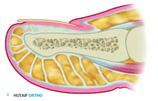
The stability of the glenohumeral joint is dictated by a highly orchestrated synergy between static and dynamic restraints. The static restraints include the osseous geometry of the glenoid and humeral head, the glenoid labrum, the articular capsule, the glenohumeral ligaments, and the negative intra-articular pressure. The glenoid cavity is inherently shallow, providing a surface area that covers only approximately one-third of the humeral head. The glenoid labrum, a fibrocartilaginous ring, effectively deepens this concavity by 50% and serves as the critical attachment site for the glenohumeral ligaments. The inferior glenohumeral ligament (IGHL) complex is the most robust and biomechanically significant static stabilizer against anterior and inferior translation, particularly when the arm is in the abducted and externally rotated (ABER) position. The IGHL consists of an anterior band, a posterior band, and an intervening axillary pouch, functioning much like a hammock to cradle the humeral head during terminal ranges of motion.
Dynamic stability is primarily conferred by the rotator cuff musculature (supraspinatus, infraspinatus, teres minor, and subscapularis), which compresses the humeral head into the glenoid concavity throughout the arc of motion—a concept known as "concavity compression." The long head of the biceps tendon also contributes to dynamic anterior stability, particularly when the arm is abducted and externally rotated. The periscapular musculature plays a vital, albeit indirect, role by dynamically positioning the glenoid in space to maintain an optimal platform for the humeral head. Dysfunction in this dynamic network, whether through primary muscle weakness, fatigue, or reflex inhibition secondary to pain (as seen in the "dead arm" syndrome), severely compromises joint stability and exacerbates the mechanical load placed upon the static capsulolabral restraints.
The characteristics of instability episodes are directly tied to these biomechanical vectors. The surgeon must elicit the specific arm positions that provoke instability, the frequency of events, and the ease of reduction. Anterior instability typically occurs with the arm in abduction and external rotation (ABER), which places maximal tension on the anterior band of the IGHL. Posterior instability typically occurs with the arm in flexion, adduction, and internal rotation (FADIR), often combined with an axial load, which stresses the posterior capsule and the posterior band of the IGHL. Inferior instability is provoked by carrying heavy objects at the side or during hyperabduction, stressing the superior glenohumeral ligament (SGHL) and the coracohumeral ligament (CHL).
Recurrence with minimal trauma in the midrange of motion is a highly concerning historical feature. Mid-range instability implies a catastrophic loss of the static osseous restraints. It is almost exclusively associated with significant bony lesions (large glenoid defects or engaging Hill-Sachs lesions) that have compromised the "glenoid track." These structural deficits must be treated surgically, often requiring bone-grafting procedures. Furthermore, dislocations that occur during sleep or with the arm resting in an overhead position are hallmark indicators of profound structural incompetence. These events are often associated with a significant anterior glenoid defect or severe capsular insufficiency. The inability of the shoulder to maintain concentric reduction at rest or during unconscious, low-stress states precludes successful non-operative management and mandates surgical reconstruction.
Internal impingement is a distinct pathological entity characterized by the abutment of the undersurface of the posterior rotator cuff (supraspinatus and infraspinatus) against the posterior-superior glenoid and labrum. It is caused by anterior humeral micro-subluxation with the shoulder in extreme external rotation and abduction (the late cocking phase of throwing). This secondary impingement is significantly more common than primary subacromial impingement in patients younger than 35 years old who are involved in upper extremity–dominant sports (e.g., baseball pitchers, volleyball players, tennis players). Treating the rotator cuff without addressing the underlying anterior capsular laxity will inevitably lead to surgical failure, as the primary biomechanical derangement remains uncorrected.
Exhaustive Indications and Contraindications
The decision to proceed with surgical intervention for recurrent shoulder instability is highly individualized, relying on a synthesis of the patient's chronological age, activity level, sport-specific demands, and the precise pathoanatomy identified during the clinical evaluation and advanced imaging. The historical classification systems, such as TUBS (Traumatic, Unilateral, Bankart, Surgery) and AMBRII (Atraumatic, Multidirectional, Bilateral, Rehabilitation, Inferior capsular shift, Interval closure), provide a foundational framework, but modern operative indications require a far more granular approach. Surgery is unequivocally indicated for young athletes (under 25 years of age) participating in high-demand, collision, or overhead sports who sustain a first-time traumatic anterior dislocation, as the natural history in this demographic demonstrates a recurrence rate exceeding 80% with non-operative management.
For patients with recurrent instability, the choice between an arthroscopic soft-tissue repair (e.g., Bankart repair with or without capsular plication) and an open osseous augmentation (e.g., Latarjet procedure, iliac crest bone graft, or distal tibial allograft) is dictated primarily by the presence and magnitude of bipolar bone loss. Arthroscopic Bankart repair is indicated for patients with recurrent anterior instability, a demonstrable anterior labral tear, and subcritical glenoid bone loss (typically defined as less than 13.5% to 15% of the inferior glenoid diameter) with an "on-track" Hill-Sachs lesion. Conversely, osseous augmentation is strictly indicated for patients with critical glenoid bone loss (greater than 15% to 20%), an "off-track" Hill-Sachs lesion that engages the anterior glenoid rim in functional positions, or those who present with highly concerning historical features such as mid-range instability or dislocations occurring during sleep.
Contraindications to surgical stabilization must be rigorously respected to prevent catastrophic postoperative failures. Absolute contraindications include active local or systemic infection, voluntary dislocators with underlying psychiatric overlay who can subluxate their shoulders at will without trauma, and patients with medically unoptimized seizure disorders. In the setting of uncontrolled epilepsy, any soft-tissue or osseous repair is virtually guaranteed to fail during a subsequent tonic-clonic event; thus, seizure control must be achieved prior to elective reconstruction. Relative contraindications include profound, untreated multidirectional instability (MDI) in a non-compliant patient who has not exhausted a minimum of six months of dedicated, periscapular-focused physical therapy. Operating prematurely on an AMBRII-type shoulder without addressing the dynamic neuromuscular control deficits often results in recurrent instability or profound postoperative stiffness.
| Surgical Procedure | Primary Clinical Indications | Historical / Pathoanatomic Findings | Absolute & Relative Contraindications |
|---|---|---|---|
| Arthroscopic Bankart Repair | Young athletes, first-time traumatic dislocators, recurrent instability without severe bone loss. | TUBS presentation, subcritical glenoid bone loss (<15%), "on-track" Hill-Sachs lesion, competent capsular tissue. | Critical bone loss (>20%), engaging "off-track" Hill-Sachs, voluntary instability, uncontrolled seizures. |
| Latarjet Procedure (Coracoid Transfer) | Collision athletes, revision stabilization, significant osseous defects. | Mid-range instability, sleep dislocations, critical glenoid bone loss (>15-20%), "off-track" Hill-Sachs lesion. | Advanced glenohumeral osteoarthritis, active infection, inadequate coracoid bone stock. |
| Arthroscopic Capsular Shift / Plication | Multidirectional instability (MDI), atraumatic subluxators failing extensive rehab. | AMBRII presentation, generalized ligamentous laxity (Beighton score >4), positive sulcus sign, inferior instability. | Failure to complete a >6-month dedicated periscapular rehab program, voluntary psychiatric dislocators. |
| Arthroscopic Remplissage | Adjunct to Bankart repair for moderate, engaging humeral head defects. | "Off-track" Hill-Sachs lesion with subcritical glenoid bone loss, preventing engagement over the anterior rim. | Massive glenoid bone loss requiring Latarjet, overhead athletes where loss of external rotation is career-ending. |
Pre-Operative Planning, Templating, and Patient Positioning
Thorough pre-operative planning is the cornerstone of successful instability surgery, heavily reliant on advanced imaging modalities dictated by the patient's clinical history. Standard radiographs, including a true anteroposterior (Grashey), axillary lateral, and Stryker notch view, are mandatory for initial assessment of joint concentricity, glenoid version, and large osseous defects. However, a non-contrast 3D computed tomography (CT) scan with digital subtraction of the humeral head is the gold standard for quantifying glenoid bone loss. Utilizing the "Pico" or best-fit circle method on the en face sagittal view of the glenoid allows the surgeon to precisely calculate the percentage of anterior-inferior bone loss. Concurrently, the width and depth of the Hill-Sachs lesion are measured to determine the "glenoid track," a critical biomechanical concept that dictates whether a combined osseous defect will engage during abduction and external rotation.
Magnetic resonance arthrography (MRA) remains the imaging modality of choice for evaluating the capsulolabral complex and the rotator cuff. The intra-articular gadolinium distends the capsule, allowing for exquisite visualization of subtle labral pathology, including anterior labroligamentous periosteal sleeve avulsions (ALPSA), Perthes lesions, and humeral avulsions of the glenohumeral ligament (HAGL). The MRA is also critical in younger patients presenting with internal impingement or "dead arm" syndrome, as it can identify partial-thickness articular-sided rotator cuff tears (PASTA lesions) and superior labrum anterior and posterior (SLAP) tears that frequently co-occur with micro-instability. The surgeon must synthesize the findings from the 3D CT and the MRA to formulate a definitive surgical plan, ensuring that both the osseous and soft-tissue components of the instability are addressed.
Patient positioning for arthroscopic stabilization is a subject of ongoing debate, with both the lateral decubitus and beach chair positions offering distinct advantages. The lateral decubitus position is frequently preferred for complex capsulolabral reconstructions and multidirectional instability. It utilizes longitudinal and lateral traction to distract the joint, providing unparalleled visualization of the inferior and posterior capsule. This position facilitates the precise placement of low anteroinferior anchors (at the 5:30 or 6 o'clock position on a right shoulder) and allows for a more aggressive, symmetric capsular shift. Care must be taken to pad all bony prominences and limit traction weight (typically 10-15 lbs) to prevent devastating brachial plexus neurapraxia.
Conversely, the beach chair position places the patient in a semi-upright, seated posture. This position is highly favored for its anatomic orientation, which simplifies the transition to an open procedure (such as a Latarjet or open capsular shift) if required. The beach chair position also avoids the risk of traction-induced nerve injuries and allows for dynamic intra-operative assessment of joint stability and range of motion. However, visualization of the inferior recess can be challenging, and meticulous attention must be paid to cerebral perfusion pressure, as the upright posture combined with general anesthesia can lead to hypotensive bradycardic events and cerebral ischemia. Regardless of the chosen position, an interscalene regional nerve block is strongly recommended to minimize intra-operative anesthetic requirements and provide robust post-operative analgesia.
Step-by-Step Surgical Approach and Fixation Technique
The surgical execution of an arthroscopic anterior stabilization (Bankart repair) demands meticulous attention to detail, beginning with a comprehensive diagnostic arthroscopy. Utilizing a standard posterior viewing portal, the surgeon systematically evaluates the biceps anchor, the superior labrum, the anterior and posterior labrum, the capsular recesses, and the articular surfaces of the glenoid and humeral head. A dynamic examination is performed, placing the arm in abduction and external rotation to directly visualize the engagement of any Hill-Sachs lesion with the anterior glenoid rim. If an "off-track" lesion is confirmed intra-operatively in the setting of subcritical glenoid bone loss, the surgical plan must be immediately adapted to include an arthroscopic remplissage (insetting the infraspinatus tendon and posterior capsule into the humeral defect) alongside the anterior repair.
The foundation of a successful Bankart repair is the complete and aggressive mobilization of the anterior-inferior capsulolabral complex. In chronic instability, the torn labrum and the attached inferior glenohumeral ligament often heal in a medially and inferiorly displaced position on the glenoid neck (an ALPSA lesion). Using an elevator or a specialized tissue liberator through an anterior-inferior working portal, the surgeon must elevate the capsulolabral tissue off the glenoid neck until the subscapularis muscle belly is clearly visualized. This release must extend inferiorly to at least the 6 o'clock position to ensure the tissue can be mobilized superiorly and laterally to restore the native bumper and re-tension the IGHL hammock. Failure to adequately mobilize the tissue is the most common technical error leading to recurrent instability.
Following mobilization, the anterior glenoid rim and the adjacent glenoid neck are meticulously prepared. A motorized burr or a hand rasp is used to decorticate the bone, creating a bleeding, cancellous bed that is critical for robust soft-tissue healing. Care must be taken to preserve the osseous architecture of the glenoid rim; over-resection will inadvertently create an iatrogenic bony defect, further destabilizing the joint. The goal is to remove all fibrous scar tissue and expose healthy bone while maintaining the natural concavity of the glenoid articular surface.
Fixation is achieved using a minimum of three, and ideally four, suture anchors to create a secure, biomechanically sound construct. The first and most critical anchor is placed at the lowest point of the defect, typically at the 5:30 or 6 o'clock position (on a right shoulder), directly on the articular margin. Modern techniques emphasize placing the anchors precisely on the articular face rather than medially on the glenoid neck, which ensures the labrum is recreated as a prominent bumper. A suture passing device is used to take a substantial bite of the inferior capsule and labrum, shifting the tissue superiorly and laterally. Subsequent anchors are placed sequentially superiorly (e.g., at the 4:30 and 3:00 positions), progressively plicating the capsule and restoring the anterior restraint. The knots (or knotless mechanisms) are secured, and the final construct is probed to confirm the restoration of the labral bumper and the elimination of the anterior-inferior drive-through sign.
Complications, Incidence Rates, and Salvage Management
Despite meticulous surgical technique, the management of recurrent shoulder instability is fraught with potential complications. The most devastating and common complication following surgical stabilization is the recurrence of instability. Following an isolated arthroscopic Bankart repair, recurrence rates range from 5% to 15% in the general population, but can skyrocket to over 25% in high-risk demographics, such as male collision athletes under the age of 20. Recurrence is almost universally linked to the failure to recognize and address critical bipolar bone loss, inadequate capsulolabral mobilization, or premature return to high-risk activities. When an arthroscopic repair fails, salvage management typically mandates an open osseous augmentation, most commonly the Latarjet procedure, to restore the anterior glenoid arc and provide the dynamic sling effect of the conjoined tendon.
Neurological injury, while relatively rare, represents a catastrophic complication. The axillary nerve is at the highest risk during both arthroscopic and open stabilization procedures. During arthroscopic capsular plication, the nerve can be tethered or sutured if passes are made too deeply into the inferior capsule at the 6 o'clock position. During the open Latarjet procedure, the musculocutaneous nerve (entering the coracobrachialis) and the axillary nerve are vulnerable to aggressive retraction or direct laceration. Axillary nerve neuropraxia presents as numbness over the lateral deltoid (regimental badge area) and weakness in shoulder abduction. While most traction neuropraxias resolve spontaneously over 3 to 6 months, baseline documentation is medicolegally and clinically mandatory. Iatrogenic laceration requires immediate microsurgical exploration and repair or grafting.
Post-operative stiffness, specifically a profound loss of external rotation, is a frequent complication, particularly following over-tensioning of the anterior capsule or an overzealous remplissage procedure. While a mild loss of terminal external rotation (5 to 10 degrees) is often an intentional and acceptable trade-off for stability, severe restriction can lead to significant functional impairment and secondary early-onset glenohumeral osteoarthritis (capsulorrhaphy arthropathy). Management of stiffness begins with aggressive, prolonged physical therapy emphasizing prolonged low-load stretching. If conservative measures fail after 6 to 9 months, an arthroscopic anterior capsular release may be required to restore functional motion, though this must be balanced against the risk of re-creating instability.
| Complication | Estimated Incidence | Etiology / Risk Factors | Salvage Management / Treatment Strategy |
|---|---|---|---|
| Recurrent Instability | 5% - 15% (Arthroscopic) 2% - 5% (Latarjet) |
Unrecognized critical bone loss, poor tissue quality, premature return to sport, technical failure. | Revision surgery with osseous augmentation (Latarjet, Iliac Crest Bone Graft, or Distal Tibial Allograft). |
| Axillary Nerve Injury | < 1% - 2% | Deep inferior capsular suture passes (6 o'clock), aggressive retraction during open procedures. | Observation and EMG at 6 weeks for neuropraxia; microsurgical exploration and grafting for complete laceration. |
| Post-Operative Stiffness (Loss of ER) | 10% - 20% | Over-tensioning of the anterior capsule, excessive remplissage, inadequate post-op rehabilitation. | Aggressive physical therapy; arthroscopic capsular release if refractory > 6-9 months. |
| Hardware Complications / Osteolysis | 1% - 5% | Anchor pullout, prominent hardware causing chondral damage, bioabsorbable anchor osteolysis. | Arthroscopic hardware removal, chondroplasty, and revision stabilization if symptomatic. |
Phased Post-Operative Rehabilitation Protocols
The post-operative rehabilitation following shoulder stabilization is as critical to the ultimate clinical outcome as the surgical execution itself. The protocol must be meticulously phased to protect the healing capsulolabral repair while progressively restoring range of motion, neuromuscular control, and dynamic strength. Phase I (Protection Phase, Weeks 0-4) is characterized by strict immobilization in a neutral or slightly externally rotated sling to minimize tension on the anterior repair. Therapeutic interventions are limited to passive range of motion (PROM) in a safe zone, typically restricting external rotation to 0-20 degrees and forward elevation to 90 degrees. Cryotherapy, scapular retractions, and pendulum exercises are initiated to prevent profound stiffness and manage post-operative pain.
Phase II (Intermediate Range of Motion Phase, Weeks 4-8) commences with the discontinuation of the sling and the initiation of active-assisted range of motion (AAROM) and active range of motion (AROM). The safe zones are progressively expanded, aiming for full forward elevation and gradual restoration of external rotation. It is critical to avoid aggressive, forced stretching into extreme abduction and external rotation, as the healing tissue remains vulnerable to plastic deformation. Submaximal, pain-free isometric strengthening of the rotator cuff and periscapular musculature is introduced to combat disuse atrophy and re-establish the baseline dynamic concavity compression.
Phase III (Strengthening Phase, Weeks 8-16) focuses on robust isotonic strengthening and the restoration of advanced neuromuscular control. As full, symmetric range of motion is achieved, resistance exercises targeting the dynamic stabilizers are intensified. Rhythmic stabilization drills, closed kinetic chain exercises, and proprioceptive neuromuscular facilitation (PNF) patterns are heavily utilized. For patients with a history of multidirectional instability (AMBRII), this phase is extensively prolonged, as the ultimate success of the procedure relies heavily on maximizing the dynamic compensatory mechanisms of the periscapular and rotator cuff musculature.
Phase IV (Return to Sport/Activity Phase, Weeks 16-24+) is a highly individualized, criteria-based progression rather than a strict timeline. Patients must demonstrate full, pain-free range of motion, symmetric rotator cuff strength (at least 90% of the contralateral extremity on isokinetic testing), and the ability to perform sport-specific functional movements without apprehension. Overhead athletes begin a progressive throwing program, while collision athletes initiate controlled contact drills. Premature clearance to high-risk activities before the restoration of dynamic stability and complete biological healing of the repair site is the primary driver of early post-operative failure.
Summary of Landmark Literature and Clinical Guidelines
The contemporary management of recurrent shoulder instability is built upon a foundation of landmark biomechanical and clinical studies that have continuously refined our understanding of pathoanatomy and surgical indications. The historical dichotomy of TUBS and AMBRII, originally popularized by Thomas Matsen, provided
