
العربية
Comprehensive Master Guide · Medically Reviewed

Hallux Valgus: Surgical Anatomy, Biomechanics, and Soft Tissue Release Principles

Master hallux valgus surgical anatomy, biomechanics, and soft tissue release principles. Discover key insights on etiology, indications, and correction.

15 Detailed Chapters
24 min read
Updated: Jun 2026
Dr. Mohammed Hutaif Clinic
Medically Reviewed by
Prof. Dr. Mohammed Hutaif Clinic
Verified Content Expert Reviewed

Quick Medical Answer

Soft tissue release in hallux valgus surgery targets contracted lateral structures like the adductor hallucis tendon, lateral capsule, and lateral sesamoid ligament, addressing medial capsule attenuation. These procedures correct biomechanical imbalances and reduce deformity, serving as crucial adjuncts to osteotomies for both mild and severe bunion correction.

Illustration of soft tissue release - Dr. Mohammed Hutaif
🎓

FRCS Masterclass: Clinical Viva

Interactive Examiner Scenario • Test your knowledge before revealing the answers.

👨‍⚕️ Examiner Scenario

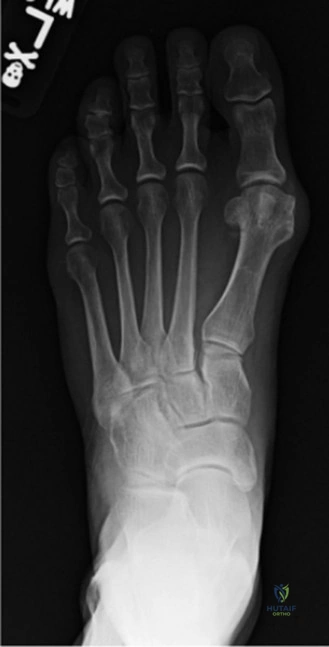
A 45-year-old female presents with a symptomatic hallux valgus. She has failed a six-month course of appropriate conservative management. You are planning a distal metatarsal osteotomy. During your clinical and radiographic assessment, you identify that the hallux valgus angle (HVA) is 28 degrees and the intermetatarsal angle (IMA) is 14 degrees. You are now considering the role of lateral soft tissue release in this procedure. Explain the biomechanical rationale for this step and identify the key structures involved.

Candidate: Lateral soft tissue release is necessary because the deforming forces pull the toe laterally. I would release the adductor hallucis and the lateral capsule. This helps to center the sesamoids under the metatarsal head. It is vital to avoid damaging the neurovascular bundle in the first web space during this part of the surgery.

❌ Common Pitfall (Poor Answer)

Failing to mention the lateral sesamoid ligament (a primary constraint) or the fibular sesamoid specifically. Candidates often forget to mention the rotational component (pronation) of the hallux or the distinction between the transverse and oblique heads of the adductor hallucis.

⭐ The Gold Standard (Perfect Answer)

The biomechanical rationale is to neutralize the lateral deforming forces that have become pathologically advantaged. The key structures to release include: 1) The adductor hallucis tendon (both oblique and transverse heads), 2) The lateral joint capsule, and 3) The lateral (fibular) sesamoid-phalangeal ligament. Addressing these is essential to allow the fibular sesamoid to recenter beneath the first metatarsal head, correcting the pronation of the hallux and preventing recurrence. I would perform this meticulously to avoid injury to the common digital nerve in the first web space.

👨‍⚕️ Examiner Scenario

During the procedure, you are assessing the lateral structures. Please look at the image provided and describe the pathological anatomy and the surgical targets for your release.

Clinical Image
Anatomical structures of the first MTP joint

Candidate: In a hallux valgus deformity, the fibular sesamoid subluxes laterally. This subluxation makes the adductor hallucis a more potent deforming force. The image highlights the lateral complex. My surgical goal is to release the tight lateral capsule and the ligamentous connections to the fibular sesamoid to restore balance.

❌ Common Pitfall (Poor Answer)

Focusing only on the "bunion" (medial eminence) and ignoring the plantar and lateral soft tissue complex. Candidates who do not link the sesamoid position to the degree of soft tissue contracture demonstrate a lack of deep understanding regarding the progression of the deformity.

⭐ The Gold Standard (Perfect Answer)

The image illustrates the 'bowstringing' effect caused by lateral sesamoid displacement. Pathologically, the lateral ligamentous complex, specifically the lateral collateral ligament and the sesamoid-phalangeal ligament, becomes chronically contracted, resisting reduction. My target is to perform a controlled release of these structures until the hallux can be passively corrected to a neutral or slightly varus position without tension. This release is the 'key to the lock'—without it, bony correction alone often fails to maintain the sesamoids in their physiological position.

👨‍⚕️ Examiner Scenario

Post-operatively, the patient develops a complication. She complains of persistent numbness in the first web space and has developed a painful, tender nodule at the site of the lateral release incision. How do you manage this?

Candidate: This sounds like a post-operative neuroma of the first dorsal digital nerve. I would start with conservative management, such as nerve blocks or desensitization, but if the pain persists, I would consider surgical excision of the neuroma.

❌ Common Pitfall (Poor Answer)

Offering surgical excision as a first-line treatment. Failing to mention the specific nerve at risk (the medial branch of the deep peroneal nerve or the dorsal proper digital nerve).

⭐ The Gold Standard (Perfect Answer)

This is a classic presentation of a symptomatic neuroma of the dorsal proper digital nerve to the hallux, often injured during the lateral release in the first web space. I would manage this initially with a conservative 3-6 month trial: local desensitization, shoe modifications (e.g., wider toe box), and targeted corticosteroid/anesthetic injections. If the patient remains functionally impaired, I would perform a formal neuroma excision with burying of the nerve end into deep, well-vascularized tissue (e.g., muscle) to prevent recurrence.

Detailed Chapters & Topics

Dive deeper into specialized chapters regarding foot-and-ankle-cases-2

15 Chapters
01
Chapter 1 18 min

Mau Osteotomy: A Masterclass in Hallux Valgus Correction

Join our masterclass on the Mau osteotomy for moderate to severe hallux valgus correction. Explore bunion etiology, pat…

02
Chapter 2 16 min

Distal Chevron Osteotomy: A Masterclass in Hallux Valgus Correction

Master the distal chevron osteotomy for hallux valgus correction. This comprehensive guide covers surgical anatomy, int…

03
Chapter 3 16 min

Ludloff Osteotomy Masterclass: Precision Correction for Hallux Valgus

Master the modified Ludloff osteotomy for symptomatic hallux valgus. Learn surgical anatomy, meticulous techniques, and…

04
Chapter 4 13 min

Scarf Osteotomy Masterclass: Precision Correction of Hallux Valgus

Master the Scarf osteotomy for symptomatic hallux valgus correction. Explore expert preoperative planning, biomechanics…

05
Chapter 5 23 min

The Dorsolateral Approach for Hallux Valgus: Anatomy, Biomechanics, and Surgical Principles

Master the dorsolateral approach for hallux valgus surgery. Discover key surgical anatomy, biomechanics, and soft tissu…

06
Chapter 6 17 min

Masterclass in the Scarf Osteotomy: Biomechanics, Technique, and Outcomes

Master the Scarf osteotomy with our expert guide. Explore biomechanics, surgical techniques, unparalleled stability, an…

07
Chapter 7 24 min

Comprehensive Guide to Medial Eminence Resection in Hallux Valgus Correction

Master medial eminence resection in hallux valgus correction. Discover expert insights on modern bunionectomy technique…

08
Chapter 8 28 min

Hallux Varus: Epidemiology, Surgical Anatomy, and Biomechanics Review

Discover the epidemiology, surgical anatomy, and biomechanics of hallux varus. Learn about iatrogenic causes, clinical …

09
Chapter 9 14 min

Medial Cuneiform and Proximal Phalangeal Osteotomies in Hallux Valgus Surgery

Master the indications, biomechanics, and surgical techniques for medial cuneiform and Akin proximal phalangeal osteoto…

10
Chapter 10 13 min

Revision Hallux Valgus Correction

Discover expert insights on revision hallux valgus correction. Learn about anatomy, natural history, and treatments for…

11
Chapter 11 19 min

Mastering Juvenile Bunion Correction: A Mitchell Osteotomy Intraoperative Guide

Master juvenile bunion correction with our intraoperative guide to the Mitchell osteotomy. Learn to restore biomechanic…

12
Chapter 12 15 min

Minimally Invasive SERI Hallux Valgus Correction: An Intraoperative Masterclass

Master the minimally invasive SERI technique for hallux valgus correction. This intraoperative guide covers forefoot bi…

13
Chapter 13 10 min

Preoperative Management and Surgical Decision-Making in Hallux Valgus Reconstruction

Comprehensive guide on the preoperative management, clinical evaluation, radiographic assessment, and surgical algorith…

14
Chapter 14 11 min

Masterclass in Plantar Fascia and Nerve Release: Open and Endoscopic Techniques

Master open and endoscopic surgical techniques for chronic heel pain. Learn to perform plantar fascia and Baxter's nerv…

15
Chapter 15 10 min

First Cuneiform Osteotomy and Management of Hallux Valgus in Cerebral Palsy

A comprehensive surgical guide on the first cuneiform osteotomy for hypermobile first ray and the management of hallux …

Dr. Mohammed Hutaif Clinic
Medically Verified Content by
Prof. Dr. Mohammed Hutaif Clinic
Consultant Orthopedic & Spine Surgeon
Guide Overview