AAOS Upper Extremity MCQs (Set 3): Shoulder & Elbow Injuries | 2008 Board Review
Key Takeaway
This high-yield question set (Set 3) for AAOS/ABOS exams focuses on crucial Upper Extremity topics. It covers the diagnosis and management of shoulder fractures, various elbow injuries, and common hand & wrist pathologies, providing comprehensive preparation for orthopedic board review and OITE.
AAOS Upper Extremity MCQs (Set 3): Shoulder & Elbow Injuries | 2008 Board Review
Comprehensive 100-Question Exam
00:00
Start Quiz
Question 1
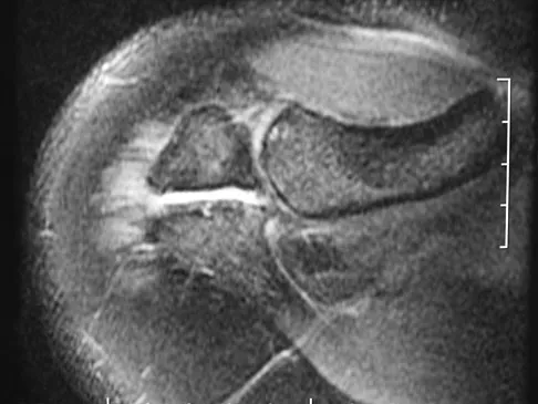
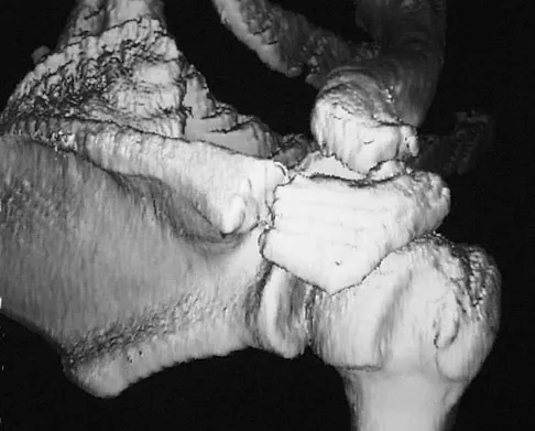
A 22-year-old right hand-dominant man who fell off his motorcycle onto the tip of his right shoulder 2 weeks ago now reports pain and difficulty raising his right arm. Examination reveals tenderness and gross movement over the lateral scapular spine and severe weakness during resisted abduction. A radiograph and 3D-CT scan are shown in Figures 24a and 24b. What is the next most appropriate step in management?
Explanation
Question 2
A 20-year-old minor league baseball pitcher is diagnosed with a symptomatic torn ulnar collateral ligament (UCL) in his pitching elbow. Nonsurgical management consisting of rest and physical therapy aimed at elbow strengthening has failed to provide relief. He has concomitant cubital tunnel symptoms that worsen while throwing. What is his best surgical option?
Explanation
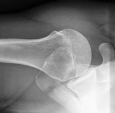
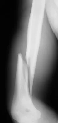
Question 3
A 30-year-old man has pain in the left arm after a motor vehicle accident. His neurovascular examination is intact, and radiographs are shown in Figures 25a and 25b. What is the best course of management?
Explanation
Question 4
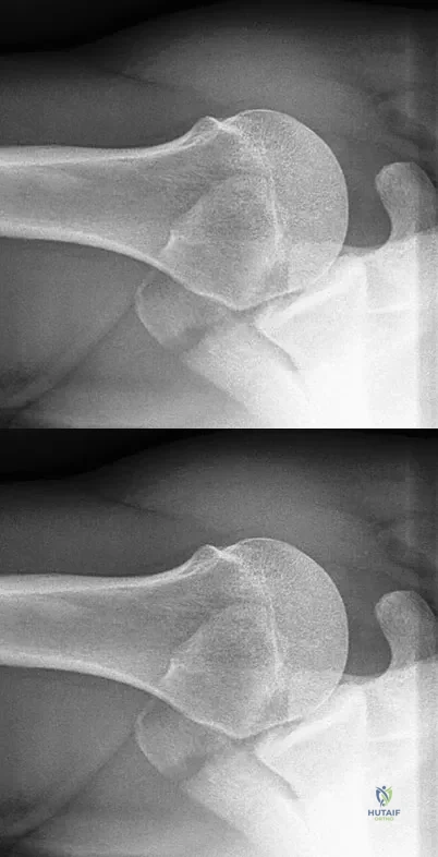
A patient who underwent open reduction and internal fixation of an olecranon fracture 2 months ago now reports painless limitation of motion. Examination reveals a well-healed incision and a flexion-extension arc from 40 degrees to 80 degrees. The patient has been performing home exercises. Radiographs are shown in Figures 26a and 26b. What is the most appropriate treatment?
Explanation
Question 5
A 23-year-old professional baseball pitcher reports shoulder pain and decreased velocity while pitching. Physical examination reveals a side-to-side internal rotation deficit of 25 degrees. The O'Brien sign is negative; Neer and Hawkins signs are negative. Rotator cuff strength is full. Radiographs are unremarkable. What is the next step in management?
Explanation
Question 6
A 72-year-old woman who is right hand-dominant has severe pain in the right shoulder that has failed to respond to nonsurgical management. She reports night pain and significant disability. Examination reveals 30 degrees of active forward elevation. An AP radiograph is shown in Figure 27. Which of the following treatment options will provide the best functional improvement?
Explanation
Question 7
A healthy 64-year-old man just underwent an uncomplicated shoulder arthroplasty for severe glenohumeral osteoarthritis. Intraoperatively, 60 degrees of external rotation was obtained. Postoperatively, he starts on a range-of-motion program. What limitations are recommended?
Explanation
Question 8
A 64-year-old man who was involved in a high-speed motor vehicle accident 6 weeks ago has been in the ICU with a closed head injury. Examination reveals that his range of motion for external rotation to the side is -30 degrees. Radiographs are shown in Figures 28a and 28b. What is the most likely diagnosis?
Explanation
Question 9
A 17-year-old high school football player reports wrist pain 5 months after the conclusion of the football season. A radiograph and MRI scan are shown in Figures 29a and 29b. What is the recommended intervention?
Explanation
Question 10
A 58-year-old woman with a history of severe asthma and long-term prednisone use reports a progression of chronic shoulder pain for the past 6 months. Radiographs and MRI scans are shown in Figures 30a through 30d. What is the most likely diagnosis?
Explanation
Question 11
A 28-year-old man sustained a shoulder dislocation 2 years ago. It remained dislocated for 3 weeks and required an open reduction. He now reports constant pain and has only 60 degrees of forward elevation and 10 degrees of external rotation. He desires to return to some sporting activities. An AP radiograph and intraoperative photograph (a view of the humeral head through a deltopectoral approach) are shown in Figures 31a and 31b. What is the best treatment option to decrease pain and improve function?
Explanation
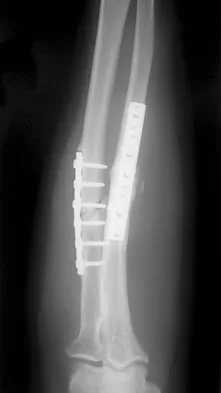
Question 12
A 34-year-old man underwent open reduction and internal fixation of a closed both bones forearm fracture 11 months ago. The radiographs shown in Figures 32a and 32b reveal a 3-mm gap and loose screws. What is the best treatment option?
Explanation
Question 13
A football lineman who sustained a traumatic injury while blocking during a game now reports that his shoulder is slipping while pass blocking. Examination reveals no apprehension in abduction and external rotation; however, he reports pain with posterior translation of the shoulder. He has full strength in external rotation, internal rotation, and supraspinatus testing. What is the pathology most likely responsible for his symptoms?
Explanation
Question 14
A 17-year-old girl has multidirectional instability of the shoulder. What is the most appropriate initial management?
Explanation
Question 15
In surgically treating hand and finger infections in patients with diabetes mellitus, what factor is associated with higher amputation rates?
Explanation
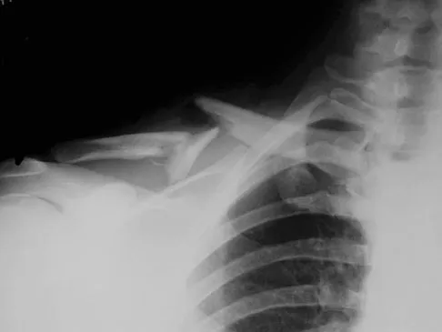
Question 16
A 40-year-old unrestrained passenger reports chest wall pain after a motor vehicle accident. Which of the following structures is most important in preventing the injury shown in Figure 33?
Explanation
Question 17
Figures 34a and 34b show the axial and sagittal MRI scans of a 36-year-old man who reports the insidious onset of pain in the right shoulder. What is the most appropriate description of the acromial morphology?
Explanation
Question 18
What is the primary indication for performing a total wrist arthroplasty in a patient with painful rheumatoid arthritis?
Explanation
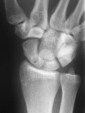
Question 19
What is the most likely cause of the lesion shown in Figures 35a and 35b?
Explanation
Question 20
During treatment of rupture of the subscapularis tendon with associated biceps instability, treatment of the biceps tendon should include which of the following?
Explanation
Question 21
What is the most common bacteria cultured from dog and cat bites to the upper extremity?
Explanation
Question 22
A previously healthy 65-year-old woman has a closed fracture of the right clavicle after falling down the basement stairs. Examination reveals good capillary refill in the digits of her right hand. Radial and ulnar pulses are 1+ at the right wrist compared with 2+ on the opposite side. In the arteriogram shown in Figure 36, the arrow is pointing at which of the following arteries?
Explanation
Question 23
Which of the following structures may help maintain radial length after a radial head fracture?
Explanation
Question 24
An adult patient has a closed humeral fracture that was treated nonsurgically and a concomitant radial nerve injury. Six weeks after injury, electromyography shows no evidence of recovery. Management should now consist of
Explanation
Question 25
A 55-year-old man who works as a carpenter reports chronic right anterior shoulder pain and weakness. Examination reveals 90 degrees of external rotation (with the arm at the side) compared to 45 degrees on the left side. His lift-off examination is positive, along with a positive belly press finding. An MRI scan reveals a chronic, retracted atrophied subscapularis tendon. What is the most appropriate management of his shoulder pain and weakness?
Explanation
Question 26
A 45-year-old male construction worker presents with persistent anterior shoulder pain. He had a prior SLAP repair 20 years ago. An MRI demonstrates a failed Type II SLAP repair and advanced biceps tendinopathy. What is the most appropriate surgical treatment?
Explanation
Question 27
According to the Hertel criteria for proximal humerus fractures, which of the following radiographic findings is most strongly associated with subsequent humeral head ischemia?
Explanation
Question 28
A 35-year-old male sustains a terrible triad injury to the elbow after a fall from a ladder. During the standard surgical reconstruction, what is the generally recommended sequence of repair?
Explanation
Question 29
A 40-year-old recreational weightlifter feels a sudden pop in his anterior elbow during a deadlift. Clinical examination reveals a reverse Popeye deformity and weakness in resisted supination. If a single anterior incision approach is utilized for repair, which nerve is at greatest risk of iatrogenic injury?
Explanation
Question 30
A 75-year-old female sustains a highly comminuted, osteoporotic intra-articular distal humerus fracture.
What is the primary advantage of total elbow arthroplasty (TEA) over open reduction and internal fixation (ORIF) in this specific patient population?
Explanation
Question 31
A 28-year-old male falls onto his shoulder while snowboarding. Radiographs reveal a completely displaced midshaft clavicle fracture with 2.5 cm of shortening.
Compared to nonoperative management, surgical fixation of this injury is associated with:
Explanation
Question 32
A 30-year-old female presents with acute wrist and elbow pain after a high-energy fall. She is diagnosed with an Essex-Lopresti injury. What is the appropriate surgical management of the radial head in this scenario?
Explanation
Question 33
A 22-year-old rugby player with recurrent anterior shoulder instability and 25% glenoid bone loss undergoes a Latarjet procedure. During the coracoid transfer, the musculocutaneous nerve must be identified and protected. What is its approximate distance from the tip of the coracoid process?
Explanation
Question 34
A 45-year-old woman falls on an outstretched hand and sustains an elbow dislocation, radial head fracture, and coronoid process fracture. She is taken to the operating room for surgical stabilization. What is the most widely accepted sequence for reconstructing the elbow in this 'terrible triad' injury?
Explanation
Question 35
A 42-year-old woman falls on an outstretched hand and sustains a terrible triad injury of the elbow. Which of the following describes the most appropriate sequence of surgical reconstruction?
Explanation
Question 36
A 28-year-old man sustains a closed midshaft humeral fracture
. On initial emergency department presentation, he is unable to extend his wrist or fingers. What is the most appropriate initial management of this patient's neurologic deficit?
Explanation
Question 37
A 35-year-old male bodybuilder reports a sudden, painful "pop" in his right antecubital fossa while performing biceps curls. Physical examination reveals a "Popeye" deformity and weakness in forearm supination. If a single-incision anterior surgical approach is chosen for repair, the patient is at highest risk for injury to which of the following structures?
Explanation
Question 38
A 78-year-old woman with severe osteoporosis presents with a 4-part proximal humerus fracture after a fall. She has a history of severe rotator cuff arthropathy with pseudo-paralysis of the shoulder prior to the injury. What is the most appropriate surgical intervention?
Explanation
Question 39
An 18-year-old male football player sustains a direct blow to his medial clavicle. He presents with severe chest pain, shortness of breath, and dysphagia. Examination shows a depression of the medial clavicle. What is the most appropriate next step in management?
Explanation
Question 40
A 45-year-old man falls onto an outstretched hand and sustains a 'terrible triad' injury of the elbow.
During surgical reconstruction to restore stability, which of the following represents the most widely accepted standard sequence of repair?
Explanation
Question 41
A 35-year-old male weightlifter experiences a sudden 'pop' and sharp pain in his dominant anterior elbow while attempting to lift a heavy box. Examination reveals a positive Hook test. If a single-incision anterior surgical approach is chosen for repair, which of the following nerves is at greatest risk of injury?
Explanation
Question 42
A lateral elbow radiograph of a 28-year-old woman who fell on an outstretched arm demonstrates a 'double arc' sign.
What does this radiographic finding indicate?
Explanation
Question 43
A 68-year-old woman sustains a displaced 4-part proximal humerus fracture. According to Hertel's radiographic criteria, which of the following findings is the strongest predictor for the subsequent development of humeral head avascular necrosis (AVN)?
Explanation
Question 44
A 19-year-old rugby player presents to the emergency department after a direct blow to the medial clavicle. He complains of severe pain, mild shortness of breath, and dysphagia. Examination reveals a palpable void at the sternoclavicular joint. What is the most appropriate initial management?
Explanation
Question 45
A 24-year-old competitive weightlifter feels a tearing sensation in his anterior axilla during the eccentric phase of a heavy bench press. He has weakness in internal rotation and adduction. Which anatomic portion of the affected muscle is most likely ruptured?
Explanation
Question 46
A 32-year-old motorcyclist is involved in a high-speed collision.
Chest radiographs demonstrate marked lateral displacement of the left scapula. The patient's left upper extremity is flaccid. What is the most critical next step in management?
Explanation
Question 47
A 40-year-old woman presents with severe wrist pain and elbow stiffness 3 months after undergoing an isolated radial head excision for a comminuted radial head fracture. She did not receive a radial head arthroplasty. Radiographs of the wrist now show positive ulnar variance. Which of the following undetected injuries was most likely present at the time of her initial trauma?
Explanation
Question 48
A 26-year-old elite volleyball attacker complains of chronic posterior shoulder pain and isolated weakness in external rotation. Forward elevation and internal rotation are full and 5/5 in strength. MRI reveals a paralabral cyst. At which anatomic location is the nerve compression most likely occurring?
Explanation
Question 49
A 50-year-old man requires an open capsular release for severe post-traumatic elbow stiffness. During a lateral column procedure, the anterior capsule is sharply elevated off the anterior humerus. Which neurovascular structure is at highest risk during this specific step?
Explanation
Question 50
A 34-year-old man sustains an oblique fracture of the distal third of the humeral shaft (Holstein-Lewis fracture).
He demonstrates an inability to extend his wrist and fingers. The injured nerve is most susceptible to tethering and injury at which specific anatomical site?
Explanation
Question 51
A 7-year-old boy sustains a Bado Type I Monteggia fracture-dislocation.
Intraoperatively, after open reduction and internal fixation of the ulnar shaft fracture, the surgeon notes that the radial head remains anteriorly dislocated. What is the most appropriate next step in management?
Explanation
Question 52
A 13-year-old male baseball pitcher presents with 3 months of lateral shoulder pain during the acceleration phase of throwing. Radiographs demonstrate widening and irregularity of the proximal humeral physis. What is the primary pathophysiology of this condition?
Explanation
Question 53
A 22-year-old cyclist falls directly onto his shoulder, sustaining a completely displaced, midshaft clavicle fracture with 2.5 cm of shortening. Compared to nonoperative management with a sling, operative plate fixation of this specific injury pattern is proven to significantly decrease the risk of which complication?
Explanation
Question 54
A 25-year-old gymnast reports clicking, pain, and a feeling of instability in her elbow when pushing herself out of a chair, months after a posterior elbow dislocation. The pivot-shift test of the elbow is positive. Which ligamentous structure is primarily deficient?
Explanation
Question 55
A 45-year-old man falls on an outstretched hand and sustains a 'terrible triad' injury of the elbow. What is the most appropriate surgical sequence to effectively restore elbow stability?
Explanation
Question 56
A 25-year-old cyclist falls, sustaining a midshaft clavicle fracture with 100% displacement and 2 cm of shortening. Compared to nonoperative management, open reduction and internal fixation (ORIF) of this fracture is associated with which of the following outcomes?
Explanation
Question 57
A 38-year-old bodybuilder feels a sudden 'pop' in his anterior arm while deadlifting. Examination reveals a positive hook test and loss of the distal biceps contour. If this injury is managed nonoperatively, which of the following functional deficits is most expected?
Explanation
Question 58
During a deltoid-splitting approach for proximal humerus fracture fixation, the surgeon must be careful to avoid the axillary nerve. At approximately what distance distal to the lateral edge of the acromion does the axillary nerve typically cross the deep surface of the deltoid?
Explanation
Question 59
A 14-year-old elite baseball pitcher presents with right shoulder pain that occurs exclusively during the late cocking phase of throwing. Radiographs demonstrate widening of the proximal humeral physis. What is the most appropriate initial management?
Explanation
Question 60
A 19-year-old male is tackled during a football game and presents with severe chest pain, dyspnea, dysphagia, and diminished pulses in his left arm. A posterior sternoclavicular dislocation is suspected. What is the most appropriate next step in management after securing the airway?
Explanation
Question 61
A 35-year-old man presents with a stiff elbow 6 months after a complex fracture-dislocation. His range of motion is 30 to 90 degrees, and radiographs show heterotopic ossification (HO). When considering surgical release and HO excision, what is the most important factor dictating the timing of surgery?
Explanation
Question 62
A 28-year-old motorcyclist is involved in a high-speed collision. He presents with massive shoulder swelling, a pulseless left upper extremity, and a completely flail limb. Radiographs show significant lateral displacement of the scapula. This condition (scapulothoracic dissociation) is most highly associated with which of the following injuries?
Explanation
Question 63
A 40-year-old woman reports recurrent clicking and a sense of 'giving way' in her lateral elbow, particularly when she pushes herself out of a chair. What ligament is primarily deficient, and what is the typical mechanism of the initial injury?
Explanation
Question 64
A 32-year-old man falls from a height onto his outstretched hands, sustaining a highly comminuted radial head fracture, diffuse forearm pain, and distal radioulnar joint (DRUJ) instability. To prevent longitudinal radioulnar dissociation, what is the most appropriate surgical management for the radial head?
Explanation
Question 65
A 72-year-old man with a chronic massive rotator cuff tear presents with pseudoparalysis. Radiographs show severe superior migration of the humeral head and acetabularization of the acromion. If a reverse total shoulder arthroplasty is performed, the center of rotation is altered in which direction compared to the native anatomy?
Explanation
Question 66
A 24-year-old woman falls on her outstretched hand. Radiographs demonstrate a coronal shear fracture of the capitellum extending medially to involve the lateral trochlear ridge (Type IV or McKee modification). What is the preferred surgical approach and fixation strategy?
Explanation
Question 67
A 21-year-old collegiate baseball pitcher presents with anterior shoulder pain. Physical examination of his throwing shoulder reveals 25 degrees less internal rotation compared to his non-throwing shoulder, but his total arc of motion is symmetric. What is the primary underlying anatomic pathology associated with this condition?
Explanation
Question 68
A 29-year-old weightlifter feels a tearing sensation in his chest while performing a heavy bench press. Examination reveals bruising and loss of the normal anterior axillary fold contour. What is the most common anatomic location for a pectoralis major rupture?
Explanation
Question 69
A 30-year-old volleyball player presents with vague posterior shoulder pain and weakness in external rotation. MRI reveals a paralabral cyst compressing the nerve at the spinoglenoid notch. Which muscle(s) will most likely demonstrate denervation changes on electromyography (EMG)?
Explanation
Question 70
A 60-year-old woman sustains a highly comminuted intra-articular distal humerus fracture (AO type 13-C). The surgeon plans an open reduction and internal fixation via a posterior approach with an olecranon osteotomy. Which osteotomy technique is associated with the highest intrinsic stability and lowest rate of hardware complications?
Explanation
Question 71
A 45-year-old man presents with persistent numbness in the ring and small fingers and intrinsic weakness. EMG confirms severe ulnar neuropathy at the elbow. During an anterior submuscular transposition of the ulnar nerve, which structure must be meticulously released to prevent secondary compression of the nerve as it exits the elbow?
Explanation
Question 72
A 40-year-old man falls on an outstretched hand and sustains a posterior elbow dislocation, a radial head fracture, and a coronoid fracture. During surgical management, what is the standard recommended sequence of repair to restore elbow stability?
Explanation
Question 73
A 28-year-old male volleyball player presents with insidious onset of right shoulder weakness. Examination reveals isolated weakness in external rotation with the arm at the side, but normal forward elevation and internal rotation. MRI shows a paralabral cyst at the spinoglenoid notch. Which of the following labral pathologies is most commonly associated with this finding?
Explanation
Question 74
Which of the following radiographic findings is the most reliable predictor of future avascular necrosis (AVN) following a displaced proximal humerus fracture?
Explanation
Question 75
A 45-year-old man undergoes a two-incision technique for repair of a distal biceps tendon rupture. Six months postoperatively, he complains of a severe progressive loss of forearm rotation, though his elbow flexion arc is fully preserved. What is the most likely complication he has developed?
Explanation
Question 76
A 78-year-old woman with severe osteoporosis falls and sustains a highly comminuted, displaced intra-articular fracture of the distal humerus. The articular fragments are too small for rigid fixation. What is the most appropriate surgical treatment that allows early mobilization?
Explanation
Question 77
A 22-year-old collegiate baseball pitcher presents with right shoulder pain. Physical examination shows a 25-degree loss of internal rotation compared to the contralateral side, with an equivalent increase in external rotation. Which of the following anatomic structures is most likely contracted?
Explanation
Question 78
A 24-year-old male cyclist sustains a midshaft clavicle fracture. Which of the following is considered an absolute indication for open reduction and internal fixation?
Explanation
Question 79
A 35-year-old man presents to the emergency department after a first-time generalized seizure. Radiographs demonstrate a posterior shoulder dislocation with an anteromedial humeral head defect (reverse Hill-Sachs lesion) involving 25% of the articular surface. The shoulder is unstable in internal rotation after closed reduction. What is the most appropriate surgical management?
Explanation
Question 80
A 42-year-old carpenter presents with chronic lateral elbow pain exacerbated by lifting objects with the forearm pronated. Nonoperative management has failed after 12 months. He undergoes surgical debridement. Histologic examination of the excised pathological tissue is most likely to show which of the following?
Explanation
Question 81
A 45-year-old woman falls on an outstretched hand, sustaining a terrible triad injury of the elbow. During surgical reconstruction, what is the most appropriate sequence of repair to restore elbow stability?
Explanation
Question 82
A 35-year-old male undergoes a two-incision surgical repair of a distal biceps tendon rupture. Postoperatively, he presents with an inability to extend his thumb and fingers at the metacarpophalangeal joints, but his wrist extension is preserved with radial deviation. Which nerve was most likely injured?
Explanation
Question 83
A 40-year-old man presents with severe shoulder pain and limited external rotation after a seizure. Radiographs demonstrate a locked posterior shoulder dislocation with an anteromedial humeral head impression fracture (reverse Hill-Sachs lesion) involving 30% of the articular surface. What is the most appropriate surgical management?
Explanation
Question 84
A 24-year-old cyclist falls directly onto his left shoulder, sustaining a closed midshaft clavicle fracture. Which of the following radiographic or clinical findings is the strongest indication for open reduction and internal fixation (ORIF) to decrease the rate of symptomatic malunion?
Explanation
Question 85
A 72-year-old woman presents with severe shoulder pain and an inability to raise her arm above 40 degrees of forward elevation. Radiographs show an acromiohumeral interval of 3 mm and severe glenohumeral osteoarthritis. If she undergoes the most appropriate arthroplasty procedure, what is its primary biomechanical advantage?
Explanation
Question 86
A 32-year-old female presents with lateral elbow pain after a fall. Imaging reveals a displaced coronal shear fracture of the capitellum with extension into the lateral trochlear ridge. What is the preferred surgical approach and fixation strategy?
Explanation
None
