العربية
Part of the Master Guide

100 High-Yield Orthopedic MCQs: Shoulder & Elbow | Mock Exam 251

AAOS | ABOS Upper Extremity MCQs (Set 2): Shoulder, Elbow & Wrist Trauma Review

23 Apr 2026 58 min read 93 Views
Upper Extremity 2008 MCQs - Part 2

Key Takeaway

This high-yield Set 2 question bank for AAOS/ABOS exams focuses on critical upper extremity topics. It covers the diagnosis, classification, and management of shoulder girdle injuries, elbow fractures, wrist ligamentous pathology, and common hand conditions, offering an essential review for board preparation.

AAOS | ABOS Upper Extremity MCQs (Set 2): Shoulder, Elbow & Wrist Trauma Review

Comprehensive 100-Question Exam


00:00

Start Quiz

Question 1

A 60-year-old right hand-dominant women fell on her outstretched arm and sustained an anterior shoulder dislocation. The shoulder is reduced in the emergency department and she is seen for follow-up 1 week later wearing a sling. Examination reveals that she has significant difficulty raising her arm in forward elevation and has excessive external rotation compared to the contralateral shoulder. What is the next most appropriate step in management?


Explanation

In patients older than age 40 years, a high suspicion of a rotator cuff tear should be kept in those patients with weakness after shoulder dislocation. Both posterior rotator cuff and subscapularis injuries have been documented. The next most appropriate step in management should be MRI. If the findings are negative, suspicion of nerve injury should lead to electromyography. Stayner LR, Cumming J, Andersen J, et al: Shoulder dislocations in patients older than 40 years of age. Orthop Clin North Am 2000;31:231-239.

Question 2

A 65-year-old woman fell onto her outstretched right arm and immediately had pain. She has a history of osteoporosis. Examination of the right arm reveals lateral arm swelling, ecchymosis, and she is unable to move the elbow due to pain. Her neurovascular status is intact. Radiographs are shown in Figures 14a and 14b. Appropriate treatment should include


Explanation

Comminuted, displaced radial head fractures (Hotchkiss type 3) require anatomic metallic radial head arthroplasty to regain function. Radial head excision has led to catastrophic sequelae including chronic wrist pain, elbow instability, and proximal radius migration. Immobilization, internal fixation, or anconeus arthroplasty are not recommended at this time because of the potentially poorer outcomes. Hotchkiss RN: Displaced fractures of the radial head: Internal fixation or excision? J Am Acad Orthop Surg 1997;5:1-10.


Question 3

A 68-year-old woman with serologically proven rheumatoid arthritis underwent an open synovectomy and radial head resection 10 years ago. She now has severe pain that has failed to respond to nonsurgical management. Examination reveals a flexion arc of greater than 90 degrees. Radiographs are shown in Figures 15a and 15b. What is the most appropriate management?


Explanation

The radiographs reveal severe arthritic changes with no joint space, and the AP view shows a progressive malalignment secondary to the radial head resection. A prosthetic arthroplasty is indicated given the severe arthritis (Larsen grade III). Unconstrained arthroplasties have not performed as well as semiconstrained arthroplasties after previous radial head resections. However, both types of arthroplasties performed better in native elbows. Synovectomies should be reserved for less advanced disease states. Whaley A, Morrey BF, Adams R: Total elbow arthroplasty after previous resection of the radial head and synovectomy. J Bone Joint Surg Br 2005;87:47-53. Maenpaa HM, Kuusela PP, Kaarela KK, et al: Reoperation rate after elbow synovectomy in rheumatoid arthritis. J Shoulder Elbow Surg 2003;12:480-483.


Question 4

Which of the following conditions is associated with palmoplantar pustulosis?


Explanation

Sternoclavicular hyperotosis is a seronegative and HLA-B27 negative rheumatic disease. In this condition, hyperostosis may appear in the spine, long bones, sacroiliac joints, and the sternoclavicular region. This entity is also associated with palmoplantar pustulosis. Wirth MA, Rockwood CA: Disorders of the sternoclavicular joint, in Rockwood CA, Matsen FA, Wirth MA, et al (eds): The Shoulder. Philadelphia, PA, WB Saunders, 2004, vol 2, pp 608-609.


Question 5

A 38-year-old left hand-dominant bodybuilder reports ecchymosis in the left axilla and anterior brachium after sustaining an injury while bench pressing 3 weeks ago. Coronal and axial MRI scans are shown in Figures 16a and 16b. What treatment method yields the best long-term results?


Explanation

The MRI scans show a rupture of the sternocostal portion of the pectoralis major tendon. This is the most common site of rupture and bench pressing is the most common etiology. Surgical repair yields better functional outcomes and patient satisfaction for tears not only at the tendon/bone interface but also at the myotendinous junction. Bak K, Cameron EA, Henderson IJ: Rupture of the pectoralis major: A meta-analysis of 112 cases. Knee Surg Sports Traumatol Arthrosc 2000;8:113-119.


Question 6

A patient sustained a sharp laceration to the base of his left, nondominant thumb 4 months ago. Examination reveals no active flexion but full passive motion of the interphalangeal joint. What is the best treatment option?


Explanation

The patient has a chronic flexor tendon laceration. There are options to restore motion and strength; therefore, fusion is not necessary. Full range of motion is present so the soft tissues are suitable for a tendon transfer. A transfer of the flexor digitorum superficialis of the ring finger to the insertion of the flexor pollicis longus on the distal phalanx provides good results with a one-stage operation. Schneider LH, Wiltshire D: Restoration of flexor pollicis longus function by flexor digitorum superficialis transfer. J Hand Surg Am 1983;8:98-101.


Question 7

A 17-year-old javelin thrower reports medial-sided elbow pain and diminished grip strength while throwing. He has decreased sensation in the little and ring fingers of his throwing hand only while throwing. The sensory deficits resolve at rest. Examination of the elbow reveals no instability and full motion. He has a positive Tinel's sign over the cubital tunnel and a positive elbow flexion test. Radiographs are normal. What is the next most appropriate step in management?


Explanation

The patient's symptoms and examination findings are consistent with ulnar neuritis/cubital tunnel syndrome, most probably exacerbated by javelin throwing. The first step includes rest and extension splinting. Surgical intervention should only be considered after failure of nonsurgical management. Posner MA: Compressive neuropathies of the ulnar nerve at the elbow and wrist. Instr Course Lect 2000;49:305-317.


Question 8

What are the most likely symptoms and examination findings related to the mass in zone 2 of Guyon's canal seen in Figure 17?


Explanation

The lesion lies in zone II of the ulnar tunnel. In that zone the deep motor branch of the ulnar nerve is susceptible to compression. Distal to the hook of the hamate, the motor branch of the ulnar nerve dives deep to innervate the interossei as it begins to move from an ulnar to radial direction. Because of its course, it has little or no give in response to a mass effect from the floor of Guyon's canal. Ganglions are the most common cause of ulnar nerve entrapment in the wrist. Lesions in zone I can affect both sensory and motor aspects of the ulnar nerve as well as the motor innervation of the hypothenar muscles. Lesions at the elbow or mid-to-proximal forearm are associated with dorsal hand numbness and tingling. Kuschner SH, Gelberman RH, Jennings C: Ulnar nerve compression at the wrist. J Hand Surg Am 1988;13:577-580.


Question 9

A football player sustains a traumatic anterior inferior dislocation of the shoulder in the last game of the season. It is reduced 20 minutes later in the locker room. The patient is neurologically intact and has regained motion. If the patient undergoes arthroscopic evaluation, what finding is seen most consistently?


Explanation

In an acute first-time dislocation, arthroscopy has been shown to reveal a Bankart lesion in most shoulders. The classic finding of labral detachment from the anterior inferior glenoid along with occasional hemorrhage within the inferior glenohumeral ligament is the most common sequelae of a traumatic anterior inferior dislocation. Acute treatment, if chosen, is repair of the labral tissue back to the glenoid plus or minus any capsular plication to address potential plastic deformation of the glenohumeral ligament. Acute treatment of a patient sustaining a first-time dislocation remains controversial. The potential indications may be patients whose dislocation occurs at the end of a season and when the desire to minimize risk of future instability outweighs the risks of surgical intervention. Taylor DC, Arciero RA: Pathologic changes associated with shoulder dislocations: Arthroscopic and physical examination findings in first-time, traumatic anterior dislocations. Am J Sports Med 1997;25:306-311. DeBerardino TM, Arciero RA, Taylor DC, et al: Prospective evaluation of arthroscopic stabilization of acute, initial anterior shoulder dislocations in young athletes: Two- to five-year follow-up. Am J Sports Med 2001;29:586-592.


Question 10

Examination of a hand with compartment syndrome is most likely to reveal which of the following?


Explanation

In a study of 19 patients with compartment syndrome of the hand, all had tense swollen hands with elevated compartment pressures. Most patients were neurologically compromised so pain with passive stretch may be difficult to illicit. Arterial inflow is present in the arch and thus pallor is not present. The typical posture of the hand is not clenched, rather it is an intrinsic minus posture of metacarpophalangeal joint extension and flexion of the proximal and distal interphalangeal joints. Oullette EA, Kelly R: Compartment syndromes of the hand. J Bone Joint Surg Am 1996;78:1515-1522.


Question 11

A cord-like middle glenohumeral ligament and absent anterosuperior labrum complex can be a normal anatomic capsulolabral variant. If this normal variation is repaired during arthroscopy, it will cause


Explanation

If the Buford complex is mistakenly reattached to the neck of the glenoid, severe painful restriction of external rotation will occur. Williams MM, Snyder SJ, Buford D Jr: The Buford complex - the "cord-like" middle glenohumeral ligament and absent anterosuperior labrum complex: A normal anatomic capsulolabral variant. Arthroscopy 1994;10:241-247.


Question 12

Figures 18a through 18c show the clinical photograph, radiograph, and CT scan of a 21-year-old man who reports persistent pain after injuring his right shoulder 4 months ago. What is the most likely factor associated with this patient's diagnosis?


Explanation

The more severe the trauma, the higher the rate of subsequent clavicular nonunion. Neither duration nor type of immobilization has been clearly demonstrated to be a causative factor in the development of nonunion. Similarly, closed reduction has not been found to alter the healing course in midshaft clavicular fractures. Lazarus MD, Seon C: Fractures of the clavicle, in Bucholz RW, Heckman JD, Court-Brown C (eds): Fractures in Adults. Philadelphia, PA, Lippincott Williams and Wilkins, 2006, vol 2, pp 1241-1242.


Question 13

A 72-year-old woman with diabetes mellitus who underwent a total shoulder arthroplasty for degenerative arthritis 5 years ago now reports the sudden onset of shoulder pain following recent hospitalization for pneumonia. Laboratory values show a WBC count of 11,400/mm3 and an erythrocyte sedimentation rate of 52mm/h. What is the most appropriate action?


Explanation

The patient has the preliminary diagnosis of an infected shoulder arthroplasty; therefore, shoulder radiographs and joint aspiration for organism identification should be the first steps in the work-up. The patient is at risk for hematogenous spread given the recent history of pneumonia and her history of diabetes mellitus. Although she has stiffness, a stretching program is not indicated with the possibility of infection. Scheduling for revision arthroplasty, or irrigation and debridement will depend on multiple factors including identification of the infecting organism, the organism's susceptibility to antibiotics, and implant stability. An MRI scan to evaluate for a rotator cuff tear is not indicated at this time. Matsen FA III, Rockwood CA Jr, Wirth MA, et al: Glenohumeral arthritis and its management, in Rockwood CA Jr, Matsen FA III (eds): Rockwood and Matsen The Shoulder, ed 2. Philadelphia, PA, WB Saunders, 1998, pp 953-954.


Question 14

The usual presentation of traumatic subscapularis tears is most often seen after forced


Explanation

The typical mechanism of injury is a fall and the patient grasps something to prevent the fall. This maneuver forces the arm into external rotation against resistance. Kreuz PC, Remiger A, Erggelet C, et al: Isolated and combined tears of the subscapularis tendon. Am J Sports Med 2005;33:1831-1837.


Question 15

A 25-year-old left hand-dominant man has severe left shoulder pain after being involved in a high-speed motor vehicle accident. Examination reveals that he is unable to move the left shoulder. His neurovascular status is intact in the entire left upper extremity. A radiograph is shown in Figure 19. What is the most appropriate surgical management of this injury?


Explanation

In this young patient, every attempt must be made to retain the native proximal humerus; therefore, open reduction and internal fixation should be attempted of both the articular segment and tuberosities to the humeral shaft. This is best accomplished through an open approach. Shoulder arthroplasty should be reserved for the elderly and for failed internal fixation. Ko JY, Yamamoto R: Surgical treatment of complex fractures of the proximal humerus. Clin Orthop Relat Res 1996;327:225-237.


Question 16

A 42-year-old patient undergoes resection of the medial clavicle for painful sternoclavicular degenerative joint disease. The postoperative course is complicated by an increase in symptoms, a medial bump, and subjective tingling in the digits. A clinical photograph and radiograph are shown in Figures 20a and 20b. What is the most appropriate procedure at this time?


Explanation

Improved peak-to-load failure data have been demonstrated by reconstruction of the sternoclavicular joint using a semitendinosis graft in a figure-of-eight pattern through the clavicle and manubrium. Resection of the medial clavicle, which compromises the integrity of the costoclavicular ligament, results in medial clavicular instability. Rockwood CA, Wirth MA: Disorders of the sternoclavicular joint, in Rockwood CA, Matsen FA, Wirth MA, et al (eds): The Shoulder. Philadelphia, PA, WB Saunders, 2004, vol 2, pp 608-609.


Question 17

Patients who have osteonecrosis of the humeral head and who have the best prognosis are those with which of the following conditions?


Explanation

The natural history of nontraumatic osteonecrosis varies greatly, so it is difficult to predict which patients will have severe arthrosis develop. Patients with sickle cell disease tend to have the most benign course. The most commonly reported cause of nontraumatic osteonecrosis is corticosteroid therapy. Fortunately, the incidence of osteonecrosis among patients treated with long-term systemic corticosteroids has fallen from more than 25% to less than 5% in recent years, owning to judicious steroid use and dosing. The interval between corticosteroid administration and the onset of shoulder symptoms is also variable, ranging from 6 to 18 months in one large series. This is comparable to the interval leading up to the onset of hip symptoms, which ranges from 6 months to 3 years or longer. The incidence of humeral head involvement has not been shown to vary with the underlying indication for steroid use. Hasan SS, Romeo AA: Nontraumatic osteonecrosis of the humeral head. J Shoulder Elbow Surg 2002;11:281-298.


Question 18

A 26-year-old right hand-dominant man has had right shoulder pain for the past 6 months. History reveals that he was the starting pitcher for his high school team. Activity modification, physical therapy, cortisone injection, and anti-inflammatory drugs have failed to improve his symptoms. He has a positive O'Brien's active compression test. What is the next most appropriate step in the diagnosis of this patient?


Explanation

MRI-arthrography has been shown to be an accurate technique for assessing the glenoid labrum in patients with suspected labral tears. Often standard MRI technique will not identify labral lesions. The use of MRI-arthrography with an intra-articular injection of gadolinium provides improved visualization of labral lesions. Bencardino and associates demonstrated a sensitivity of 89%, a specificity of 91%, and an accuracy of 90% in detecting labral lesions. SLAP lesions can be visualized on coronal oblique sequences as a deep cleft between the superior labrum and the glenoid that extends well around and below the biceps anchor. Often, contrast will diffuse into the labral fragment, causing it to appear ragged or indistinct. Applegate GR, Hewitt M, Snyder SJ, et al: Chronic labral tears: Value of magnetic resonance arthrography in evaluating the glenoid labrum and labral-bicipital complex. Arthroscopy 2004;20:959-963. Bencardino JT, Beltran J, Rosenberg ZS, et al: Superior labrum anterior-posterior lesions: Diagnosis with MR arthrography of the shoulder. Radiology 2000;214:267-271.


Question 19

A 32-year-old woman sustained an elbow dislocation, and management consisted of early range of motion. Examination at the 3-month follow-up appointment reveals that she has regained elbow motion but has a weak pinch. A clinical photograph is shown in Figure 21. What is the most likely diagnosis?


Explanation

The photograph shows the characteristic attitude of the hand when an anterior interosseous nerve palsy is present. The patient is unable to flex the interphalangeal joint to the joint of the thumb. Anterior interosseous nerve palsies are often misdiagnosed as tendon ruptures. Schantz K, Reigels-Nielsen P: The anterior interosseous nerve syndrome. J Hand Surg Br 1992;17:510-512.


Question 20

A 59-year-old man underwent interposition arthroplasty for osteoarthritis of the elbow 9 years ago. Over the past year the patient has had increasing pain and elbow instability. There is no clinical evidence of infection, and radiographs show no new bony process. What is the best option for this patient?


Explanation

In a series reported by Blaine and associates, 12 patients were converted from interposition to total elbow arthroplasty. This procedure was successful in 10 out of 12 patients. Blaine TA, Adams R, Morrey BF: Total elbow arthroplasty after interposition arthroplasty for elbow arthritis. J Bone Joint Surg Am 2005;87;286-292.


Question 21

What are the proposed biomechanical advantages of the Grammont reverse total shoulder arthroplasty when compared to a standard shoulder arthroplasty?


Explanation

The Grammont reverse total shoulder arthroplasty is designed to medialize the center of rotation, thereby increasing the deltoid moment arm and lengthening the deltoid. Werner CM, Steinmann PA, Gilbert M: Treatment of painful pseudoparesis due to irreparable rotator cuff dysfunction with the Delta III reverse-ball-and-socket total shoulder prosthesis. J Bone Joint Surg Am 2005;87:1476-1486.


Question 22

A 17-year-old high school football player reports wrist pain after being tackled. Radiographs are shown in Figures 22a through 22c. What is the recommended intervention?


Explanation

The patient has an acute fracture of the proximal pole. A 100% healing rate has been reported for open reduction and internal fixation of proximal pole fractures via a dorsal approach. This allows for direct viewing of the fracture line, facilitates reduction, and bone grafting can be done through the same incision if necessary. A vascularized or corticocancellous graft is reserved for nonunions. Proximal fractures are very slow to heal with a cast, if they heal at all. As a small fragment, percutaneous fixation is very difficult and has been reported for waist fractures. Rettig ME, Raskin KB: Retrograde compression screw fixation of acute proximal pole scaphoid fractures. J Hand Surg Am 1999;24:1206-1210.


Question 23

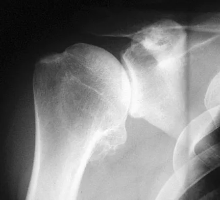
A 74-year-old woman with rheumatoid arthritis reports shoulder pain that has failed to respond to nonsurgical management. AP and axillary radiographs are shown in Figures 23a and 23b. Examination reveals active forward elevation to 120 degrees and external rotation to 30 degrees. What treatment option results in the most predictable pain relief and function?


Explanation

Most studies have shown that total shoulder arthroplasties yield better pain relief and improved forward elevation when compared to hemiarthroplasty in patients with rheumatoid arthritis. Although rotator cuff tears are more common in this patient population, this patient has good forward elevation and no significant superior migration of the humeral head; therefore, a reverse arthroplasty is not indicated. The arthritis is too advanced in this patient to consider arthroscopy, but in less advanced cases it can improve range of motion and decrease pain. Metal-backed glenoid components have shown higher rates of loosening. Collin DN, Harryman DT II, Wirth MA: Shoulder arthroplasty for the treatment of inflammatory arthritis. J Bone Joint Surg Am 2004;86:2489-2496. Baumgarten KM, Lashgari CM, Yamaguchi K: Glenoid resurfacing in shoulder arthroplasty: Indications and contraindications. Instr Course Lect 2004;53:3-11.


Question 24

A 69-year-old woman has just undergone an uncomplicated total shoulder arthroplasty for glenohumeral osteoarthritis. A press-fit humeral stem and a cemented all-polyethylene glenoid component were placed. At this point, what is the postoperative rehabilitation plan?


Explanation

The patient needs to immediately begin an active assisted range-of-motion program emphasizing forward elevation and external rotation to the side. Sling immobilization without stretching for either 3 or 6 weeks will result in severe stiffness that will compromise her ultimate range of motion. Since she has a good quality subscapularis tendon, there is no need to avoid beginning external rotation to the side. However, starting a strengthening program at 3 weeks risks tearing the subscapularis tendon repair. Active strengthening should not begin for 6 weeks postoperatively to allow the subscapularis tendon repair time to heal. Boardman ND III, Cofield RH, Bengston KA, et al: Rehabilitation after total shoulder arthroplasty. J Arthroplasty 2001;16:483-486.


Question 25

A 27-year-old woman reports the acute atraumatic onset of burning pain in her right shoulder followed a week later by significant weakness and the inability to abduct her shoulder. One week prior to this incident she had recovered from a flu-like syndrome. Examination reveals full passive motion of the shoulder and the inability to actively raise the arm. Sensation in the right upper extremity is normal. Cervical spine examination is normal. Radiographs of the shoulder and cervical spine are normal. What is the most likely diagnosis?


Explanation

The patient has symptoms and examination findings of acute brachial neuritis which is often a diagnosis of exclusion. The recent viral flu-like symptoms have shown a correlation with the development of this disorder. The acute, severe shoulder weakness excludes calcific tendinitis, impingement, and poliomyelitis. A normal cervical spine examination makes cervical disk disease unlikely. Turner JW, Parsonage MJ: Neuralgic amyotrophy (paralytic brachial neuritis). Lancet 1957;2:209-212.


Question 26

A 24-year-old man falls on an outstretched hand. Radiographs and subsequent MRI confirm a displaced proximal pole scaphoid fracture. What is the most appropriate surgical approach and fixation method?


Explanation

Proximal pole scaphoid fractures are best approached dorsally to preserve the tenuous blood supply entering distally. This also allows for a straight-line screw trajectory perpendicular to the fracture.

Question 27

A 45-year-old man sustains a terrible triad injury of the elbow. During surgical reconstruction, what is the generally recommended sequence of repair?


Explanation

The standard sequence for terrible triad reconstruction is deep to superficial. This means coronoid fixation first, followed by radial head fixation or replacement, and finally LCL repair.

Question 28

A 78-year-old woman with severe rheumatoid arthritis sustains a comminuted intra-articular distal humerus fracture. Radiographs show osteopenic bone and extensive articular fragmentation. What is the most appropriate surgical treatment?


Explanation

Total elbow arthroplasty is the treatment of choice for highly comminuted, intra-articular distal humerus fractures in elderly patients. It allows for early mobilization in patients with poor bone quality or pre-existing inflammatory arthritis.

Question 29

A 35-year-old woman sustains a highly comminuted radial head fracture and reports right wrist pain. Examination reveals tenderness over the distal radioulnar joint (DRUJ). If the radial head is deemed unreconstructible, what is the most appropriate management?


Explanation

This is an Essex-Lopresti injury characterized by a radial head fracture and DRUJ disruption. Radial head resection alone leads to proximal radial migration, requiring radial head arthroplasty and DRUJ stabilization.

Question 30

Six months after undergoing volar locked plating for a distal radius fracture, a 55-year-old woman presents with an inability to flex the interphalangeal joint of her thumb. What is the most likely cause of this complication?


Explanation

Flexor pollicis longus (FPL) tendon rupture is a known complication of volar plating for distal radius fractures. It often occurs when the plate is positioned distal to the watershed line, causing frictional wear.

Question 31

A 40-year-old man sustains an anterior shoulder dislocation with an associated displaced greater tuberosity fracture. Following successful closed reduction of the glenohumeral joint, radiographs show the greater tuberosity is displaced 8 mm superiorly. What is the most appropriate next step?


Explanation

Following shoulder reduction, greater tuberosity fractures displaced >5 mm in active patients generally require surgical fixation. This prevents subacromial impingement and restores rotator cuff function.

Question 32

A 42-year-old bodybuilder feels a pop in his anterior elbow while lifting weights. Examination reveals an abnormal hook test and weakness in supination. During a single-incision anterior repair of the distal biceps tendon, which nerve is at greatest risk of injury?


Explanation

The lateral antebrachial cutaneous nerve (LABCN) is at highest risk during the anterior single-incision approach to the distal biceps. This is due to its superficial location in the surgical field.

Question 33

A 30-year-old woman presents with elbow pain after a fall. Imaging reveals a capitellum fracture consisting primarily of articular cartilage with very little subchondral bone. How is this fracture classified?


Explanation

A Kocher-Lorenz (Type II) capitellum fracture involves an articular cartilage shear fragment with minimal attached subchondral bone. Hahn-Steinthal (Type I) involves a large piece of subchondral bone.

Question 34

A 38-year-old man presents with a locked posterior shoulder dislocation following a seizure. CT scan confirms an anteromedial humeral head defect (reverse Hill-Sachs lesion) involving 35% of the articular surface. What is the most appropriate surgical management?


Explanation

For reverse Hill-Sachs defects between 20-40%, transfer of the subscapularis or lesser tuberosity into the defect is indicated. This modified McLaughlin procedure restores anterior stability and prevents engagement.

Question 35

Which of the following is a widely accepted relative indication for open reduction and internal fixation of a midshaft clavicle fracture?


Explanation

Shortening >2 cm, complete displacement, and severe Z-deformity are relative indications for operative fixation of midshaft clavicle fractures. Surgery decreases the risk of nonunion and symptomatic malunion.

Question 36

A 28-year-old construction worker falls from a height and sustains a wrist injury. Radiographs show the lunate is displaced and rotated volar to the radius, while the capitate remains aligned with the radius. What is this injury pattern?


Explanation

A volar lunate dislocation represents the final stage (Mayfield IV) of perilunate instability. The lunate is displaced volarly (the "spilled teacup" sign), while the capitate falls back into alignment with the radius.

Question 37

A 6-year-old boy falls off monkey bars and sustains a diaphyseal fracture of the proximal third of the ulna with an associated anterior dislocation of the radial head. What Bado classification does this represent?


Explanation

A Bado Type I Monteggia fracture is characterized by a fracture of the proximal or middle third of the ulna with an anterior dislocation of the radial head. It is the most common Monteggia type in children.

Question 38

A 50-year-old man sustains a transverse, non-comminuted fracture of the olecranon. He undergoes tension band wiring. What is the primary biomechanical principle of this fixation construct?


Explanation

Tension band wiring converts the tensile forces on the dorsal cortex caused by the pull of the triceps into compressive forces at the articular surface. This promotes primary bone healing in transverse fractures.

Question 39

A 35-year-old man sustains a distal third spiral fracture of the humerus. On examination, he is unable to extend his wrist or fingers. Which nerve is most commonly injured in this specific fracture pattern?


Explanation

A Holstein-Lewis fracture is a spiral fracture of the distal third of the humeral shaft. It has a high association with radial nerve entrapment or injury as the nerve passes through the intermuscular septum.

Question 40

A 32-year-old man falls on an extended wrist. Radiographs reveal a scapholunate gap of 4 mm and a cortical ring sign of the scaphoid. What is the most appropriate management for an acute, repairable scapholunate ligament tear?


Explanation

Acute scapholunate ligament tears with diastasis should be treated surgically with open reduction and primary ligament repair. This is often augmented with a dorsal capsulodesis or K-wire pinning to restore carpal kinematics.

Question 41

A 45-year-old woman sustained a nondisplaced distal radius fracture treated in a cast. Six weeks later, she suddenly notices an inability to extend her thumb at the interphalangeal joint. She denies any new trauma. What is the most likely cause of this finding?


Explanation

Extensor pollicis longus (EPL) tendon rupture is a known complication following nondisplaced distal radius fractures due to ischemia or attrition at Lister's tubercle. It typically presents several weeks post-injury with an inability to actively extend the thumb interphalangeal joint.

Question 42

A 72-year-old woman with severe osteoporosis presents with a closed, displaced 3-part proximal humerus fracture involving the surgical neck and greater tuberosity. The humeral head is varus and severely retroverted. What is the most appropriate surgical treatment?


Explanation

Reverse total shoulder arthroplasty (RTSA) provides more reliable outcomes and improved active elevation compared to ORIF or hemiarthroplasty for displaced 3- and 4-part fractures in elderly patients with poor bone quality. It mitigates the risk of fixation failure and relies less on tuberosity healing for overhead function.

Question 43

A 35-year-old man falls on an outstretched hand and sustains a 'terrible triad' injury of the elbow. To optimally restore elbow stability, which of the following sequences is recommended during surgical reconstruction?


Explanation

The standard surgical sequence for a terrible triad injury works from deep to superficial, or inside-out. This involves fixing the coronoid first, followed by repairing or replacing the radial head, and finally repairing the lateral ulnar collateral ligament (LUCL).

Question 44

A 22-year-old man falls on a hyperextended wrist. Radiographs reveal a displaced fracture of the proximal pole of the scaphoid. What is the primary arterial supply to the proximal pole of the scaphoid, which places this injury at high risk for avascular necrosis?


Explanation

The dorsal carpal branch of the radial artery provides the predominant blood supply to the scaphoid, entering distally and flowing retrogradely to the proximal pole. Fractures at the proximal pole disrupt this retrograde flow, heavily increasing the risk of avascular necrosis.

Question 45

A 28-year-old man sustains a fracture of the middle third of the radius with associated distal radioulnar joint (DRUJ) dislocation. Following rigid open reduction and internal fixation of the radius, the DRUJ easily reduces but subluxates in pronation while remaining completely stable in supination. What is the next best step in management?


Explanation

If the DRUJ is stable in supination following anatomic fixation of a Galeazzi fracture, the treatment of choice is immobilization in supination for 4-6 weeks. Operative intervention (pinning or TFCC repair) is reserved for cases that are unstable in all positions of rotation.

Question 46

A 25-year-old cyclist sustains a completely displaced, shortened (>2 cm) midshaft clavicle fracture. Compared to nonoperative management, operative fixation with plate and screws is primarily associated with a lower incidence of which of the following?


Explanation

Operative fixation of completely displaced, shortened midshaft clavicle fractures significantly reduces the rate of symptomatic nonunion and symptomatic malunion compared to nonoperative treatment. However, operative management does carry risks of infection and hardware irritation requiring subsequent removal.

Question 47

A 40-year-old man sustains a Bado Type I Monteggia fracture-dislocation. Intraoperatively, following rigid plate fixation of the ulna fracture, the radial head remains anteriorly dislocated. What is the most appropriate next step in management?


Explanation

In Monteggia fractures, the radial head should spontaneously reduce once the ulnar length and alignment are anatomically restored. If the radial head remains dislocated after ulnar fixation, the surgeon must first assume that the ulnar reduction is imperfect and address any residual angulation or shortening.

Question 48

When utilizing a posterior approach with an olecranon osteotomy for open reduction and internal fixation of a highly comminuted intra-articular distal humerus fracture, a chevron osteotomy is often preferred over a transverse osteotomy because it:


Explanation

A chevron or V-shaped olecranon osteotomy provides inherent bony rotatory stability when reduced, and increases the surface area for healing compared to a simple transverse osteotomy. It facilitates anatomic restoration of the articular surface.

Question 49

A 55-year-old woman is treated with a volar locking plate for a displaced volar Barton's fracture. Postoperatively, she develops severe paresthesias and burning pain in her thumb, index, and middle fingers that fail to improve after two weeks. Which of the following is the most appropriate next step?


Explanation

Post-traumatic carpal tunnel syndrome can occur after distal radius fracture or its fixation. Severe, unrelenting, or progressive symptoms of median neuropathy following ORIF should prompt surgical median nerve decompression to prevent permanent nerve injury.

Question 50

A 19-year-old male presents with severe medial chest pain, dysphagia, and a choking sensation following a rugby tackle. CT scan confirms a posterior sternoclavicular dislocation. Which vascular structure is located immediately posterior to the sternoclavicular joint and is most at risk of injury?


Explanation

The brachiocephalic (innominate) vein is located immediately posterior to the sternoclavicular joint, making it the most vulnerable vascular structure in a posterior sternoclavicular dislocation. The trachea and esophagus are also at risk, leading to symptoms like dysphagia or shortness of breath.

Question 51

A 30-year-old man presents with a markedly displaced scapular body fracture following a high-speed motorcycle accident. Which of the following associated injuries is statistically most commonly seen in patients with this high-energy fracture?


Explanation

Scapula fractures generally require high-energy trauma, and associated injuries are present in over 80-90% of cases. Ipsilateral rib fractures are the most common associated injury, occurring in approximately 50% of patients.

Question 52

A Bryan and Morrey Type I (Hahn-Steinthal) fracture of the capitellum is best described radiographically and anatomically as:


Explanation

The Hahn-Steinthal (Type I) fracture involves a large osseous fragment of the capitellum containing a significant amount of subchondral bone. Type II (Kocher-Lorenz) is a thin articular cartilage shell, Type III is comminuted, and Type IV (McKee modification) involves the trochlea.

Question 53

A 45-year-old man felt a sudden 'pop' in his anterior elbow while lifting a heavy box. On examination, a positive Hook test is noted. Which of the following physical examination findings is most functionally characteristic of this specific injury?


Explanation

A positive Hook test indicates a complete distal biceps tendon rupture. The biceps brachii is the primary supinator of the forearm, especially with the elbow flexed, so its rupture results in profound weakness in supination, along with mild-to-moderate weakness in elbow flexion.

Question 54

A 28-year-old man falls backward on an extended, ulnarly deviated wrist. Radiographs reveal a volar lunate dislocation. According to Mayfield's progressive stages of perilunate instability, what is the exact sequence of ligamentous failure leading to this end-stage injury?


Explanation

Mayfield described a four-stage progression of perilunate instability starting radially and progressing ulnarly. Stage I disrupts the scapholunate joint, Stage II the capitolunate joint, Stage III the lunotriquetral joint, and Stage IV involves dorsal radiocarpal disruption allowing the lunate to dislocate volarly.

Question 55

A 32-year-old man sustains a spiral fracture of the distal third of the humeral shaft (Holstein-Lewis fracture). On presentation, he exhibits a complete inability to extend his wrist or digits. If surgical exploration is indicated, where is the radial nerve most likely to be entrapped or injured?


Explanation

Holstein-Lewis fractures are spiral fractures of the distal one-third of the humerus. The radial nerve is at high risk of entrapment or laceration as it is tethered piercing the lateral intermuscular septum to pass from the posterior to the anterior compartment.

Question 56

A 26-year-old hockey player sustains a direct downward blow to his shoulder. Radiographs demonstrate superior displacement of the clavicle relative to the acromion by approximately 200%. The coracoclavicular distance is more than twice that of the uninjured side. According to the Rockwood classification, what type of acromioclavicular injury is this?


Explanation

A Rockwood Type V acromioclavicular dislocation involves severe superior displacement of the clavicle (100% to 300%) relative to the acromion, accompanied by significant stripping of the deltotrapezial fascia. Type III involves up to 100% displacement, and Type IV is displaced posteriorly into the trapezius.

Question 57

A 45-year-old man falls from a ladder and sustains a 'terrible triad' injury to his elbow. Which of the following represents the most widely accepted surgical sequence for reconstructing this injury to restore stability?


Explanation

The standard deep-to-superficial approach for a terrible triad injury involves fixing the coronoid first, followed by the radial head, and finally repairing the lateral ulnar collateral ligament (LUCL).

Question 58

A 35-year-old woman presents with severe elbow and wrist pain after a fall. Radiographs demonstrate a comminuted radial head fracture. On examination, she has marked tenderness and instability over the distal radioulnar joint (DRUJ). Which of the following treatments is absolutely contraindicated?


Explanation

This patient has an Essex-Lopresti injury (radial head fracture, interosseous membrane tear, and DRUJ disruption). Radial head excision alone is contraindicated as it will lead to proximal migration of the radius and chronic wrist pain.

Question 59

A 28-year-old man sustains a closed fracture of the distal third of the humeral shaft. On initial presentation, his radial nerve function is intact. Following closed reduction and splinting, he immediately loses the ability to actively extend his wrist and fingers. What is the most appropriate next step in management?


Explanation

A secondary radial nerve palsy that develops immediately after closed manipulation of a distal third humeral shaft fracture (Holstein-Lewis pattern) is an indication for immediate surgical exploration, as the nerve may be entrapped in the fracture site.

Question 60

A 78-year-old woman with a history of severe osteoporosis sustains a displaced 4-part proximal humerus fracture. She lives independently and is active. What surgical option provides the most predictable return of forward elevation and is least dependent on tuberosity healing?


Explanation

RTSA relies on deltoid function rather than an intact rotator cuff. It provides more predictable functional outcomes for forward elevation in elderly patients with 4-part fractures, even if the tuberosities fail to heal.

Question 61

A 22-year-old collegiate rugby player presents with recurrent anterior shoulder instability. CT imaging demonstrates a 30% anterior glenoid bone defect. Which of the following is the most appropriate surgical management?


Explanation

Critical glenoid bone loss (>20-25%) in a young collision athlete is a classic indication for a bony augmentation procedure, such as the Latarjet procedure, due to the unacceptably high failure rate of soft-tissue stabilization alone.

Question 62

A 21-year-old male presents with radial-sided wrist pain after falling on an outstretched hand. Radiographs show a displaced fracture of the proximal pole of the scaphoid. Which of the following blood supplies is most at risk, and what is the optimal surgical approach?


Explanation

The proximal pole of the scaphoid relies on retrograde blood flow from the dorsal carpal branch of the radial artery. A dorsal surgical approach is preferred for proximal pole fractures to achieve direct access without further jeopardizing this blood supply.

Question 63

A 42-year-old man sustains a fracture of the proximal third of the ulna with an associated radial head dislocation. During operative management, the ulna is anatomically reduced and rigidly plated, but the radial head remains dislocated. What is the most appropriate next step?


Explanation

In a Monteggia fracture-dislocation, if the radial head fails to reduce after anatomic fixation of the ulna, soft tissue interposition (such as the annular ligament or joint capsule) blocking reduction must be suspected and openly explored.

Question 64

A 34-year-old male presents to the ER after a generalized tonic-clonic seizure. His shoulder is locked in internal rotation. A modified axillary radiograph reveals a posterior shoulder dislocation with an anteromedial humeral head defect involving 30% of the articular surface. Which procedure is most appropriate?


Explanation

For a reverse Hill-Sachs lesion involving 20% to 40% of the articular surface associated with posterior instability, a modified McLaughlin procedure (transfer of the lesser tuberosity or subscapularis into the defect) is indicated to prevent engagement.

Question 65

A 28-year-old motorcyclist is involved in a high-speed collision. Lateral wrist radiographs show the lunate is displaced and rotated volar to the radius, while the capitate remains aligned with the longitudinal axis of the radius. What is the diagnosis?


Explanation

This classic 'spilled teacup' sign on the lateral radiograph, where the lunate is volarly displaced while the capitate remains colinear with the radius, defines a lunate dislocation. In a perilunate dislocation, the lunate remains seated in the radial fossa while the capitate is dislocated.

Question 66

Which of the following is considered an absolute indication for operative fixation of an acute midshaft clavicle fracture?


Explanation

Absolute indications for ORIF of a clavicle fracture include open fractures, neurovascular compromise, and severe skin tenting threatening skin necrosis. Displacement and shortening are relative indications.

Question 67

A 45-year-old man undergoes a modified two-incision repair for a distal biceps tendon rupture. Postoperatively, he presents with the inability to actively extend his fingers and thumb at the metacarpophalangeal joints. Wrist extension is preserved but deviates radially. Which nerve is most likely injured?


Explanation

The posterior interosseous nerve (PIN) is highly vulnerable during the lateral exposure of a two-incision distal biceps repair. PIN palsy results in loss of digit extension and radial deviation during wrist extension due to extensor carpi ulnaris weakness.

Question 68

Six weeks after undergoing nonoperative management of a nondisplaced distal radius fracture, a 60-year-old woman suddenly loses the ability to extend her thumb interphalangeal joint. What is the most appropriate surgical management?


Explanation

Delayed EPL rupture after a distal radius fracture is secondary to attrition or ischemia in the third dorsal compartment. Primary repair is typically impossible due to tendon retraction and degeneration, making EIP-to-EPL transfer the gold standard.

Question 69

A 30-year-old male sustains a Galeazzi fracture-dislocation. Following rigid open reduction and internal fixation of the radius, the distal radioulnar joint (DRUJ) is found to be irreducibly dislocated. What is the most likely interposed structure?


Explanation

The extensor carpi ulnaris (ECU) tendon is the most common soft-tissue structure to block reduction of the DRUJ in a Galeazzi fracture-dislocation, requiring open exploration for removal.

Question 70

Tension band wiring is most appropriate and biomechanically sound for which of the following olecranon fracture patterns?


Explanation

Tension band wiring converts tension forces at the dorsal cortex into compression forces at the articular surface. This mechanism relies on an intact volar cortex to act as a buttress, making it ideal for simple transverse, non-comminuted fractures.

Question 71

A 35-year-old man falls on his outstretched arm, sustaining an elbow dislocation with associated radial head and coronoid process fractures. After operative restoration of the coronoid and radial head, the elbow remains unstable in extension. What is the most appropriate next step in management?


Explanation

The standard surgical sequence for a terrible triad injury involves coronoid fixation, radial head repair or replacement, and LUCL repair. If the elbow remains unstable after these steps, MCL repair or a hinged external fixator may be considered.

Question 72

A 75-year-old woman with severe glenohumeral osteoarthritis and a known massive irreducible rotator cuff tear sustains a severely displaced 3-part proximal humerus fracture. What is the most appropriate surgical treatment?


Explanation

Reverse total shoulder arthroplasty is indicated for complex proximal humerus fractures in elderly patients with pre-existing rotator cuff arthropathy or massive rotator cuff tears. It relies on the deltoid for overhead function, bypassing the deficient rotator cuff.

Question 73

A 45-year-old man undergoes volar locked plating for a displaced distal radius fracture. Six months later, he presents with an inability to actively extend his thumb interphalangeal joint. This complication is most likely due to rupture of which structure?


Explanation

Prominent dorsal screws extending past the dorsal cortex in a volar plate construct are a common cause of extensor tendon irritation and EPL rupture. The EPL is particularly vulnerable as it curves around Lister's tubercle.

Question 74

A 28-year-old woman falls from a height, sustaining a highly comminuted, unsalvageable radial head fracture and positive ulnar variance at the wrist with distal radioulnar joint (DRUJ) instability. What is the most appropriate management of the radial head in this setting?


Explanation

This is an Essex-Lopresti injury involving longitudinal radioulnar dissociation. Excision of the radial head without replacement leads to proximal radial migration; therefore, radial head arthroplasty combined with DRUJ stabilization is required.

Question 75

A 25-year-old cyclist falls and sustains a midshaft clavicle fracture. Which of the following is considered an absolute indication for immediate operative fixation of this injury?


Explanation

Absolute indications for operative fixation of clavicle fractures include open fractures, neurovascular compromise, and severe skin tenting threatening to progress to an open fracture. Shortening of greater than 2 cm is generally considered a relative indication.

Question 76

A 20-year-old man presents with anatomic snuffbox tenderness following a fall on an outstretched hand. Initial radiographs are negative, but an MRI confirms a non-displaced scaphoid waist fracture. What is the most appropriate definitive management?


Explanation

Non-displaced scaphoid waist fractures can be successfully treated nonoperatively with a short-arm cast for 6-8 weeks. Operative fixation is generally reserved for displaced fractures, proximal pole fractures, or athletic patients desiring quicker return to play.

Question 77

A 32-year-old man requires open reduction and internal fixation of a highly comminuted intercondylar distal humerus fracture (OTA type 13-C3). To provide the most extensive exposure to the articular surface, which surgical approach is preferred?


Explanation

A chevron olecranon osteotomy provides the widest exposure to the articular surface of the distal humerus, which is critical for restoring complex, comminuted intra-articular fractures. The osteotomy must be anatomically repaired at the end of the procedure.

Question 78

A 40-year-old woman falls on her outstretched hand and sustains a capitellum fracture. Radiographs show a large anterior osteochondral fragment that includes the capitellum and the lateral half of the trochlea. This describes which type of fracture pattern?


Explanation

A Type 4 capitellum fracture (McKee modification of the Bryan and Morrey classification) involves a coronal shear fracture that includes the capitellum and the lateral half of the trochlea. It often requires stable anatomic fixation to prevent post-traumatic arthrosis.

Question 79

A 30-year-old male presents after a motorcycle crash with a swollen, painful wrist and numbness in his thumb, index, and long fingers. The lateral radiograph shows the lunate displaced palmar to the radius, while the capitate remains aligned with the radius. What is the diagnosis?


Explanation

In a lunate dislocation, the lunate is displaced and tilted palmarly (creating a 'spilled teacup' sign on the lateral radiograph), while the capitate remains aligned with the radius. Median nerve symptoms are common due to acute carpal tunnel compression.

Question 80

A 28-year-old professional athlete sustains a direct blow to the shoulder. Radiographs demonstrate a 150% superior displacement of the clavicle relative to the acromion. He is diagnosed with a Rockwood Type III AC joint dislocation. What is the generally recommended initial management?


Explanation

Type III AC joint separations are generally treated nonoperatively initially, yielding functional results comparable to surgery with fewer complications. Surgery may be considered later for persistent symptoms, or acutely in highly demanding overhead athletes.

Question 81

A 75-year-old right-hand-dominant woman presents with a 4-part proximal humerus fracture after a ground-level fall. Radiographs demonstrate significant osteopenia and a severely comminuted calcar. She has a documented history of severe rotator cuff arthropathy in the affected shoulder. What is the most appropriate surgical management?


Explanation

Reverse total shoulder arthroplasty is the treatment of choice for elderly patients with 4-part proximal humerus fractures and pre-existing rotator cuff dysfunction. It provides more predictable functional outcomes compared to hemiarthroplasty when tuberosity healing is uncertain.

Question 82

A 25-year-old male cyclist sustains a midshaft clavicle fracture. Which of the following is considered an absolute indication for operative fixation?


Explanation

Absolute indications for operative fixation of a clavicle fracture include open fractures, neurovascular compromise, and impending skin ischemia (tenting). Displacement, shortening, and comminution are considered relative indications.

Question 83

During a coracoclavicular (CC) ligament reconstruction for a chronic Type V acromioclavicular joint separation, the surgeon must recreate the anatomical footprints. Which of the following accurately describes the native CC ligament anatomy?


Explanation

The conoid ligament is located posteromedial to the trapezoid ligament and inserts approximately 4.5 cm medial to the distal end of the clavicle. The trapezoid is anterolateral and inserts approximately 3 cm medial to the distal clavicle.

Question 84

A 42-year-old male sustains a severely displaced fracture of the scapular body and neck following a high-speed motor vehicle collision. Which of the following radiographic findings represents an absolute indication for surgical intervention?


Explanation

Glenohumeral instability or subluxation is an absolute indication for operative fixation of scapula fractures. Articular step-off >4 mm, medialization >20 mm, and angulation >45 degrees are generally considered relative indications.

Question 85

An 18-year-old male rugby player presents with a posterior sternoclavicular dislocation. His neurovascular status is intact, and he has no respiratory distress. What is the most appropriate next step in management?


Explanation

Posterior sternoclavicular dislocations carry a significant risk of injury to the great vessels and trachea. Closed reduction should be attempted in the operating room under general anesthesia with cardiothoracic surgery on standby.

Question 86

A 78-year-old female with long-standing rheumatoid arthritis sustains a severely comminuted intra-articular distal humerus fracture. Her bone quality is exceptionally poor. What is the preferred surgical treatment to maximize her early functional outcome?


Explanation

Total elbow arthroplasty is the preferred treatment for comminuted, intra-articular distal humerus fractures in elderly patients with pre-existing inflammatory arthritis and poor bone quality, allowing for immediate postoperative range of motion.

Question 87

A 35-year-old female presents with a 'terrible triad' injury of the elbow consisting of an elbow dislocation, radial head fracture, and coronoid fracture. What is the standard sequence of repair during surgical reconstruction?


Explanation

The standard surgical sequence for terrible triad injuries addresses structures from deep to superficial: coronoid fixation first, followed by radial head repair or replacement, and finally lateral ulnar collateral ligament (LUCL) repair.

Question 88

A patient presents with an anteromedial facet fracture of the coronoid process. This specific fracture pattern is the hallmark of varus posteromedial rotatory instability (VPMRI) of the elbow. Which ligamentous complex is consistently injured and requires evaluation in this setting?


Explanation

Anteromedial facet fractures of the coronoid are pathognomonic for varus posteromedial rotatory instability (VPMRI). This injury mechanism inherently involves a disruption of the lateral collateral ligament (LCL) complex, specifically avulsing from the lateral epicondyle.

Question 89

A 40-year-old man underwent excision of a comminuted radial head fracture 6 weeks ago. He now presents with progressive ulnar-sided wrist pain and weakness. Radiographs demonstrate proximal migration of the radius. This complication is a result of an unrecognized injury to which structure?


Explanation

Proximal migration of the radius following radial head excision indicates an unrecognized disruption of the interosseous membrane, known as an Essex-Lopresti lesion. This results in longitudinal radioulnar dissociation and requires radial head replacement.

Question 90

A 65-year-old woman is seen 6 months after undergoing volar locked plating for a distal radius fracture. She complains of a sudden inability to flex the interphalangeal joint of her thumb. What is the most likely etiology?


Explanation

Flexor pollicis longus (FPL) tendon rupture is a well-documented complication of volar plating of the distal radius. It typically occurs due to attritional wear when the plate is positioned distal to the watershed line (Soong Grade 2).

Question 91

A 22-year-old male sustains a fracture through the proximal pole of the scaphoid. This fracture pattern carries a high risk of avascular necrosis because the primary intraosseous blood supply to the proximal pole enters at which anatomical location?


Explanation

The primary blood supply to the scaphoid arises from the dorsal carpal branch of the radial artery, which enters the dorsal ridge at the distal pole. Blood then flows in a retrograde fashion to supply the proximal pole, predisposing it to avascular necrosis.

Question 92

A 28-year-old male presents with a volar lunate dislocation following a fall on an extended wrist. According to the Mayfield sequence of perilunate instability, what is the first intercarpal ligament to rupture?


Explanation

The Mayfield sequence describes the progressive disruption of perilunate ligaments from radial to ulnar. Stage I begins with the rupture of the scapholunate interosseous ligament.

Question 93

A 7-year-old boy sustains a Bado Type III Monteggia fracture-dislocation (lateral dislocation of the radial head with proximal ulnar fracture). Which of the following neurologic injuries is most commonly associated with this specific fracture pattern?


Explanation

Posterior interosseous nerve (PIN) palsy is the most common neurologic deficit associated with Monteggia fracture-dislocations, and it is particularly prevalent in Bado Type III (anterolateral/lateral radial head dislocation) injuries.

Question 94

During open reduction and internal fixation of a Galeazzi fracture, rigid anatomic fixation of the radius has been achieved. What is the most appropriate intraoperative method to assess the stability of the distal radioulnar joint (DRUJ)?


Explanation

Following stabilization of the radius in a Galeazzi fracture, the DRUJ must be evaluated. This is done by ranging the forearm through full supination and pronation while manually assessing for excessive anteroposterior translation of the ulnar head.

Question 95

A 40-year-old male presents with an unprovoked seizure resulting in a right posterior shoulder dislocation. Imaging reveals an anteromedial humeral head defect (reverse Hill-Sachs lesion) involving 30% of the articular surface. What is the most appropriate surgical management to prevent recurrent instability?


Explanation

For a reverse Hill-Sachs lesion involving 20% to 40% of the articular surface, filling the defect via transfer of the lesser tuberosity or subscapularis tendon (McLaughlin or modified McLaughlin procedure) is the recommended treatment to prevent engagement.

None

Clinic OS
Medically Verified Content by
Prof. Clinic OS
Consultant Orthopedic & Spine Surgeon
Chapter Index