Calcaneonavicular Coalition Resection: An Adult Surgical Masterclass with EDB Interposition
Key Takeaway
This masterclass guides orthopaedic fellows through calcaneonavicular coalition resection in adult patients. We cover comprehensive anatomy, meticulous preoperative planning, and a step-by-step intraoperative execution of the Ollier approach with extensor digitorum brevis interposition. Emphasis is placed on neurovascular protection, precise osteotomy, and managing potential complications for optimal outcomes.
Comprehensive Introduction and Patho-Epidemiology
Tarsal coalitions represent a fascinating, complex, and historically challenging subset of foot and ankle pathology. A tarsal coalition is defined as an abnormal structural fusion—whether fibrous (syndesmosis), cartilaginous (synchondrosis), or osseous (synostosis)—between two or more adjacent tarsal bones. Embryologically, this condition arises from a failure of normal segmentation and cleavage of the primitive undifferentiated mesenchyme during the first trimester of fetal development. While the true incidence in the general population is difficult to ascertain due to a high prevalence of asymptomatic individuals, prevailing literature suggests an incidence ranging from 1% to 13%, with calcaneonavicular and talocalcaneal coalitions comprising the vast majority (approximately 90%) of these anomalies.
The calcaneonavicular coalition is unique. Unlike talocalcaneal fusions, which bridge an existing, anatomically normal joint space, the calcaneonavicular coalition forms a bridge between two bones that, in a normal physiologic state, do not possess a true articular interface. Specifically, the abnormal connection spans from the anterior process of the calcaneus to the inferolateral aspect of the navicular. In the pediatric and adolescent populations, these coalitions often present as a rigid, painful flatfoot deformity, classically termed "peroneal spastic flatfoot," as the bridge ossifies and restricts subtalar motion. However, the adult presentation of a calcaneonavicular coalition demands a distinct paradigm of clinical reasoning and surgical management.
In the adult patient, the coalition has typically completed its ossification process, presenting as a rigid, mature synostosis. Many of these adult patients traverse their early decades entirely asymptomatic, only to present in their twenties, thirties, or beyond with acute-on-chronic hindfoot pain. This delayed presentation is frequently unmasked by a traumatic event, such as a severe inversion ankle sprain or an acute midfoot crush injury. The trauma disrupts the tenuous, asymptomatic biomechanical equilibrium of the foot, fracturing the coalition or irritating the surrounding soft tissue envelope, thereby tipping the patient into a state of chronic, recalcitrant pain.
The natural history of an untreated, symptomatic calcaneonavicular coalition in an adult is characterized by progressive biomechanical failure. The rigid tethering of the transverse tarsal (Chopart) joint complex severely limits the normal coupled kinematics of the hindfoot and midfoot. Consequently, the requisite motion for ambulation on uneven terrain is forcefully shunted to adjacent joints, primarily the talonavicular, calcaneocuboid, and subtalar articulations. Over time, this aberrant stress distribution inevitably accelerates articular cartilage degradation, precipitating early-onset secondary osteoarthrosis. Therefore, the surgical objective in the adult is not merely pain relief through resection, but the crucial restoration of hindfoot kinematics to preserve the longevity of the adjacent tarsal joints.
Detailed Surgical Anatomy and Biomechanics
A profound comprehension of the regional osteology, soft tissue intervals, and neurovascular topography is the bedrock of a successful calcaneonavicular coalition resection. The primary osseous landmarks dictate the boundaries of our surgical field. The coalition itself extends from the anterior process of the calcaneus—a structure that normally serves as the origin for the bifurcate ligament—to the inferolateral margin of the navicular. The surgeon must acutely recognize the proximity of the true articular surfaces. Just posterior and superior to the coalition lies the anterior facet of the subtalar joint; immediately distal is the calcaneocuboid joint; and medial to the navicular insertion is the talonavicular joint. Inadvertent violation of any of these pristine articular surfaces during aggressive resection will lead to catastrophic post-operative arthrosis.
Biomechanically, the normal foot relies on the subtalar joint to act as a directional torque transmitter, converting the internal and external rotation of the tibia into pronation and supination of the foot. During the gait cycle, as the subtalar joint everts, the axes of the talonavicular and calcaneocuboid joints become parallel, "unlocking" the midfoot to accommodate uneven terrain. Conversely, subtalar inversion diverges these axes, "locking" the midfoot to provide a rigid lever arm for terminal stance and push-off. A calcaneonavicular coalition rigidly locks the midfoot complex, obliterating this vital transition. The peroneal musculature often goes into secondary spasm (not true spasticity, but an adaptive, protective guarding mechanism) in an attempt to splint the painful, restricted hindfoot, leading to the classic planovalgus appearance.
The soft tissue envelope overlying the lateral hindfoot is dense with critical structures that must be meticulously preserved. The Extensor Digitorum Brevis (EDB) muscle is the central soft tissue landmark for this procedure. Originating from the dorsal and lateral surfaces of the anterior calcaneus, the EDB muscle belly lies directly superficial to the calcaneonavicular coalition. It is this exact anatomical relationship that allows us to elegantly elevate the EDB as a distally based flap and subsequently interpose it into the resection void. Deep and slightly plantar to the EDB origin run the peroneus longus and brevis tendons within their common synovial sheath, which must be retracted inferiorly and protected from the oscillating saw or osteotome.
Neurovascularly, the lateral approach places two primary sensory nerves at significant risk. The sural nerve, providing sensation to the lateral heel and lateral border of the foot, courses posterior to the lateral malleolus and runs superficially along the peroneal tendons. More dorsally, the dorsal intermediate cutaneous branch of the superficial peroneal nerve traverses the surgical field to supply the dorsolateral midfoot and forefoot. Injury to either of these nerves can result in debilitating neuromas or Complex Regional Pain Syndrome (CRPS), complications that often eclipse the initial coalition pain. Meticulous, blunt dissection within the subcutaneous tissues is mandatory to identify, mobilize, and protect these neural structures throughout the entirety of the procedure.
Exhaustive Indications and Contraindications
The decision to proceed with surgical resection of a calcaneonavicular coalition in an adult must be predicated on a rigorous evaluation of the patient's symptoms, radiographic findings, and response to conservative modalities. Surgery is never prophylactic for an asymptomatic coalition discovered incidentally. A minimum of three to six months of dedicated, non-operative management—encompassing judicious use of NSAIDs, activity modification, physical therapy, customized rigid orthoses (such as a UCBL device to restrict subtalar motion), and potentially a period of immobilization in a controlled ankle motion (CAM) boot or short-leg cast—must be exhausted prior to surgical consideration.
When conservative measures fail to yield sustainable relief, surgical intervention becomes warranted. However, the presence of a coalition does not automatically dictate a resection. The surgeon must critically evaluate the adjacent joints for degenerative changes. In the presence of advanced, symptomatic arthrosis of the subtalar, talonavicular, or calcaneocuboid joints, a simple resection will fail to relieve the patient's pain and may, in fact, exacerbate it by increasing motion across a degenerated joint. In such scenarios, a primary arthrodesis (e.g., subtalar or triple arthrodesis) is the procedure of choice.
| Category | Specific Clinical Parameters |
|---|---|
| Absolute Indications | - Persistent, debilitating lateral/deep hindfoot pain localized to the coalition site. - Failure of a comprehensive 3-to-6-month non-operative treatment protocol. - Radiographic confirmation of a calcaneonavicular coalition without adjacent joint arthrosis. - Recurrent ankle instability or frequent sprains directly attributable to hindfoot rigidity. |
| Relative Indications | - Mild, asymptomatic radiographic narrowing of the posterior subtalar facet (requires careful patient counseling regarding potential future arthrodesis). - Co-existing flexible flatfoot deformity (may require concomitant procedures like medializing calcaneal osteotomy or spring ligament repair). |
| Absolute Contraindications | - Advanced, symptomatic degenerative joint disease of the subtalar, talonavicular, or calcaneocuboid joints. - Active local or systemic infection. - Severe peripheral vascular disease compromising wound healing. - Neuropathic arthropathy (Charcot foot) involving the hindfoot/midfoot. |
| Relative Contraindications | - Concomitant talocalcaneal coalition (often necessitates a more complex, combined resection or primary fusion). - Poorly controlled diabetes mellitus or profound immunosuppression. - Patient inability or unwillingness to adhere to strict post-operative rehabilitation protocols. |
Pre-Operative Planning, Templating, and Patient Positioning
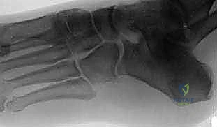
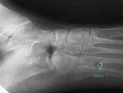
Surgical success is architected long before the incision is made. Pre-operative planning for an adult calcaneonavicular coalition resection demands a comprehensive radiographic analysis. Standard weight-bearing plain radiographs of the foot (Anteroposterior, Lateral, and 45-degree internal oblique views) are mandatory. The 45-degree oblique view is the definitive plain film for visualizing the calcaneonavicular interval, often revealing the classic "anteater sign"—a tubular prolongation of the anterior process of the calcaneus approaching the navicular. However, plain films consistently underestimate the true three-dimensional volume and osseous maturity of the coalition.
Therefore, a fine-cut Computed Tomography (CT) scan is considered the gold standard and is absolutely essential for all adult patients contemplating resection. The CT scan provides a high-resolution, multi-planar roadmap. It allows the surgeon to precisely quantify the width, depth, and exact anatomical boundaries of the bony bridge. Furthermore, the CT scan is invaluable for ruling out a subtle, concomitant talocalcaneal coalition (which occurs in up to 20% of cases) and for definitively assessing the articular cartilage integrity of the adjacent Chopart and subtalar joints. Magnetic Resonance Imaging (MRI) is generally reserved for cases where the coalition is suspected to be purely fibrous or cartilaginous (less common in adults) or when concurrent soft tissue pathology, such as peroneal tendinopathy or a spring ligament tear, requires evaluation.
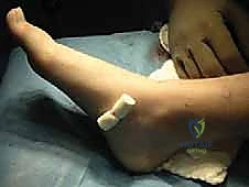
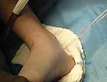
In the operating theater, meticulous attention to patient positioning optimizes surgical ergonomics and visualization. The patient is positioned supine on a standard radiolucent operating table. A crucial maneuver is the placement of a well-padded bump under the ipsilateral hip/sacrum. This internally rotates the lower extremity, bringing the lateral aspect of the foot directly into the surgeon's line of sight and preventing the natural tendency of the leg to externally rotate. A pneumatic tourniquet is applied to the proximal thigh to ensure a bloodless surgical field, which is critical for the precise identification of cutaneous nerves and the meticulous execution of the osteotomies.
Fluoroscopy is an indispensable tool during this procedure. The C-arm must be positioned to allow for unimpeded, orthogonal Anteroposterior, Lateral, and Oblique imaging of the foot throughout the case. The surgical team must ensure that the C-arm can be maneuvered into the field without compromising the sterility of the draped extremity. Prophylactic intravenous antibiotics (typically a first-generation cephalosporin) are administered 30 to 60 minutes prior to tourniquet inflation and skin incision, adhering to standard surgical infection prevention protocols.
Step-by-Step Surgical Approach and Fixation Technique
With the patient optimally positioned and the tourniquet inflated, the surgical masterclass commences. The objective is a wide, complete resection of the osseous bridge, followed by the interposition of a biologic spacer to prevent recurrence.
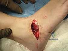
1. Incision and Initial Exposure
The procedure utilizes a standard Ollier approach, which provides direct, anatomic access to the lateral hindfoot and Chopart joint complex.
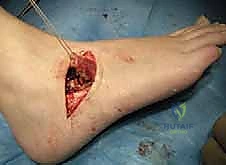
The incision is curvilinear, approximately 4 to 6 centimeters in length, centered directly over the palpable prominence of the anterior process of the calcaneus. It is designed along the resting skin tension lines (Langer's lines) to optimize cosmetic healing. The incision begins plantarly, just superior to the palpable peroneal tendon sheath, and curves dorsally toward the most lateral tendon of the Extensor Digitorum Longus (EDL).
Sharp dissection is carried through the dermis, followed by meticulous, blunt dissection through the subcutaneous adipose tissue. Pre-emptive electrocautery of traversing superficial veins is performed to maintain a pristine field. The surgeon must immediately identify and mobilize the sural nerve plantarly and the dorsal intermediate cutaneous branch of the superficial peroneal nerve dorsally. These neural structures are gently retracted with vessel loops. Deep to the subcutaneous layer, the inferior extensor retinaculum is incised in line with the skin incision, exposing the underlying muscular fascia.
2. Extensor Digitorum Brevis (EDB) Flap Elevation
The Extensor Digitorum Brevis muscle is now visualized, serving as the biologic key to preventing coalition recurrence.
Using a scalpel and a periosteal elevator (such as a Cobb or Key elevator), the EDB muscle is sharply detached from its broad origin on the dorsal and lateral aspects of the anterior calcaneus. It is imperative to elevate the muscle as a single, contiguous unit, taking exceptional care to preserve the overlying, glistening epimysial fascia. This fascia provides the necessary tensile strength for the sutures that will later anchor the flap into the resection defect.
Once the proximal origin is fully liberated, a heavy, absorbable suture (e.g., #0 or #1 Vicryl) is woven through the proximal stump of the EDB using a modified Mason-Allen or Krackow locking technique. This suture provides a robust handle for manipulation and will eventually serve to pull the muscle belly deep into the resected bone void.
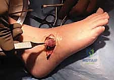
3. Coalition Exposure and Resection
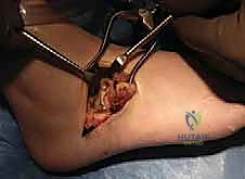
With the EDB flap reflected distally and held in traction, the underlying calcaneonavicular coalition is brought into stark relief.
The coalition typically presents as a thick, continuous mass of cortical and cancellous bone obliterating the normal interval between the anterior calcaneus and the navicular. The surgeon must clearly define the margins of the proposed resection. The resection must be essentially rectangular, extending from the calcaneus to the navicular, ensuring a minimum gap of 1.0 to 1.5 centimeters is achieved.
Resection is initiated using broad, sharp osteotomes or a sagittal saw. The cuts are made perpendicular to the long axis of the coalition. Extreme caution must be exercised to avoid penetrating the adjacent, normal articulations. The posterior cut must stay distal to the anterior facet of the subtalar joint; the distal cut must remain proximal to the talonavicular joint; and the plantar cut must not violate the calcaneocuboid joint.
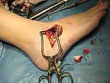
Following the initial parallel cuts, the central block of the osseous coalition is levered out and removed.
The resulting void is then meticulously debrided using rongeurs and a high-speed motorized burr. The surgeon must aggressively burr back the bony margins until healthy, bleeding cancellous bone is exposed on both the calcaneal and navicular sides. A smooth, rectangular defect of at least 1.5 cm is the goal. Fluoroscopy is utilized at this juncture to confirm the adequacy of the resection and to ensure no residual plantar or medial osseous bridging remains. The surgeon should also manually invert and evert the hindfoot; a successful resection will yield immediate, noticeable improvement in subtalar and Chopart mobility.
4. EDB Interposition and Closure
To prevent the highly osteogenic cancellous surfaces from fusing post-operatively, the previously prepared EDB muscle flap is interposed into the defect.
To secure the flap deep within the void, drill holes are created through the remaining navicular bone, directing the drill bits from the lateral resection margin exiting plantarly and medially. Alternatively, modern surgical techniques often utilize a suture anchor placed deep within the medial aspect of the navicular or the plantar calcaneus.
The two limbs of the Vicryl suture previously placed in the EDB flap are passed through these drill holes (using a suture passer or Keith needles) or tied directly to the suture anchor. As the sutures are tensioned and tied over the medial bony bridge, the EDB muscle belly is drawn forcefully into the depths of the resected gap, acting as a highly vascularized, biologic spacer.
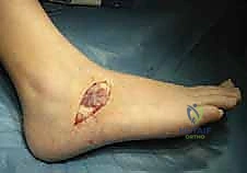
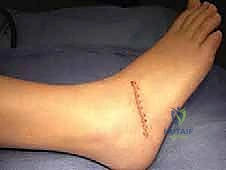
Once the interposition is secured, the wound is copiously irrigated. The inferior extensor retinaculum is loosely reapproximated over the defect if possible, followed by a meticulous, layered closure of the subcutaneous tissue and skin. A sterile, bulky compressive dressing and a well-padded posterior splint are applied with the foot held in a neutral position.
Complications, Incidence Rates, and Salvage Management
While calcaneonavicular coalition resection with EDB interposition is a highly successful procedure, it is not devoid of complications. The adult population, possessing mature, dense bone and a prolonged history of altered biomechanics, faces unique post-operative challenges. The surgeon must be intimately familiar with these potential pitfalls and possess the armamentarium to manage them effectively.
The most catastrophic complication is the recurrence of the coalition, which manifests as a return of rigid, painful hindfoot mechanics. Recurrence is almost exclusively due to inadequate initial resection (leaving a gap of less than 1 cm) or failure to properly secure a robust biologic spacer. Nerve injuries, particularly to the sural nerve, are also distressingly common and can lead to debilitating neuropathic pain.
| Complication | Estimated Incidence | Etiology / Risk Factors | Salvage Management & Treatment Strategy |
|---|---|---|---|
| Recurrence of Coalition (Synostosis) | 5% - 15% | Inadequate resection gap (< 1.5cm); failure of EDB interposition; delayed mobilization. | Revision resection (if adjacent joints remain pristine, though technically demanding) vs. Primary Arthrodesis (Subtalar or Triple Arthrodesis is the definitive salvage). |
| Nerve Injury / Neuroma (Sural or SPN) | 10% - 20% | Aggressive retraction; blind subcutaneous dissection; entrapment in scar tissue. | Initial: Gabapentinoids, targeted nerve blocks, desensitization therapy. Surgical: Neuroma excision with proximal nerve burying into muscle or bone. |
| Continued Pain / Adjacent Joint Arthrosis | 15% - 25% | Pre-existing undiagnosed arthrosis; irreversible biomechanical damage prior to surgery. | Intra-articular corticosteroid injections; orthotic bracing; eventual progression to isolated or multiple hindfoot arthrodesis. |
| Complex Regional Pain Syndrome (CRPS) | 2% - 5% | Neural irritation; prolonged immobilization; exaggerated inflammatory response. | Aggressive, early physical therapy; sympathetic nerve blocks; pain management consultation; Vitamin C prophylaxis post-operatively. |
| Wound Dehiscence / Infection | 2% - 4% | Poor soft tissue handling; smoking; diabetes mellitus. | Local wound care; oral/IV antibiotics; rarely requires surgical debridement unless deep infection occurs. |
Phased Post-Operative Rehabilitation Protocols
The post-operative rehabilitation protocol is as critical to the ultimate success of the procedure as the surgical resection itself. The primary objective is a delicate balancing act: protecting the soft tissue healing and the EDB interposition while simultaneously initiating early, aggressive range of motion (ROM) to prevent the formation of rigid scar tissue across the resection gap. Immobilizing the patient for an extended period is a historical error that drastically increases the risk of recurrence.
Phase 1: Immediate Post-Operative (Weeks 0 - 2)
The patient is placed in a well-padded, short-leg posterior splint in a neutral position immediately post-operatively. The patient is strictly Non-Weight-Bearing (NWB) on the operative extremity, utilizing crutches or a knee scooter. Elevation above the level of the heart is mandatory to mitigate edema and promote wound healing. At the two-week mark, the patient returns to the clinic for splint removal, wound inspection, and suture removal.
Phase 2: Early Mobilization and Protection (Weeks 2 - 6)
Once the incision is adequately healed, the patient is transitioned into a removable Controlled Ankle Motion (CAM) boot. Weight-bearing status is gradually progressed from NWB to Partial Weight-Bearing (PWB), depending on patient tolerance and surgeon preference. Crucially, it is during this phase that early, active, and active-assisted Range of Motion (ROM) exercises are initiated. The patient is instructed to remove the boot multiple times a day to perform aggressive subtalar inversion/eversion and ankle dorsiflexion/plantarflexion exercises. Physical therapy is formally commenced, focusing on edema control, intrinsic foot muscle activation, and gentle mobilization of the Chopart and subtalar joints.
Phase 3: Strengthening and Proprioception (Weeks 6 - 12)
By week six, assuming radiographic and clinical progress is satisfactory, the patient is weaned out of the CAM boot and transitioned into a supportive athletic shoe, often with a custom orthotic or an over-the-counter arch support. The patient is advanced to Full Weight-Bearing (FWB). Physical therapy intensifies, shifting focus to strengthening the peroneal musculature, which has often been chronically weakened or inhibited by the pre-operative "spastic" guarding. Proprioceptive training (e.g., BAPS board, single-leg stance) is critical to restore dynamic stability to the hindfoot.
Phase 4: Return to Unrestricted Activity (Months 3+)
Beyond the three-month mark, the focus is on returning the patient to their pre-injury level of activity, occupational demands, and athletic pursuits. Patients may continue to experience mild, intermittent aching or swelling for up to six to twelve months post-operatively as the soft tissues remodel and the foot adapts to its newly restored kinematics. Impact activities and high-level agility sports are gradually reintroduced as strength and confidence permit.
Summary of Landmark Literature and Clinical Guidelines
The surgical management of calcaneonavicular coalitions has evolved significantly, heavily influenced by several landmark studies that have shaped our current standard of care.
The concept of resecting the coalition and utilizing the Extensor Digitorum Brevis as an interpositional biologic spacer was popularized and refined by Cowell in the 1970s. His seminal work demonstrated that simply resecting the bone without addressing the empty void led to unacceptably high rates of hematoma organization, ossification, and ultimately, recurrence. The EDB flap became the gold standard, providing a vascularized barrier that reliably prevented synostosis.
Further defining the adult treatment paradigm, Gonzalez and Kumar published critical outcome data differentiating pediatric from adult resections. Their research underscored that while adults have a longer duration of altered biomechanics, meticulous, wide resection (ensuring a gap greater than 1 cm) combined with EDB interposition yields good to excellent functional outcomes in the majority of patients, provided that pre-existing degenerative joint disease is absent. They highlighted that age alone is not a contraindication to resection.
More recently, comprehensive reviews by Mubarak and others have solidified the clinical guidelines. The consensus dictates that CT imaging is non-negotiable for pre-operative planning in adults to map the 3D anatomy and rule out talocalcaneal involvement. Furthermore, contemporary literature consistently emphasizes the vital importance of early, aggressive post-operative mobilization. The historical practice of prolonged casting has been universally abandoned in favor of early ROM protocols, which have drastically reduced the incidence of post-operative stiffness and recurrence, cementing the resection and EDB interposition as a highly effective, joint-preserving procedure in the appropriately selected adult patient.
