Mastering Uncemented Total Hip Arthroplasty: A Complete Guide

Key Takeaway
Here are the crucial details you must know about Mastering Uncemented Total Hip Arthroplasty: A Complete Guide. An uncemented total hip is an arthroplasty procedure for symptomatic degenerative hip disease. It involves cementless components for the acetabular and femoral parts. Initial fixation relies on a press-fit, and component surfaces are designed to promote bone in- or on-growth, ensuring long-term stability without the use of bone cement. This approach yields excellent mid- to long-term results.
Introduction and Epidemiology
Total hip arthroplasty represents one of the most successful surgical interventions in modern medicine, functioning as the definitive standard of care for symptomatic degenerative joint disease of the hip that remains unresponsive to optimized nonoperative treatment. The evolution of total hip arthroplasty has seen a paradigm shift from predominantly cemented fixation, pioneered by Sir John Charnley, to cementless (uncemented) fixation. This transition was initially driven by the need to address aseptic loosening and osteolysis observed in younger, more active patients with cemented components.
Cementless total hip arthroplasty has demonstrated excellent mid- to long-term survivorship, with registry data frequently reporting greater than 95% survivorship at 15 to 20 years. The fundamental principle of cementless fixation relies on achieving immediate mechanical stability (press-fit) followed by secondary biologic fixation through osseointegration. The acetabular component typically achieves this initial fixation via an interference fit within the reamed hemipelvis, utilizing a highly porous surface—such as sintered beads, titanium plasma spray, or trabecular metal—to facilitate bone in-growth or on-growth. Similarly, the femoral component obtains initial stability through a press-fit mechanism in either the metaphysis or diaphysis. Modern femoral stem designs vary geometrically, primarily categorized into metaphyseal fit-and-fill, wedge-shaped (single or double taper), and fully porous-coated diaphyseal engaging stems.

Pathogenesis of Degenerative Joint Disease
Degenerative joint disease of the hip is the common endpoint of various distinct pathologic processes. Primary osteoarthritis is the most prevalent etiology, characterized by progressive articular cartilage degradation, subchondral sclerosis, and osteophyte formation. Secondary causes include inflammatory arthropathies (e.g., rheumatoid arthritis, ankylosing spondylitis), developmental dysplasia of the hip, osteonecrosis (avascular necrosis), post-traumatic arthritis, and sequelae of childhood hip disorders such as slipped capital femoral epiphysis or Legg-Calvé-Perthes disease. The natural history of hip degeneration follows a variable and often unpredictable symptomatic course. The progression of joint space narrowing and the severity of clinical symptoms do not always correlate linearly, necessitating a highly individualized approach to surgical timing.
Global Epidemiology and Utilization Trends
The utilization of total hip arthroplasty has grown exponentially worldwide. Epidemiological projections indicate a continued surge in procedural volume, driven by aging populations, rising obesity rates, and expanding indications for younger, highly active patients. Uncemented fixation has become the dominant technique in North America and many parts of Europe, accounting for over 90% of all primary total hip arthroplasties performed in the United States. This epidemiological shift reflects growing confidence in modern porous coating technologies and highly cross-linked polyethylene bearing surfaces, which have dramatically reduced the incidence of wear-induced osteolysis that historically plagued earlier implant generations.
Surgical Anatomy and Biomechanics
A profound understanding of hip anatomy and biomechanics is critical for executing a successful cementless total hip arthroplasty. The surgical exposure must provide unhindered visualization of the acetabulum, including the anterior and posterior walls, the superior dome, the posterior rim, and the inferior teardrop (cotyloid notch).
Acetabular Anatomy
The acetabulum is a hemispherical cavity formed by the confluence of the ilium, ischium, and pubis. The transverse acetabular ligament spans the inferior notch and serves as a critical intraoperative landmark for determining the true floor of the acetabulum and establishing appropriate component version. The optimal positioning of the acetabular component typically targets 40 to 45 degrees of inclination and 15 to 20 degrees of anteversion, often referred to as the "safe zone" of Lewinnek, though modern concepts emphasize functional pelvic tilt and spinopelvic mobility.
Proximal Femoral Anatomy
The proximal femur consists of the femoral head, neck, and the greater and lesser trochanters. The vascular supply to the femoral head, primarily derived from the medial femoral circumflex artery, is routinely sacrificed during the femoral neck osteotomy. The internal architecture of the proximal femur is defined by the primary compressive and tensile trabecular systems (Ward's triangle), which dictate the patterns of stress transfer and influence the design rationale for metaphyseal-engaging versus diaphyseal-engaging cementless stems. The calcar femorale, a dense vertical plate of bone originating from the posteromedial aspect of the femoral shaft and radiating proximally toward the greater trochanter, serves as a critical structural buttress for load transmission.
Hip Joint Biomechanics and Offset
The hip functions as a classic ball-and-socket joint, acting as a class I lever system where the center of rotation (the femoral head) serves as the fulcrum. The abductor musculature (primarily the gluteus medius and minimus) provides the effort force, counterbalancing the body weight acting through the center of gravity. Femoral offset is defined as the perpendicular distance from the center of rotation of the femoral head to the anatomical axis of the femur. Restoring or slightly increasing femoral offset during arthroplasty is paramount; it optimizes the abductor moment arm, reduces the joint reaction force, enhances soft tissue tension, and minimizes the risk of impingement and subsequent dislocation.
Failure to restore offset can result in abductor weakness, a persistent Trendelenburg gait, and increased reactive forces across the articular bearing surface, accelerating wear. Conversely, excessive offset may lead to trochanteric bursitis or undue strain on the abductor insertion.
Spinopelvic Parameters and Functional Safe Zones
Modern biomechanical understanding has transcended the static Lewinnek safe zone, recognizing the dynamic interplay between the lumbar spine and the pelvis. Spinopelvic mobility dictates how the pelvis tilts during postural transitions (e.g., from standing to sitting). Pelvic incidence, a fixed morphological parameter, equals the sum of pelvic tilt and sacral slope.

In a normal spine, transitioning from standing to sitting results in posterior pelvic tilt, which functionally increases acetabular anteversion, accommodating hip flexion and preventing anterior impingement. Patients with a stiff spine (e.g., due to multilevel lumbar fusion, ankylosing spondylitis, or severe spondylosis) fail to undergo this compensatory posterior pelvic tilt. Consequently, their acetabular version remains relatively static, placing them at a significantly higher risk for anterior impingement and posterior dislocation during sitting. Surgeons must preoperatively identify spinopelvic stiffness and adjust the targeted acetabular component position accordingly, often requiring increased operative anteversion or the use of dual mobility constructs.
Indications and Contraindications
The decision to proceed with uncemented total hip arthroplasty relies on a comprehensive clinical and radiographic assessment. The primary indication remains debilitating pain that restricts activities of daily living and is refractory to exhaustive nonoperative measures.
Operative Indications
Operative intervention is indicated for advanced joint degeneration where radiographic evidence correlates strongly with clinical symptoms. While chronologic age was historically a limiting factor for cementless fixation due to concerns over osteopenia, physiologic age and bone quality are now considered more relevant metrics. Cementless components require adequate host bone stock to achieve the requisite primary mechanical stability. In cases of severe osteoporosis (e.g., Dorr Type C bone), cemented femoral fixation may still be preferable to mitigate the risk of intraoperative periprosthetic fracture and failure of osseointegration.
| Condition | Operative Indication for THA | Non Operative Management Strategy |
|---|---|---|
| Primary Osteoarthritis | Severe joint space narrowing, subchondral cysts, refractory pain. | NSAIDs, weight loss, physical therapy, intra-articular corticosteroid injections. |
| Osteonecrosis | Ficat Stage III or IV with subchondral collapse and secondary arthritis. | Core decompression (early stages), protected weight bearing, bisphosphonates. |
| Inflammatory Arthritis | Advanced articular destruction, profound functional limitation. | Disease-modifying antirheumatic drugs (DMARDs), biologics, joint rest. |
| Femoral Neck Fracture | Displaced intracapsular fracture in an active, independent older adult. | Rare for displaced fractures; non-operative only if medically unfit for anesthesia. |
| Developmental Dysplasia | Secondary osteoarthritis with subluxation or structural deformity. | Activity modification, periacetabular osteotomy (if performed prior to arthritis onset). |

Absolute and Relative Contraindications
Absolute contraindications to total hip arthroplasty include active local or systemic infection, severe medical comorbidities precluding safe anesthesia, and profound neuromuscular disease lacking the muscular control necessary to maintain joint stability. Relative contraindications encompass severe untreated osteoporosis, morbid obesity (BMI > 40), active intravenous drug use, and advanced neuroarthropathy (Charcot joint). Poor dental hygiene or active cutaneous infections must be optimized prior to elective arthroplasty to minimize the risk of hematogenous seeding and periprosthetic joint infection.
Pre Operative Planning and Patient Positioning
Thorough preoperative planning is the cornerstone of reproducible success in cementless total hip arthroplasty. Planning begins with a meticulous clinical evaluation and culminates in precise radiographic templating.
Clinical Evaluation and Leg Length Assessment
Preoperative clinical assessment must rigorously evaluate leg length discrepancy. It is vital to differentiate true leg length discrepancy (structural differences in the femur or tibia) from apparent leg length discrepancy (caused by pelvic obliquity, adduction/abduction contractures, or spinal deformity). True leg length is measured from the anterior superior iliac spine to the medial malleolus, while apparent leg length is measured from the umbilicus to the medial malleolus. Accurate documentation of preoperative leg lengths sets patient expectations and guides intraoperative restoration.
Radiographic Templating
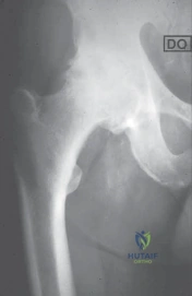
Standard radiographic evaluation requires an anteroposterior (AP) view of the pelvis, an AP view of the affected hip, and a cross-table lateral view. A magnification marker, typically a 25 mm radiopaque sphere positioned at the level of the greater trochanter, is essential for calibrating digital templating software.

The templating sequence follows a systematic progression:
1. Determination of the Center of Rotation: The normal center of rotation is identified on the contralateral, unaffected hip (if normal) and transposed to the affected side.
2. Acetabular Sizing and Positioning: The acetabular template is positioned at a 40-degree inclination. The medial border should rest adjacent to the radiographic teardrop, preserving the subchondral bone of the cotyloid notch. The superolateral margin indicates the degree of required host bone coverage.
3. Femoral Sizing and Neck Resection Level: The femoral template is sized to achieve cortical contact and fill the metaphysis or diaphysis, depending on the stem design. The distance from the lesser trochanter to the planned neck osteotomy is measured to guide the intraoperative cut.
4. Offset and Leg Length Restoration: The offset of the templated stem is compared to the native anatomy. Modular head lengths are selected to fine-tune leg length and offset simultaneously.

Patient Positioning and Operating Room Setup
Patient positioning depends entirely on the chosen surgical approach. The posterior and anterolateral approaches are predominantly performed in the lateral decubitus position. Rigid pelvic fixation using peg boards or specialized pelvic positioners is mandatory to prevent intraoperative rolling, which can severely distort the surgeon's perception of acetabular version. The direct anterior approach is performed supine, often utilizing a specialized traction table to facilitate femoral elevation and exposure, though it can also be performed on a standard radiolucent operating table.

Detailed Surgical Approach and Technique
The execution of a cementless total hip arthroplasty requires precise soft tissue handling, accurate bone preparation, and meticulous component implantation. The choice of surgical approach depends on surgeon training, patient anatomy, and specific implant requirements.

Surgical Approaches to the Hip
The Posterior Approach (Kocher-Langenbeck) remains the most universally utilized approach globally. It involves splitting the gluteus maximus in line with its fibers and detaching the short external rotators (piriformis, superior gemellus, obturator internus, inferior gemellus) and the posterior capsule to access the joint. It provides unparalleled expansile access to both the acetabulum and the femur but historically carried a higher risk of posterior dislocation prior to the advent of meticulous capsular repair techniques and larger femoral heads.
The Direct Anterior Approach (Smith-Petersen) utilizes a true internervous and intermuscular plane between the tensor fasciae latae (superior gluteal nerve) and the sartorius (femoral nerve) superficially, and the gluteus medius and rectus femoris deep. Proponents cite earlier functional recovery and lower dislocation rates, though it carries specific risks such as lateral femoral cutaneous nerve neuropraxia and intraoperative femur fractures during elevation.
The Anterolateral Approach (Watson-Jones) exploits the intermuscular interval between the tensor fasciae latae and the gluteus medius. It provides excellent acetabular exposure and inherent posterior stability but may result in abductor weakness or a prolonged limp due to disruption or traction injury to the anterior third of the gluteus medius.

The Posterior Approach Step by Step
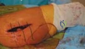
For the posterior approach, the patient is secured in the lateral decubitus position. A curvilinear incision is made centered over the posterior third of the greater trochanter. The fascia lata and the iliotibial band are incised longitudinally. The gluteus maximus fibers are bluntly separated. The sciatic nerve is protected but typically not routinely exposed unless distorted by prior surgery or significant deformity.

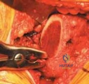
The short external rotators are identified, tagged with heavy non-absorbable suture, and transected near their insertion on the greater trochanter. The posterior capsule is incised, often in a T-shaped or L-shaped fashion, and retained for later repair. The hip is then dislocated via internal rotation, flexion, and adduction. A precise femoral neck osteotomy is performed utilizing an oscillating saw, guided by preoperative templating measurements referenced from the lesser trochanter.
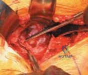
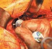
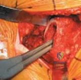
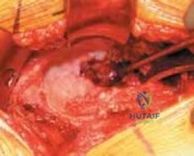
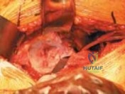
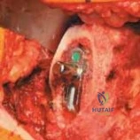
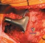
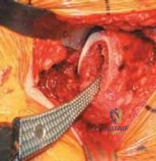
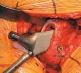
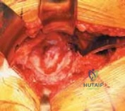
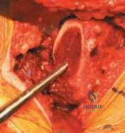
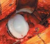
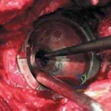
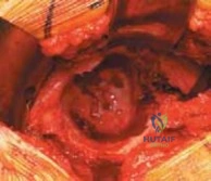
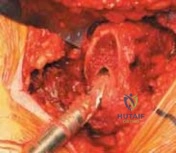
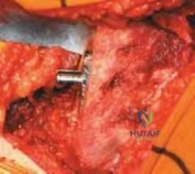
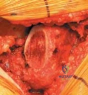
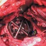
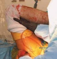
Acetabular
Clinical & Radiographic Imaging