The Painful Total Hip Arthroplasty: Etiology, Anatomy, Biomechanics, and Management
Key Takeaway
A painful total hip arthroplasty demands a systematic evaluation, differentiating mechanical failure, infection (PJI), biological reactions, and anatomical issues. Causes include aseptic loosening, component malposition, leg length discrepancy, abductor dysfunction, and nerve impingement. Thorough understanding of anatomy and biomechanics is critical for diagnosis and effective management in orthopedic practice.
You are presented with a 68-year-old patient who is 18 months post-primary total hip arthroplasty (THA). They complain of progressive, activity-related anterior groin pain that is beginning to limit their walking distance. Please describe your systematic approach to evaluating this "painful THA."
Candidate: I would start with a thorough history and physical exam. I'd order bloods (ESR/CRP) and radiographs. If those are inconclusive, I’d consider aspiration or MRI, and rule out referred pain like the spine.
Providing a "shopping list" of tests without a logical structure. Failing to differentiate between "intrinsic" (prosthesis-related) and "extrinsic" (e.g., spinal, vascular) causes. Neglecting to prioritize infection, which must be ruled out in every painful THA.
A structured, algorithmic approach: 1. History: Onset (start-up vs. constant/nocturnal), radiation, and associated mechanical symptoms (e.g., clicking/instability). 2. Physical Exam: Assessing gait, local tenderness (trochanteric bursitis vs. deep groin pain), range of motion, and neurovascular status. 3. Laboratory: ESR and CRP to screen for PJI. 4. Imaging: Standard AP/Lateral films looking for loosening, migration, or lucency. Use MARS-MRI for soft tissue (trunnionosis/pseudotumor) and CT for component orientation. 5. Differential Diagnosis: Categorize into Intrinsic (Aseptic loosening, infection, component malposition, iliopsoas impingement) vs. Extrinsic (Lumbar spine, hip abductor pathology, referred vascular pain).
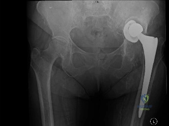
Look at this radiograph of a patient presenting with persistent lateral-sided hip pain and a Trendelenburg gait following a primary THA. What biomechanical principles are at play, and what do you see?
Candidate: The prosthesis looks okay, but maybe the offset is wrong. The patient has a limp, which suggests an abductor problem. I would check the offset and leg length.
Failing to link the biomechanics to the clinical exam. Answering "the offset is wrong" without explaining why (e.g., decreased lever arm) or missing the relationship between the center of rotation and joint reaction forces.
The candidate must define the biomechanical failure: 1. Offset: Reduced femoral offset shortens the abductor lever arm, requiring higher abductor muscle force and increasing joint reaction forces (JRF). 2. Center of Rotation: High-riding or laterally displaced centers of rotation further increase the body-weight lever arm. 3. Clinical Correlation: Explain that this leads to abductor fatigue and "trochanteric pain" despite the implant appearing stable. 4. Synthesis: State that restoring the center of rotation and femoral offset is critical to normalizing gait and reducing pain.
During a revision THA, you encounter a well-fixed cementless femoral stem that needs to be removed. What are your indications for an Extended Trochanteric Osteotomy (ETO), and what are the technical "must-dos" to ensure it heals?
Candidate: I use an ETO when the stem is really stuck. I cut the bone, take the stem out, and wire it back together.
Vague technical descriptions. Failing to mention the distal extent (must be past the porous coating), the importance of maintaining soft tissue attachments (vastus lateralis), or the need for specific postoperative weight-bearing restrictions.
Indications: Well-fixed stems, infection (to remove all cement/biofilm), or severe proximal deformity. Technique: 1. Extend at least 12-15cm distally (beyond the porous coating). 2. Utilize drill holes as stress risers to prevent propagation. 3. Maintain the abductor/vastus attachments for blood supply. 4. "Book" opening the fragment. 5. Secure with multiple heavy-duty cables. Post-op: Toe-touch weight bearing for 6-8 weeks and avoidance of active abduction to prevent non-union.
