العربية
Comprehensive Master Guide · Medically Reviewed

AAOS Pediatric Orthopedic MCQs (Set 2): DDH, SCFE & Spinal Deformities | Board Review

Master pediatric orthopedics with interactive AAOS board review MCQs. Test your knowledge on DDH, SCFE, and spinal deformities to ace exams today!

14 Detailed Chapters
64 min read
Updated: Apr 2026
Clinic OS
Medically Reviewed by
Prof. Clinic OS
Verified Content Expert Reviewed

Quick Medical Answer

This high-yield question set for the AAOS, ABOS, and OITE exams focuses on crucial pediatric orthopedic conditions. It covers the diagnosis and management of developmental dysplasia of the hip (DDH), slipped capital femoral epiphysis (SCFE), and common spinal deformities like scoliosis and kyphosis, including treatment strategies and complications.

Pediatrics 2007 MCQs - Part 2

AAOS Pediatric Orthopedic MCQs (Set 2): DDH, SCFE & Spinal Deformities | Board Review

Comprehensive 100-Question Exam


00:00

Start Quiz

Question 1

The parents of a 15-month-old child report that he is not yet walking. Further evaluation, rather than reassurance and observation, should be conducted if the child is not performing what other activity?


Explanation

A child not ambulating at age 15 months is still within normal limits. The child should be able to sit by age 9 months. The remaining milestones listed are reached later in development.

Question 2

Of the following clinical situations, which is most likely to lead to osteonecrosis associated with a slipped capital femoral epiphysis (SCFE)?


Explanation

Osteonecrosis of the femoral head is the most devastating complication of SCFE. There is a 47% incidence of ischemic necrosis associated with an unstable SCFE. By definition, the patient with an unstable SCFE is unable to bear weight even with crutches. Osteonecrosis is most likely associated with the initial femoral head displacement rather than the result of either tamponade from hemarthrosis or from gentle repositioning prior to stabilization. Age, sex, and obesity are not risk factors for osteonecrosis. Loder RT, Richards BS, Shapiro PS, et al: Acute slipped capital femoral epiphysis: The importance of physeal stability. J Bone Joint Surg Am 1993;75:1134-1140.


Question 3

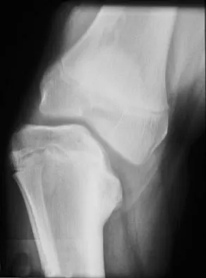
An 8-year-old boy has had pain and swelling around the right knee for the past 4 weeks. He recalls bumping it about 4 weeks ago. He has no pain in other joints, and denies any fevers, chills, or other symptoms. A radiograph is shown in Figure 13. Laboratory studies show a WBC count of 9,700/mm3, an erythrocyte sedimentation rate of 18 mm/h, and a C-reactive protein level of 3.7 mg/L. What is the next most appropriate step in management?


Explanation

The history and laboratory findings are consistent with osteomyelitis of the patella. The radiograph reveals bone destruction in the patella; therefore, the next most appropriate step is open biopsy and debridement of the site. Aspiration of the knee joint may be needed to rule out septic arthritis prior to patellar debridement. With this amount of bone destruction, surgical debridement is helpful to obtain cultures and to remove necrotic material. Administering antibiotics without any prior culture increases the risk of negative cultures later and a potentially incorrect choice of antibiotic. A neoplasm should be included in the differential. It would be inappropriate to initiate chemotherapy and radiation therapy without a biopsy-confirmed diagnosis. A bone scan is likely to demonstrate uptake, but radiographs have already localized the abnormality to the patella. Morrisy RT: Bone and joint sepsis, in Morrissy RT, Weinstein SL (eds): Lovell and Winter's Pediatric Orthopaedics, ed 5. Philadelphia, PA, Lippincott Williams & Wilkins, 2001, pp 466-470.


Question 4

A 2-year-old child is being evaluated for limb-length and girth discrepancy. As a newborn, the patient was large for gestational age and had hypoglycemia. Current examination shows enlargement of the entire right side of the body, including the right lower extremity and foot. The skin shows no abnormal markings, and the neurologic examination is normal. The spine appears normal. Radiographs confirm a 2-cm discrepancy in the lengths of the lower extremities. Additional imaging studies should include


Explanation

The patient may have Beckwith-Wiedemann syndrome (BWS), which consists of exophthalmos, macroglossia, gigantism, visceromegaly, abdominal wall defects, and neonatal hypoglycemia. Hemihypertrophy develops in approximately 15% of patients with BWS. Patients with hemihypertrophy that is the result of BWS have a 40% chance of developing malignancies such as Wilms' tumor or hepatoblastoma; therefore, frequent ultrasound screening is recommended until about age 7 years. The absence of nevi and vascular markings helps to rule out other causes of hemihypertrophy, such as neurofibromatosis, Proteus syndrome, and Klippel-Trenaunay syndrome. Bone age estimations are not accurate at this young age but may become more useful later to help predict the timing of epiphysiodesis procedures. DeBaun MR, Tucker MA: Risk of cancer during the first four years of life in children from The Beckwith-Wiedemann Syndrome Registry. J Pediatr 1998;132:398-400. Ballock RT, Wiesner GL, Myers MT, et al: Hemihypertrophy concepts and controversies. J Bone Joint Surg Am 1997;79:1731-1738.


Question 5

A 12 1/2-year-old boy reports intermittent knee pain and limping that interferes with his ability to participate in sports. He actively participates in football, basketball, and baseball. He denies any history of injury. Examination shows full range of motion without effusion. Radiographs reveal an osteochondritis dissecans (OCD) lesion on the lateral aspect of the medial femoral condyle. MRI scans are shown in Figures 14a and 14b. Initial treatment should consist of


Explanation

This skeletally immature patient has a small OCD lesion that appears stable, and he has not undergone any treatment. Therefore, a trial of immobilization until pain resolves is the best initial choice. Thereafter, cessation of sport activities for 4 to 6 months may allow healing of the lesion. Surgical treatment of juvenile OCD lesions is reserved for unstable lesions, patients who have not shown radiographic evidence of healing and are still symptomatic after 6 months of nonsurgical management, or patients who are approaching skeletal maturity. Good results with stable in situ lesions that have failed to respond to nonsurgical management have been reported with both transarticular and retroarticular drilling. Results after excision alone are poor at 5-year follow-up, and it is unclear if microfracture will improve the long-term outcome. Mosaicplasty may be the next best option for patients who remain or become symptomatic after excision of the fragment and microfracture. Wall E, Von Stein D: Juvenile osteochondritis dissecans. Orthop Clin North Am 2003;34:341-353.


Question 6

A 14-year-old boy undergoes application of a circular frame with tibial and fibular osteotomy for gradual limb lengthening. He initiates lengthening 7 days after surgery. During the first week of lengthening, he reports that turning of the distraction devices is becoming increasingly difficult. On the 9th day of lengthening, he is seen in the emergency department after feeling a pop in his leg and noting the acute onset of severe pain. What complication has most likely occurred?


Explanation

Incomplete corticotomy may result from osteotomy with limited soft-tissue stripping and exposure. When the patient begins distraction, tension develops at all wire/half-pin and bone interfaces, leading to increasing difficulty in distraction and limb pain. Sudden spontaneous completion of the osteotomy with continued tension applied by the fixator results in acute distraction of the osteotomy with severe pain. Premature consolidation is unlikely this early following the initial surgery. Birch JG, Samchukov ML: Use of the Ilizarov method to correct lower limb deformities in children and adolescents. J Am Acad Orthop Surg 2004;12:144-154.


Question 7

What is the most common primary malignant bone or cartilage tumor in children?


Explanation

Osteosarcoma is the most common primary malignant bone tumor (5.6 per 1 million children younger than age 15 years), and Ewing's sarcoma is second (2.1 per 1 million children). Giant cell tumor and chondrosarcoma are rare in children. Osteochondroma is more common than any of the above tumors in children, but it is not malignant. Himelstein BP, Dormans JP: Malignant bone tumors of childhood. Pediatr Clin North Am 1996;43:967-984. Pierz KA, Womer RB, Dormans JP: Pediatric bone tumors: Osteosarcoma, Ewing's sarcoma, and chondrosarcoma associated with multiple hereditary osteochondromatosis. J Pediatr Orthop 2001;21:412-418.


Question 8

What is the peak period of onset in children with pauciarticular juvenile rheumatoid arthritis?


Explanation

Approximately one half of patients with juvenile rheumatoid arthritis (JRA) have the pauciarticular form, which by definition includes only patients with fewer than five joints involved. The peak period of onset is between the ages of 2 and 4 years, with half of the affected children coming to medical attention before age 4 years. The knee is most often affected, with the ankle-subtalar and elbow joints next in frequency. The average duration of the disease is 2 years and 9 months, with half the cases lasting less than 2 years. Arthritis, in Herring JA (ed): Tachdjian's Pediatric Orthopaedics, ed 3. St Louis, MO, WB Saunders, 2002, pp 1811-1839.


Question 9

A 10-year-old girl who is Risser stage 0 has back deformity associated with neurofibromatosis type 1 (NF1). She has no back pain. Examination shows multiple cafe-au-lait nevi with normal lower extremity neurologic function and reflexes. Standing radiographs of the spine show a short 50-degree right thoracic scoliosis with a kyphotic deformity of 55 degrees (apex T8). A 10-degree progression in scoliosis has occurred during the past 1 year. There is no cervical deformity. MRI shows mild dural ectasia, primarily in the upper lumbar region. Management should consist of


Explanation

Scoliotic deformities in patients with NF1 are often dysplastic with short, angular curves. Posterior arthrodesis is made more difficult by the presence of kyphosis and of weak posterior elements caused by dural ectasia. Combined anterior and posterior spinal arthrodesis is generally preferred for progressive dysplastic curves to maximize deformity correction and to decrease the risk of pseudarthrosis. Anterior fusion may also prevent crankshaft phenomenon in young children. Brace treatment is not effective for large, rigid, or dysplastic curves. Kim HW, Weinstein SL: Spine update: The management of scoliosis in neurofibromatosis. Spine 1997;22:2770-2776.


Question 10

In obstetrical brachial plexus palsy, which of the following signs is associated with the poorest prognosis for recovery in a 2-month-old infant?


Explanation

Persistent Horner's sign (ptosis, myosis, and anhydrosis) is a sign of proximal injury, usually avulsion of the roots from the cord which disrupts the sympathetic chain. Root rupture or avulsion proximal to the myelin sheath has less chance of healing. Two-month-old infants with persistent weakness in the other areas described may still have a good prognosis for recovery. Concurrent clavicle fracture has been shown to have no prognostic value. Clarke HM, Curtis CG: An approach to obstetrical brachial plexus injuries. Hand Clin 1995;11:563-581.


Question 11

A 6-year-old boy with acute hematogenous osteomyelitis of the distal femur is being treated with intravenous antibiotics. The most expeditious method to determine the early success or failure of treatment is by serial evaluations of which of the following studies?


Explanation

Successful antibiotic treatment of osteomyelitis should lead to a rapid decline in the CRP. The CRP should decline after 48 to 72 hours of appropriate treatment. Imaging studies will take much longer to show resolution of bone infection. Unkila-Kallio L, Kallio MJ, Eskola J, et al: Serum C-reactive protein, erythrocyte sedimentation rate, and white blood cell count in acute hematogenous osteomyelitis of children. Pediatrics 1994;93:59-62.


Question 12

A 6-year-old girl has a painless spinal deformity. Examination reveals 2+ and equal knee jerks and ankle jerks, negative clonus, and a negative Babinski. The straight leg raising test is negative. Abdominal reflexes are asymmetrical. PA and lateral radiographs are shown in Figures 15a and 15b. What is the next most appropriate step in management?


Explanation

The patient has an abnormal neurologic exam as shown by the abnormal abdominal reflexes. Furthermore, she has a significant curve and is younger than age 10 years. These findings are not consistent with idiopathic scoliosis. MRI will best rule out syringomyelia or an intraspinal tumor. Bracing and surgery are not indicated for this small curvature prior to obtaining an MRI scan. Ginsburg GM, Bassett GS: Back pain in children and adolescents: Evaluation and differential diagnosis. J Am Acad Orthop Surg 1997;5:67-78.


Question 13

Figure 16 shows the radiograph of a 7-year-old boy who sustained a pathologic fracture of the left humerus 1 day ago. Initial management should consist of


Explanation

The radiograph shows a pathologic fracture through a unicameral (simple) bone cyst (UBC). This is the most common location and presentation of a UBC. Less than 10% of UBCs heal spontaneously following a fracture. Urgent biopsy is not indicated because the lesion appears benign and the histology of fracture callus may be misinterpreted as osteosarcoma. After the fracture heals with the use of a sling and swathe, the UBC may be treated with a minimally invasive procedure such as injection of bone marrow and/or demineralized bone matrix. The chance for success is relatively low in an active cyst located adjacent to the physis. More invasive procedures, such as curettage, Rush rod fixation, or cannulated screw decompression, have been described but are rarely necessary for treatment of upper extremity cysts. Rougraff BT, Kling TJ: Treatment of active unicameral bone cysts with percutaneous injection of demineralized bone matrix and autogenous bone marrow. J Bone Joint Surg Am 2002;84:921-929. Robosch A, Saraph V, Linhart WE: Flexible intramedullary nailing for the treatment of unicameral bone cysts in long bones. J Bone Joint Surg Am 2000;82:1447-1453.


Question 14

Figure 17 shows the AP radiograph of a 5-year old child who has mild short stature and a painless bilateral gluteus medius lurch. Initial work-up should include


Explanation

Bilateral flattening of the femoral heads suggests multiple epiphyseal dysplasia; therefore, a skeletal survey is indicated to look for involvement of other epiphyses. Unilateral flattening of the femoral head would suggest Legg-Perthes disease. Sponseller PD: Skeletal dysplasias, in Morrissy RT, Weinstein SL (eds): Lovell and Winter's Pediatric Orthopaedics, ed 5. Philadelphia, PA, Lippincott Williams & Wilkins, 2001, pp 269-270.


Question 15

A 7-year-old girl with spinal muscular atrophy (SMA) type II has popping of the left hip. Examination reveals painless subluxation of the joint in adduction with palpable reduction in abduction. Radiographs show coxa valga, subluxation of the left hip, and pelvic obliquity with elevation of the left hemipelvis. Treatment should consist of


Explanation

Observation is the treatment of choice. Hip subluxation and dislocation are not uncommon in patients with SMA type II who are unlikely to be ambulatory. Scoliosis occurs in these patients 100% of the time and frequently creates pelvic obliquity. However, in long-term follow-up, patients with SMA type II and hip dislocations had little associated pain or functional limitations because of hip instability. In addition, recurrent hip subluxation after surgical treatment has been documented. Given the rarity of symptoms from hip instability in long-term follow-up, and the possibility of recurrent dislocation, surgical intervention for hip instability may expose SMA type II patients to undue surgical risk for minimal if any functional gain. Sporer SM, Smith BG: Hip dislocation in patients with spinal muscular atrophy. J Pediatr Orthop 2003;23:10-14.


Question 16

A newborn with myelomeningocele has no movement below the waist and has bilateral hips that dislocate with provocative flexion and adduction. What is the best treatment option for the hip instability?


Explanation

The status of the hips (located or dislocated) in children with thoracic-level myelomeningocele has no effect on the functional outcome of these patients. Management of unstable hips in this population should be limited to treatment of the contractures that may lead to poor limb positioning in either braces or a wheelchair. The use of the Pavlik harness and/or spica cast is contraindicated because they would promote flexion and abduction contractures. In the past, open reduction either through an anterior or medial approach had been performed with a high incidence of redislocation and other complications, with little functional gain for the child. Gabriel KG: Natural history of hip deformity in spina bifida, in Sarwark JR, Lubicky JP (eds): Caring for the Child With Spina Bifida. Rosemont, IL, American Academy of Orthopaedic Surgeons, 2001, pp 89-103.


Question 17

A 14-year-old boy reports a 4-month history of increasing backache with difficulty walking long distances. His parents state that he walks with his knees slightly flexed and is unable to bend forward and get his hands to his knees. He denies numbness, tingling, and weakness in his legs and denies loss of bladder and bowel control. A lateral radiograph of the lumbosacral spine is shown in Figure 18. What is the best surgical management for this condition?


Explanation

The patient has a grade 4 spondylolisthesis. Optimal surgical management is posterior spinal fusion from L4 to the sacrum. The use of instrumentation is controversial. Vertebrectomy is typically reserved for spondylo-optosis (grade 5) cases. Spinal fusion from L5 to S1 usually is not successful for a slip that is greater than 50%. Isolated anterior spinal fusion has not been successful, and direct repair of the pars defect is only useful for spondylolysis without spondylolisthesis. Lenke LG, Bridwell KH: Evaluation and surgical treatment of high-grade isthmic dysplastic spondylolisthesis. Instr Course Lect 2003;52:525-532.


Question 18

Duchenne's muscular dystrophy is a genetic disorder that is transmitted by which of the following modes of inheritance?


Explanation

Patients with Duchenne's muscular dystrophy show progressive muscular weakness because of the absence of dystrophin and have the clinical picture of progressive muscle weakness. The condition is an X-linked genetic disease. Fitzgerald RH, Kaufer H, Malkani AL: Orthopaedics. St Louis, MO, Mosby Year Book, 2002, pp 1573-1583.


Question 19

A 4-month-old infant is referred for evaluation of congenital scoliosis. The child has no congenital heart anomalies, and a renal ultrasound shows that he has one kidney. Examination reveals mild scoliosis and a large hairy patch on the child's back. Neurologic evaluation is normal for his age. A clinical photograph and radiograph are shown in Figures 19a and 19b. Initial management should consist of


Explanation

Congenital anomalies of the spine, including failure of formation and failure of segmentation, are associated with other anomalies in other organ systems that develop at the same time. These include anomalies in the genitourinary system, cardiac anomalies, Sprengel's deformity, radial hypoplasia, and gastrointestinal anomalies including imperforate anus and trachealesophageal fistula. Spinal dysraphism is the most common associated abnormality. McMaster found an 18% incidence before the common use of MRI. Bradford and associates reported on 16 of 42 patients with congenital spinal anomalies and spinal dysraphism using MRI. Neural axis lesions may be associated with visible midline abnormalities such as a hairy patch or nevus. The child has already had a cardiac and renal work-up, and based on the findings of the hairy patch and congenital vertebral anomalies, MRI of the entire spine is prudent at this time. Spinal fusion is indicated for progressive congenital scoliosis or kyphosis. Physical therapy does not affect the natural history of congenital scoliosis. McMaster MJ: Occult intraspinal anomalies and congenital scoliosis. J Bone Joint Surg Am 1984;66:588-601. Bradford DS, Heithoff KB, Cohen M: Intraspinal abnormalities and congenital spine deformities: A radiographic and MRI study. J Pediatr Orthop 1991;11:36-41.


Question 20

A 12-year-old boy reports limping and chronic knee pain that is now inhibiting his ability to participate in sports. Clinical examination and radiographs of the knee are normal. Additional evaluation should include


Explanation

While all of the answers may be appropriate, radiating pain from hip pathology must be excluded. At this age, a slipped capital femoral epiphysis is likely. Therefore, the hip must be examined. Kocher MS, Bishop JA, Weed B, et al: Delay in diagnosis of slipped capital femoral epiphysis. Pediatrics 2004;113:322-325.

Question 21

In children with moderate to severe osteogenesis imperfecta (OI), intravenous pamidronate therapy has been shown to increase the thickness of cortical bone. This occurs primarily as a consequence of


Explanation

Histologic studies have shown that increased bone turnover is the rule in OI. Pamidronate (and all bisphosphonates) reduce osteoclast-mediated bone resorption. Osteoblastic new bone formation on the periosteal surface of long bones is minimally impaired. With inhibition of osteoclastic bone resorption on the endosteal surface, the cortex of the bone can begin to thicken as it does with normal growth in individuals unaffected by OI. Mineralization and collagen matrix organization are not directly affected by pamidronate. Zeitlin L, Fassier F, Glorieux FH: Modern approach to children with osteogenesis imperfecta. J Pediatr Orthop B 2003;12:77-87. Falk MJ, Heeger S, Lynch KA, et al: Intravenous bisphosphonate therapy in children with osteogenesis imperfecta. Pediatrics 2003;111:573-578.

Question 22

Split posterior tibial tendon transfer is used in the treatment of children with cerebral palsy. Which of the following patients is considered the most appropriate candidate for this procedure?


Explanation

Split posterior tibial tendon transfers are best performed in patients with spastic cerebral palsy who are between the ages of 4 and 7 years and have flexible equinovarus deformities. Rigid deformities typically require bony reconstruction procedures. Tendon transfers in patients with athetosis are unpredictable. Green NE, Griffin PP, Shiavi R: Split posterior tibial-tendon transfer in spastic cerebral palsy. J Bone Joint Surg Am 1983;65:748-754.

Question 23

Late surgical treatment of posttraumatic cubitus varus (gunstock deformity) is usually necessitated by the patient reporting problems related to


Explanation

Cubitus varus, elbow hyperextension, and internal rotation are all typical components of the gunstock deformity. This deformity results from malunion of a supracondylar fracture of the humerus. All of the problems listed above have been reported as sequelae of a gunstock deformity, although the malunion usually causes no functional limitations. Unacceptable appearance is the most common reason why patients or parents request corrective osteotomy. O'Driscoll SW, Spinner RJ, McKee MD, et al: Tardy posterolateral rotatory instability of the elbow due to cubitus varus. J Bone Joint Surg Am 2001;83:1358-1369. Gurkan I, Bayrakci K, Tasbas B, et al: Posterior instability of the shoulder after supracondylar fractures recovered with cubitus varus deformity. J Pediatr Orthop 2002;22:198-202.

Question 24

An 11-year-old boy sustained an ankle injury while playing football. Figure 20 shows an AP radiograph obtained the day of injury. Treatment should consist of


Explanation

The child has an injury involving both the growth plate and the articular surface of the ankle. Because of the significant displacement, open reduction and internal fixation is indicated to realign the physis and joint surface. The best method of fixation to avoid growth arrest is one that does not cross the physis. This is usually achieved by a transverse epiphyseal screw parallel to the physis. If the metaphyseal fragment was large enough, a transverse metaphyseal screw could be used instead. The incidence of growth arrest following physeal ankle injuries is as high as 50%, and long-term follow-up is indicated. Cass JR, Peterson HA: Salter-Harris Type-IV injuries of the distal tibial epiphyseal growth plate, with emphasis on those involving the medial malleolus. J Bone Joint Surg Am 1983;65:1059-1070.


Question 25

A 3-year-old child has bilateral genu varum and short stature. Radiographs show physeal widening and generalized osteopenia. The femora and tibiae show anterolateral bowing. Laboratory studies show low normal serum calcium values, significantly decreased serum phosphate levels, and normal parathyroid hormone (PTH), alkaline phosphatase, and vitamin-D levels. These findings are consistent with


Explanation

Children with vitamin D-resistant rickets are short in stature and have genu varum, physeal widening, and generalized osteopenia. The abnormality in inherited vitamin D-resistant rickets is the renal tubule's inability to resorb phosphate leading to hypophosphatemia. Laboratory findings in the condition are normal or near normal serum calcium values, significantly decreased serum phosphate levels, elevated alkaline phosphatase levels, and normal PTH and vitamin-D levels. The most common form is inherited as an X-linked dominant trait. Nutritional rickets has a normal or low serum phosphate levels, normal or low serum calcium values, and decreased levels of 25(OH) vitamin D and 1,25-dihydroxyvitamin D. Hypophosphatasia is a rare condition characterized by a deficiency of alkaline phosphatase in the serum and tissues, leading to generalized abnormal mineralization of bone. Primary hyperparathyroidism usually is caused by a parathyroid adenoma, and the child generally has abdominal problems and hypercalcemic crisis. Laboratory findings include elevated serum calcium values, alkaline phosphatase levels, and PTH levels, and decreased serum phosphate levels. Children with renal osteodystrophy tend to have genu valgum, and laboratory findings include elevated serum phosphate, alkaline phosphatase, and PTH levels, and low serum calcium values. Findings of renal disease include elevated BUN and creatinine. Herring JA: Metabolic and endocrine bone diseases, in Herring JA (ed): Tachdjian's Pediatric Orthopaedics, ed 3. Philadelphia, PA, WB Saunders, 2002, pp 1686-1710.

Question 26

A 6-week-old female born breech presents for evaluation of developmental dysplasia of the hip (DDH). Ultrasound of the hips reveals an alpha angle of 45 degrees and a beta angle of 78 degrees on the left side. The right hip is normal. What is the most appropriate next step in management?


Explanation

An alpha angle less than 60 degrees and a beta angle greater than 55 degrees (Graf Type IIc or worse) indicate significant acetabular dysplasia. The Pavlik harness is the gold standard for treatment of DDH in infants younger than 6 months of age.

Question 27

A 2-year-old female who recently immigrated is brought to the clinic for a noticeable limp and leg length discrepancy. Radiographs reveal an untreated, high-riding developmental dislocation of the right hip. What is the most appropriate definitive management?


Explanation

In a child older than 18 to 24 months with an untreated DDH, closed reduction has a high failure and avascular necrosis rate. Open reduction with a concomitant pelvic osteotomy (e.g., Salter or Pemberton) is generally required to address the secondary capsular and acetabular changes.

Question 28

A 13-year-old obese male presents to the emergency department with severe right thigh pain after a minor slip on the ice. He is completely unable to bear weight on the right leg. Radiographs demonstrate a slipped capital femoral epiphysis (SCFE). Which of the following complications is he at the greatest risk of developing?


Explanation

The patient's inability to bear weight even with crutches categorizes this as an unstable SCFE. Unstable slips carry a significantly higher risk of osteonecrosis (avascular necrosis), reaching up to nearly 50% in some series.

Question 29

A 12-year-old premenarchal female presents for a routine evaluation. Radiographs reveal a right thoracic adolescent idiopathic scoliosis (AIS) curve measuring 35 degrees. Her Risser stage is 0. What is the most appropriate management?


Explanation

This patient has significant remaining growth (premenarchal, Risser 0) and a progressive curve between 25 and 45 degrees. TLSO bracing is indicated to halt progression and decrease the likelihood of requiring surgical intervention.

Question 30

A 12-year-old boy undergoes in-situ screw fixation for a unilateral SCFE. Which of the following patient profiles represents an absolute indication for prophylactic pinning of the asymptomatic contralateral hip?


Explanation

Prophylactic pinning of the contralateral hip is strongly indicated in patients with endocrine disorders (e.g., hypothyroidism) or renal osteodystrophy due to the extremely high risk of bilateral involvement. Other indications often include age less than 10 or inability to follow up.

Question 31

A 4-month-old infant with DDH has been treated in a Pavlik harness for 3 weeks. At the current visit, the mother notes that the child is not kicking the left leg as much. On exam, the infant lacks active knee extension on the left. Injury to which nerve is the most likely cause?


Explanation

Femoral nerve palsy in a Pavlik harness is caused by excessive hip flexion. It presents with absent active knee extension, and the harness should be adjusted or temporarily discontinued until nerve function recovers.

Question 32

A 3-year-old boy presents with a progressive spinal deformity. Radiographs reveal a fully segmented hemivertebra at T8 with a localized scoliotic curve of 38 degrees that has progressed 10 degrees over the last year. What is the recommended treatment?


Explanation

A fully segmented hemivertebra has high growth potential and typically causes progressive congenital scoliosis. Early hemivertebra excision and short segment fusion is the treatment of choice to prevent severe structural deformity while sparing unaffected motion segments.

Question 33

In a typical patient with a slipped capital femoral epiphysis (SCFE), the relative displacement of the proximal femoral epiphysis in relation to the femoral neck is most accurately described as:


Explanation

In SCFE, the epiphysis remains structurally housed within the acetabulum while the femoral neck displaces anteriorly and superiorly. Thus, the relative position of the epiphysis to the neck is posterior and inferior.

Question 34

On an anteroposterior (AP) pelvis radiograph of a normal 6-month-old infant, the femoral head ossific nucleus should be located in the inferomedial quadrant formed by the intersection of which two radiographic lines?


Explanation

Hilgenreiner's line (horizontal through triradiate cartilages) and Perkin's line (vertical from the lateral margin of the acetabulum) divide the hip into quadrants. In a normal hip, the ossific nucleus sits in the inferomedial quadrant.

Question 35

A 14-year-old elite gymnast presents with persistent low back pain. Radiographs demonstrate a Grade II L5-S1 isthmic spondylolisthesis. She has failed 6 months of rest, bracing, and physical therapy. What is the most appropriate surgical treatment?


Explanation

For a symptomatic low-grade (Grade I or II) isthmic spondylolisthesis that fails conservative management, an in-situ L5-S1 posterior spinal fusion (with or without decompression depending on radicular symptoms) is the standard surgical treatment. Pars repair is typically reserved for L1-L4 defects without significant slip.

Question 36

A 9-year-old child with a BMI in the 10th percentile presents with bilateral, insidious-onset hip pain. Radiographs confirm bilateral mild SCFE. Given the patient's atypical presentation, which of the following laboratory tests is most critical to obtain?


Explanation

Atypical SCFE (patients < 10 years old, thin habitus, or bilateral simultaneous presentation) strongly suggests an underlying endocrine or metabolic disorder. Hypothyroidism is the most common endocrine etiology, requiring evaluation of TSH and free T4.

Question 37

A newborn presents in the nursery with bilateral severe clubfeet, knee recurvatum, and rigid, irreducible dislocated hips. Evaluation reveals multiple joint contractures and decreased muscle mass. Which of the following is the most likely diagnosis?


Explanation

Arthrogryposis multiplex congenita (amyoplasia) is characterized by multiple rigid congenital joint contractures, including teratologic hip dislocations that are typically irreducible without extensive soft tissue release.

Question 38

A 15-year-old girl is diagnosed with adolescent idiopathic scoliosis (Lenke Type 1). Her main thoracic curve measures 58 degrees and is flexible on side-bending. Her pulmonary function tests are normal. What is the standard of care for this patient?


Explanation

In a skeletally mature or nearly mature patient with a thoracic AIS curve exceeding 50 degrees, posterior spinal fusion with segmental instrumentation is the standard surgical treatment to halt progression and correct deformity.

Question 39

When evaluating an anteroposterior (AP) pelvis radiograph for a suspected SCFE, a line is drawn along the superior border of the femoral neck. In a normal hip, this line should intersect a portion of the lateral epiphysis. What is the name of this line?


Explanation

Klein's line is drawn along the superior margin of the femoral neck on the AP radiograph. In a normal hip, it intersects the lateral portion of the femoral head epiphysis; in SCFE, it often passes superior to the epiphysis (Trethowan's sign).

Question 40

A 6-week-old female with developmental dysplasia of the hip (DDH) has been treated with a Pavlik harness for 4 weeks. Repeat ultrasound reveals a persistent dislocation with an alpha angle of 35 degrees. What is the most appropriate next step in management?


Explanation

If a Pavlik harness fails to reduce a dislocated hip after 3 to 4 weeks, it should be discontinued to avoid "Pavlik harness disease" (posterior acetabular wall damage). The next appropriate step is a trial of a rigid abduction orthosis (e.g., Ilfeld) or proceeding to closed reduction and spica casting.

Question 41

Which of the following patients diagnosed with a unilateral slipped capital femoral epiphysis (SCFE) is most strongly indicated for prophylactic pinning of the contralateral hip?


Explanation

Prophylactic contralateral pinning is highly recommended for patients with underlying endocrinopathies, metabolic disorders (like renal osteodystrophy), or previous radiation therapy due to the extremely high risk of bilateral involvement. Idiopathic cases have a lower risk, and contralateral pinning is assessed on a case-by-case basis.

Question 42

A 12-year-old girl presents with adolescent idiopathic scoliosis (AIS). Her radiographs demonstrate a right thoracic curve of 35 degrees. She is premenarchal and Risser 0. Which of the following is the most appropriate management?


Explanation

In a premenarchal, Risser 0 patient with an AIS curve between 25 and 40 degrees, the risk of progression is very high (often >60%). Full-time bracing (e.g., TLSO) is indicated to alter the natural history and prevent progression to the surgical range.

Question 43

A 4-year-old girl presents with a persistently subluxated right hip following previous treatment for DDH. A pelvic osteotomy is planned to improve anterolateral acetabular coverage. Which of the following osteotomies hinges at the triradiate cartilage and decreases the volume of the acetabulum?


Explanation

The Pemberton osteotomy hinges at the flexible triradiate cartilage, allowing the acetabular roof to be hinged down, thus decreasing acetabular volume and improving anterolateral coverage. The Salter osteotomy hinges at the pubic symphysis and does not change acetabular volume.

Question 44

A 13-year-old boy presents to the emergency department unable to bear weight on his left leg after a minor fall. Radiographs confirm a severe slipped capital femoral epiphysis (SCFE). Which of the following complications is he at the highest risk for developing compared to a patient with a stable SCFE?


Explanation

An unstable SCFE (defined by the inability to bear weight even with crutches) has a significantly higher risk of osteonecrosis (up to 50%) compared to a stable SCFE. Urgent but careful reduction and fixation, or a modified Dunn procedure, is often required.

Question 45

Which of the following vertebral anomalies carries the highest risk for rapid curve progression in congenital scoliosis, often necessitating early surgical intervention?


Explanation

A unilateral unsegmented bar combined with a contralateral hemivertebra at the same level has the highest rate of progression (often 5-10 degrees per year) because growth is tethered on one side and accelerated on the other. Early surgical fusion is invariably required.

Question 46

During the ultrasound evaluation of a 4-week-old infant suspected of having DDH, the alpha angle is measured. This angle is formed by the intersection of the iliac bone line and which of the following structures?


Explanation

The alpha angle is formed by the intersection of the baseline (ilium) and the bony acetabular roof. It represents the bony coverage of the femoral head and should normally be greater than 60 degrees.

Question 47

A 12-year-old boy with a BMI of 32 complains of left knee pain for 2 months. Knee examination is normal. When his left hip is passively flexed to 90 degrees, the thigh deviates into obligatory external rotation. What is the pathomechanics of the underlying disorder?


Explanation

In a Slipped Capital Femoral Epiphysis (SCFE), the epiphysis remains securely positioned in the acetabulum while the metaphysis (femoral neck) displaces anteriorly and superiorly. This spatial shift results in the characteristic obligatory external rotation during hip flexion.

Question 48

A 15-year-old boy presents with progressive mid-back pain and a rounded posture. Standing lateral radiographs reveal a thoracic kyphosis of 60 degrees. Which of the following radiographic findings confirms the diagnosis of classic Scheuermann's disease?


Explanation

Sorensen's criteria for the diagnosis of classic Scheuermann's kyphosis require anterior wedging of at least 5 degrees in three or more consecutive vertebrae. Other common but non-diagnostic findings include Schmorl's nodes and endplate irregularities.

Question 49

An infant treated with a Pavlik harness for DDH develops a femoral nerve palsy. Which of the following positioning errors is the most likely cause?


Explanation

Excessive flexion in a Pavlik harness (typically >120 degrees) can cause compression of the femoral nerve against the inguinal ligament, leading to a temporary palsy. Excessive abduction is associated with a different severe complication: avascular necrosis.

Question 50

A 4-week-old female infant is diagnosed with developmental dysplasia of the hip (DDH) and placed in a Pavlik harness. During a follow-up visit, the parents report that the child is not actively kicking her left leg. On examination, the knee lacks active extension, but ankle motion is intact. What is the most likely cause of this finding?


Explanation

Hyperflexion of the hip in a Pavlik harness can compress the femoral nerve against the rim of the pelvis, leading to transient femoral nerve palsy. Hyperabduction is historically associated with avascular necrosis.

Question 51

Which of the following conditions is considered an absolute indication for prophylactic in situ pinning of the contralateral asymptomatic hip in a patient presenting with unilateral Slipped Capital Femoral Epiphysis (SCFE)?


Explanation

Endocrinopathies and metabolic disorders, such as renal osteodystrophy and hypothyroidism, carry a nearly 100% risk of bilateral SCFE. Prophylactic pinning of the contralateral hip is highly recommended in these populations.

Question 52

A 2-year-old child presents with a congenital scoliosis secondary to a fully segmented hemivertebra at T8. Which of the following is the most critical screening test to perform in this patient?


Explanation

Congenital scoliosis is frequently associated with VACTERL anomalies. A renal ultrasound and an MRI of the entire spine are essential to rule out genitourinary anomalies and intraspinal dysraphism.

Question 53

A 3-year-old girl presents with a painless limp. Examination reveals a positive Trendelenburg sign. Radiographs show a dislocated left hip with a false acetabulum. What is the most appropriate surgical management?


Explanation

In children older than 2-3 years with a dislocated hip, open reduction combined with a femoral shortening osteotomy (to reduce AVN risk) and a pelvic osteotomy (to correct dysplasia) is the standard of care.

Question 54

A 12-year-old obese boy presents to the emergency department with severe groin pain after a minor fall and is completely unable to bear weight. Radiographs confirm a slipped capital femoral epiphysis. According to the Loder classification, what is the primary complication associated with his inability to bear weight?


Explanation

The inability to bear weight defines an unstable SCFE in the Loder classification. Unstable slips carry a much higher risk of avascular necrosis (up to 50%) compared to stable slips.

Question 55

A 13-year-old female presents with adolescent idiopathic scoliosis. She is pre-menarchal and Risser 0. Her right thoracic curve measures 32 degrees on standing PA radiographs. What is the most appropriate management?


Explanation

TLSO bracing is indicated for growing children (Risser 0-2, pre-menarchal) with curves between 25 and 45 degrees to prevent curve progression.

Question 56

On a coronal ultrasound of a 6-week-old infant's hip, the alpha angle measures 48 degrees and the beta angle measures 70 degrees. According to the Graf classification, what does this alpha angle indicate?


Explanation

An alpha angle less than 60 degrees indicates a shallow bony acetabular roof, typical of developmental dysplasia. A normal alpha angle is 60 degrees or greater.

Question 57

A 14-year-old boy underwent in situ pinning for a stable SCFE 6 months ago. He now presents with progressive hip stiffness. Examination shows severe restriction of all hip motions. Radiographs show a 2 mm joint space symmetrically but no evidence of AVN. What is the most likely diagnosis?


Explanation

Chondrolysis is a severe complication of SCFE, characterized by progressive joint space narrowing and marked global stiffness. It is strongly associated with unrecognized pin penetration into the joint space.

Question 58

A 15-year-old gymnast presents with persistent low back pain and tight hamstrings. Radiographs reveal a Grade III isthmic spondylolisthesis at L5-S1. She has failed 6 months of nonoperative treatment. What is the most appropriate surgical intervention?


Explanation

For high-grade (Grade III or higher) isthmic spondylolisthesis failing conservative care, an instrumented posterior spinal fusion (with or without interbody support) is indicated. Pars repair is reserved for young patients with normal alignment and no slip.

Question 59

A 2-year-old boy is being followed after closed reduction and spica casting for DDH at age 8 months. Which radiographic finding is considered the earliest indicator of avascular necrosis of the femoral head in this patient?


Explanation

The earliest radiographic sign of AVN in a treated DDH patient is the failure of the ossific nucleus to appear, or its asymmetric, delayed appearance compared to the normal, unaffected side.

Question 60

Slipped capital femoral epiphysis (SCFE) represents a mechanical failure through which specific histologic layer of the physis?


Explanation

SCFE typically occurs through the zone of hypertrophy in the physis. This zone is mechanically weakened during the adolescent growth spurt, increasing susceptibility to shear stresses.

Question 61

In a 9-month-old male with a left thoracic curve measuring 25 degrees, the rib-vertebral angle difference (RVAD) of Mehta is measured at 28 degrees. What is the most likely natural history of this curve and the recommended treatment?


Explanation

An RVAD greater than 20 degrees in infantile idiopathic scoliosis signifies a high risk for severe curve progression. Serial elongation-derotation-flexion (Mehta) casting is the treatment of choice to halt progression.

Question 62

An infant is born with bilateral dislocated hips and rigid extension contractures of the knees. Genetic testing confirms arthrogryposis multiplex congenita. What is the expected success rate of Pavlik harness treatment in this patient?


Explanation

Teratologic hip dislocations, such as those associated with arthrogryposis or myelomeningocele, are rigid and have an extremely high failure rate with a Pavlik harness (<10% success). Open reduction is almost always required.

Question 63

A 25-year-old male presents with groin pain exacerbated by hip flexion and internal rotation. He has a history of mild SCFE treated with in situ pinning at age 13. Radiographs show a prominent alpha angle and a "pistol grip" deformity. What type of femoroacetabular impingement (FAI) is most likely occurring?


Explanation

SCFE commonly alters the proximal femoral anatomy, leaving a prominent metaphysis at the anterolateral head-neck junction. This leads to classic symptomatic Cam-type femoroacetabular impingement.

Question 64

A 12-year-old non-ambulatory male with Duchenne muscular dystrophy presents with a progressive thoracolumbar scoliosis of 55 degrees. His forced vital capacity (FVC) is currently 45% of predicted. What is the most appropriate management?


Explanation

Scoliosis in Duchenne muscular dystrophy is relentlessly progressive. Posterior fusion to the pelvis is indicated when curves exceed 20-30 degrees and the patient's FVC is still >30% to tolerate the procedure.

Question 65

When evaluating a 5-month-old with suspected DDH, an AP pelvis radiograph is obtained. Which of the following describes a normal spatial relationship on this imaging?


Explanation

In a normal pediatric hip radiograph, the ossified proximal femoral metaphysis should lie in the lower medial quadrant formed by the intersection of Perkin's line (vertical) and Hilgenreiner's line (horizontal).

Question 66

A 10-year-old girl with primary hypothyroidism presents with bilateral vague knee pain. Frog-leg lateral radiographs of the pelvis demonstrate widening of the bilateral proximal femoral physes without obvious slippage. What is the most appropriate next step in management?


Explanation

Physeal widening with symptoms in an endocrine patient indicates a "pre-slip" state. Due to the extremely high risk of progression to bilateral displacement, urgent prophylactic in situ pinning is warranted.

Question 67

A 15-year-old Risser 4 male with adolescent idiopathic scoliosis has a single right thoracic curve measuring 55 degrees. He is entirely asymptomatic. What is the primary indication for performing a posterior spinal fusion in this patient?


Explanation

Thoracic curves greater than 50 degrees at skeletal maturity have a high risk of continued progression (approximately 1 degree per year) throughout adulthood. Surgical fusion is indicated primarily to halt this lifelong progression.

Question 68

During an anterior open reduction for a developmental dislocation of the hip in a 14-month-old, the surgeon notes an hour-glass constriction of the joint capsule. Which structure is directly responsible for creating this specific capsular constriction?


Explanation

The iliopsoas tendon crosses the anterior aspect of the hip capsule. In a chronically dislocated DDH, it creates a tight hour-glass constriction, acting as a major mechanical block to concentric reduction.

Question 69

A 16-year-old patient who had a severe, unstable SCFE treated with in situ pinning 4 years ago now presents with severe hip pain and limited abduction. Radiographs show a collapsed, sclerotic femoral head with crescent signs. What is the most definitive surgical option for this patient?


Explanation

This patient has advanced avascular necrosis with femoral head collapse secondary to an unstable SCFE. Total hip arthroplasty provides reliable pain relief and functional restoration when head-preserving procedures are no longer viable.

Question 70

A 6-week-old infant with developmental dysplasia of the hip (DDH) is being treated with a Pavlik harness. During a follow-up visit, the parents report that the child has stopped kicking the left leg. On examination, there is an absence of active knee extension on the left side, but withdrawal to painful stimuli on the plantar foot is intact. What is the most appropriate next step in management?


Explanation

Femoral nerve palsy is a known complication of Pavlik harness treatment, typically caused by hyperflexion of the hips. Management consists of temporarily discontinuing the harness or significantly reducing flexion until the nerve recovers, which usually resolves spontaneously.

Question 71

A 13-year-old boy with a BMI in the 98th percentile presents to the emergency department unable to bear weight on his right leg after tripping over a rug. Radiographs confirm a slipped capital femoral epiphysis (SCFE). According to the Loder classification, which of the following is the most significant consequence of his inability to bear weight?


Explanation

The Loder classification defines an unstable SCFE by the inability to bear weight, even with assistive devices. Unstable SCFE has a significantly higher rate of avascular necrosis (AVN), reported to be up to 47%, compared to nearly 0% in stable slips.

Question 72

A newborn is evaluated in the nursery and noted to have a spinal asymmetry. Radiographs reveal a fully segmented hemivertebra at T8, confirming a diagnosis of congenital scoliosis. Which of the following screening evaluations is most critical in the initial workup of this patient?


Explanation

Congenital scoliosis is highly associated with VACTERL anomalies, specifically genitourinary anomalies (up to 30%) and congenital heart defects (up to 15%). Consequently, screening with a renal ultrasound and an echocardiogram is standard practice.

Question 73

A 4-week-old female infant born in breech presentation undergoes a screening hip ultrasound. The coronal view demonstrates a shallow acetabulum with an alpha angle of 45 degrees and a beta angle of 80 degrees. According to the Graf classification, what is the most appropriate management?


Explanation

An alpha angle of less than 60 degrees indicates developmental dysplasia (Graf type IIc or worse depending on specific features). A Pavlik harness is the standard of care for infants younger than 6 months with dysplastic or dislocated but reducible hips.

Question 74

An 8-year-old boy presents with bilateral groin pain and an altered gait. Radiographs demonstrate bilateral stable slipped capital femoral epiphyses (SCFE). His height is in the 5th percentile, and his weight is in the 90th percentile. Which of the following laboratory evaluations is most critical in determining the etiology of his condition?


Explanation

Patients presenting with SCFE under the age of 10 or with simultaneous bilateral presentation should be evaluated for underlying endocrine abnormalities. Hypothyroidism is the most common endocrine disorder associated with atypical SCFE.

Question 75

A 13-year-old girl with adolescent idiopathic scoliosis (AIS) presents for follow-up. She is pre-menarchal and Risser stage 0. Standing radiographs reveal a progressive right thoracic curve measuring 32 degrees. Based on the BrAIST trial, what is the most appropriate recommendation?


Explanation

The BrAIST trial established that full-time bracing (at least 18 hours daily) significantly decreases the rate of curve progression to the surgical threshold in skeletally immature patients with curves between 25 and 40 degrees.

Question 76

An 18-month-old girl undergoes an open reduction for a chronically dislocated developmental dysplasia of the hip (DDH). Intraoperatively, there is significant acetabular dysplasia with a steep, shallow acetabulum. Which pelvic osteotomy is most appropriate to provide anterolateral coverage by redirecting the acetabulum without reducing its volume?


Explanation

The Salter osteotomy is a complete, redirectional innominate osteotomy that hinges on the pubic symphysis, improving anterolateral coverage without changing the volume or shape of the acetabulum itself. Pemberton and Dega are incomplete, volume-reducing osteotomies.

Question 77

In which of the following patients presenting with a unilateral slipped capital femoral epiphysis (SCFE) is prophylactic in situ pinning of the contralateral hip most strongly indicated?


Explanation

Prophylactic pinning of the contralateral hip is strongly recommended in patients with endocrine or metabolic disorders (such as renal osteodystrophy, hypothyroidism, or panhypopituitarism) due to an exceedingly high risk of subsequent bilateral involvement.

Question 78

A 15-year-old male gymnast complains of worsening lower back pain over the past 3 weeks, exacerbated by extension. Plain radiographs show no obvious cortical break or spondylolisthesis. Which imaging modality is most sensitive for detecting an early, active pars interarticularis stress reaction?


Explanation

MRI with T2 fat-suppressed or STIR sequences is highly sensitive for detecting bone marrow edema indicative of an early pars stress reaction before a definitive fracture occurs, successfully avoiding the ionizing radiation associated with CT or SPECT scans.

Question 79

Following a closed reduction of developmental dysplasia of the hip (DDH) in a 6-month-old, the child is placed in a hip spica cast. To minimize the risk of developing iatrogenic avascular necrosis (AVN), which of the following joint positions must be strictly avoided during casting?


Explanation

Extreme abduction (>60 degrees) combined with extreme flexion places high tension on the medial circumflex femoral artery, significantly increasing the risk of avascular necrosis. The Ramsey 'safe zone' is utilized to balance hip stability with adequate perfusion.

Question 80

A 16-year-old boy presents with progressive mid-back pain and a rounded posture. Standing lateral radiographs reveal a thoracic kyphosis of 65 degrees. According to the classic Sorensen criteria, what radiographic finding is required to definitively diagnose Scheuermann's kyphosis?


Explanation

The Sorensen criteria for diagnosing Scheuermann's kyphosis require anterior wedging of 5 degrees or more in at least three consecutive apical vertebrae, typically alongside a regional kyphosis greater than 40-45 degrees.

Question 81

A 13-year-old girl underwent an uncomplicated in situ pinning for a stable SCFE 6 months ago. She now returns with a stiff, painful hip and severe limitation in all planes of motion. Radiographs demonstrate profound global joint space narrowing (less than 3 mm). What is the most likely etiology of her current condition?


Explanation

Chondrolysis is characterized by an acute loss of articular cartilage, presenting with severe multidirectional stiffness and concentric joint space narrowing (<3mm). It is strongly associated with unrecognized intra-articular hardware penetration during SCFE pinning.

Question 82

A 9-month-old boy is evaluated for a left thoracic scoliosis measuring 28 degrees. Radiographic measurement shows a rib-vertebral angle difference (RVAD) of Mehta of 25 degrees with Phase 2 rib head overlap. What is the most appropriate management strategy?


Explanation

Infantile idiopathic scoliosis with an RVAD greater than 20 degrees (especially with Phase 2 rib head overlap) has a high likelihood of progression. Serial Mehta casting is the gold standard for early-onset progressive curves to potentially cure or delay surgical needs.

Question 83

When evaluating an anteroposterior pelvis radiograph of a 12-month-old child suspected of having residual developmental dysplasia of the hip (DDH), the acetabular index is measured. What is generally considered the upper limit of normal for the acetabular index at this age?


Explanation

In a normally developing 12-month-old child, the acetabular index should be 25 degrees or less. An angle significantly greater than this indicates ongoing acetabular dysplasia that may require further intervention.

Question 84

A 14-year-old non-ambulatory boy with Duchenne muscular dystrophy (DMD) presents with a progressive 55-degree thoracolumbar scoliosis. His forced vital capacity (FVC) is currently 40% of predicted. What is the most appropriate intervention for his spinal deformity?


Explanation

Scoliosis in Duchenne muscular dystrophy is relentlessly progressive and does not respond to bracing. Posterior spinal fusion to the pelvis is indicated for curves over 20-30 degrees in non-ambulatory patients, ideally performed before FVC declines below 30% to minimize perioperative pulmonary complications.

Question 85

A 4-week-old female is treated with a Pavlik harness for developmental dysplasia of the hip (DDH). At the 2-week follow-up, she exhibits decreased spontaneous extension of the right knee and an absent patellar reflex. What is the most appropriate next step in management?


Explanation

Femoral nerve palsy is a known complication of hyperflexion in a Pavlik harness, presenting as decreased knee extension. The harness must be discontinued immediately to allow for neurologic recovery before pursuing alternative treatments.

Question 86

A 13-year-old boy presents with severe left hip pain and an inability to bear weight following a minor fall. Radiographs confirm a slipped capital femoral epiphysis (SCFE). Which of the following is the most likely severe complication directly associated with this specific presentation?


Explanation

The inability to bear weight, even with crutches, defines an unstable SCFE. Unstable SCFE has a significantly higher rate of osteonecrosis (up to 47%) compared to stable SCFE, making it the most critical complication.

Question 87

Which of the following congenital spinal anomalies carries the highest risk of rapid curve progression, typically necessitating the earliest surgical intervention?


Explanation

A unilateral unsegmented bar with a contralateral hemivertebra creates a severe growth mismatch (tethering on one side, active growth on the other). It carries the highest risk of rapid progression and usually requires early surgical fusion.

Question 88

An infant undergoes screening ultrasound for developmental dysplasia of the hip (DDH). The report notes an alpha angle of 45 degrees. Which of the following anatomic structures form the lines used to measure the alpha angle?


Explanation

The alpha angle measures the bony concavity of the acetabulum and is formed by the intersection of the baseline (ilium) and the bony roof line. An alpha angle less than 60 degrees generally indicates a shallow, dysplastic acetabulum.

Question 89

A 7-year-old boy, whose weight is in the 30th percentile, presents with a stable slipped capital femoral epiphysis (SCFE). Which of the following laboratory studies is most highly indicated for this patient?


Explanation

SCFE presenting in children under 10 years old, over 16 years old, or with atypical body habitus (weight < 50th percentile) is highly associated with endocrine disorders. Hypothyroidism is a leading cause, making TSH and free T4 essential screening tests.

Question 90

A 12-year-old premenarchal female with adolescent idiopathic scoliosis (AIS) has a right thoracic curve of 32 degrees and a Risser stage of 1. According to the Bracing in Adolescent Idiopathic Scoliosis Trial (BRAIST), what is the most significant factor determining the success of brace treatment?


Explanation

The BRAIST study conclusively demonstrated a strong dose-response relationship in bracing efficacy for AIS. Wearing the brace for more hours per day significantly increases the success rate in preventing progression to the surgical threshold.

Question 91

An 18-month-old girl undergoes a closed reduction trial for a dislocated right hip. Intraoperative arthrography reveals a medial dye pool measuring 7 mm. What is the most appropriate next step in management?


Explanation

A medial dye pool greater than 4-5 mm on an arthrogram indicates interposed tissue (such as an inverted limbus, pulvinar, or ligamentum teres) preventing concentric reduction. Open reduction is required to clear these obstacles and achieve a stable joint.

Question 92

A 14-year-old boy undergoes in situ percutaneous pinning for a stable slipped capital femoral epiphysis (SCFE). Which of the following screw configurations provides the optimal balance of biomechanical stability and minimized complication risk?


Explanation

A single screw placed in the center-center of the epiphysis provides sufficient biomechanical stability for a stable SCFE. Adding a second screw does not clinically improve stability but significantly increases the risk of articular penetration and chondrolysis.

Question 93

An 8-month-old boy is diagnosed with infantile idiopathic scoliosis. Radiographs reveal a 25-degree left thoracic curve. Which of the following radiographic parameters best predicts whether this curve will progress rather than spontaneously resolve?


Explanation

Mehta's rib-vertebral angle difference (RVAD) is the most reliable prognostic indicator for infantile idiopathic scoliosis. An RVAD greater than 20 degrees strongly predicts progressive deformity requiring intervention, whereas an RVAD less than 20 degrees often resolves spontaneously.

Question 94

A 4-year-old girl with residual acetabular dysplasia requires a pelvic osteotomy. The surgeon plans an incomplete transiliac osteotomy that hinges on the triradiate cartilage to decrease the volume and change the shape of the acetabulum. Which procedure is being described?


Explanation

The Pemberton osteotomy hinges on the open triradiate cartilage, allowing the surgeon to reshape the acetabulum and reduce its volume. In contrast, the Salter osteotomy hinges on the pubic symphysis and redirects the entire acetabular segment without altering its shape.

Question 95

Following pinning of a SCFE, a 13-year-old patient develops a stiff, painful hip with a 15-degree flexion contracture. Radiographs reveal global narrowing of the joint space to less than 3 mm. What is the most likely diagnosis?


Explanation

Chondrolysis involves the acute dissolution of articular cartilage, presenting with marked stiffness, pain, and global joint space narrowing on radiographs. It is a severe complication of SCFE, highly associated with unrecognized intra-articular hardware penetration.

Question 96

A 14-year-old female presents with back pain and is diagnosed with an L5-S1 isthmic spondylolisthesis. Which of the following radiographic findings is considered the most significant risk factor for further slip progression?


Explanation

The slip angle (sagittal roll) is the angle between the L5 inferior endplate and the posterior aspect of the S1 body. A slip angle greater than 40-50 degrees is the most significant predictor of slip progression in spondylolisthesis and often dictates the need for surgical stabilization.

Question 97

A 2.5-year-old child presents with an untreated, completely dislocated left hip (DDH) and is scheduled for an open reduction. To minimize the risk of osteonecrosis during the reduction in a child of this age, which adjunctive procedure is most commonly required?


Explanation

In children older than 2 to 3 years, chronically contracted soft tissues severely increase the joint reactive forces upon reduction. A femoral shortening osteotomy decompresses the joint, significantly reducing the risk of avascular necrosis during open reduction.

Question 98

When evaluating an AP pelvis radiograph for a suspected slipped capital femoral epiphysis (SCFE), what defines an abnormal Klein's line?


Explanation

Klein's line is drawn along the superior border of the femoral neck on an AP radiograph. In SCFE, the epiphysis is displaced posteriorly and inferiorly, causing Klein's line to intersect less of the lateral epiphysis (or miss it entirely) compared to the normal contralateral hip.

Question 99

A 15-year-old boy presents with a Lenke Type 1 (main thoracic) adolescent idiopathic scoliosis curve of 55 degrees. When planning posterior spinal fusion, what is the primary goal regarding the selection of the lowest instrumented vertebra (LIV)?


Explanation

In AIS surgery, particularly for main thoracic curves, preserving lumbar motion is a critical goal to prevent long-term functional limitation and degeneration. The lowest instrumented vertebra (LIV) is typically chosen at or near the stable vertebra to spare as many lower lumbar segments as possible.

Question 100

An infant is diagnosed with a teratologic hip dislocation associated with arthrogryposis multiplex congenita. What is the most appropriate initial management strategy for the hip?


Explanation

Teratologic dislocations involve severe, rigid soft-tissue contractures and typically do not respond to a Pavlik harness or closed reduction methods. Early open reduction, frequently combined with capsulorrhaphy and a femoral shortening osteotomy, is the standard of care to achieve a concentric reduction.

None

Detailed Chapters & Topics

Dive deeper into specialized chapters regarding pediatrics-2007-set-2-mcqs-4021

14 Chapters
01
Chapter 1 65 min

Orthopedic Pediatrics 2026 MCQs: Board Review Questions & Answers (Part 1)

Master your 2026 exams with our Orthopedic Pediatrics Board Review MCQs. Practice with high-yield questions, detailed a…

02
Chapter 2 65 min

Orthopedic Pediatrics 2026 MCQs: Board Review Questions & Answers (Part 2)

Master Orthopedic Pediatrics with our 2026 board review MCQs. Practice with Part 2 questions and answers to enhance you…

03
Chapter 3 71 min

Orthopedic Pediatrics 2026 MCQs: Board Review Questions & Answers (Part 3)

Master your exams with Part 3 of our Orthopedic Pediatrics 2026 MCQs. Practice board review questions, check your answe…

04
Chapter 4 60 min

Orthopedic Pediatrics 2026 MCQs: Board Review Questions & Answers (Part 4)

Ace your 2026 Orthopedic Pediatrics Board Review with Part 4 of our premium MCQ practice questions and detailed answers…

05
Chapter 5 62 min

Orthopedic Pediatrics 2026 MCQs: Board Review Questions & Answers (Part 1)

Ace your exams with our Orthopedic Pediatrics 2026 MCQs. Practice with interactive board review questions and answers t…

06
Chapter 6 64 min

Orthopedic Pediatrics 2026 MCQs: Board Review Questions & Answers (Part 2)

Ace your exams with Part 2 of our Orthopedic Pediatrics 2026 MCQs. Practice interactive board review questions and answ…

07
Chapter 7 60 min

Orthopedic Pediatrics 2026 MCQs: Board Review Questions & Answers (Part 3)

Ace your 2026 exams with Part 3 of our Orthopedic Pediatrics MCQs. Test your knowledge with interactive board review qu…

08
Chapter 8 95 min

Orthopedic Pediatrics 2026 MCQs: Board Review Questions & Answers (Part 4)

Ace your 2026 exams with Part 4 of our Orthopedic Pediatrics MCQs. Practice high-yield board review questions and answe…

09
Chapter 9 51 min

Orthopedic Pediatrics 2026 MCQs: Board Review Questions & Answers (Part 1)

Master your exam with our Orthopedic Pediatrics 2026 MCQs. Explore comprehensive board review questions and answers to …

10
Chapter 10 86 min

Orthopedic Pediatrics 2026 MCQs: Board Review Questions & Answers (Part 2)

Master your exams with Part 2 of our Orthopedic Pediatrics 2026 MCQs. Practice interactive board review questions and a…

11
Chapter 11 88 min

Orthopedic Pediatrics 2026 MCQs: Board Review Questions & Answers (Part 3)

Prepare for the 2026 Orthopedic Pediatrics board exam with Part 3 of our interactive MCQs. Test your knowledge with exp…

12
Chapter 12 87 min

Orthopedic Pediatrics 2026 MCQs: Board Review Questions & Answers (Part 4)

Ace your exams with Part 4 of our Orthopedic Pediatrics 2026 MCQs. Practice interactive board review questions and deta…

13
Chapter 13 67 min

AAOS Pediatric Orthopedics MCQs (Set 4): DDH, SCFE & Supracondylar Humerus | Board Review

Master AAOS pediatric orthopedics with our interactive Board Review MCQs (Set 4). Test your knowledge on DDH, SCFE, and…

14
Chapter 14 84 min

Pediatrics 2004 Practice Questions: Set 1 (Solved)

Master Pediatrics with 2004 updated practice questions (Set 1). Includes evidence-based answers for residents and surge…

Clinic OS
Medically Verified Content by
Prof. Clinic OS
Consultant Orthopedic & Spine Surgeon
Guide Overview