العربية
Part of the Master Guide

AAOS Pediatric Orthopedic MCQs (Set 2): DDH, SCFE & Spinal Deformities | Board Review

Pediatrics 2004 Practice Questions: Set 1 (Solved)

27 Apr 2026 84 min read 78 Views
Pediatrics 2004 MCQs - Part 1

Key Takeaway

Your ultimate guide to Pediatrics 2004 Practice Questions: Set 1 (Solved) starts here. Access high-yield Pediatrics questions for the 2004 board exam. This module (Set 1) covers critical topics including surgical techniques, pathology, and treatment protocols with verified answers.

Pediatrics 2004 Practice Questions: Set 1 (Solved)

Comprehensive 100-Question Exam


00:00

Start Quiz

Question 1

An 8-year-old boy sustains nondisplaced midshaft fractures of the tibia and fibula after being struck by a car while he was riding his bicycle. No other injuries are noted, but the patient reports pain with passive motion of his toes. His neurovascular examination is otherwise normal. What is the best course of action?

Pediatrics 2004 Practice Questions: Set 1 (Solved) - Figure 1


Explanation

Pain with passive motion of the toes is a recognized early sign of increased compartment pressures. At a minimum, a baseline evaluation of the leg compartment pressures should be obtained. While it is normal for the patient to have pain related to the associated muscle contusions, any significant concerns should be addressed immediately in light of the severe consequences likely when a compartment syndrome occurs. Mubarak SJ, Owen CA, Hargens AR, et al: Acute compartment syndromes: Diagnosis and treatment with the aid of the wick catheter. J Bone Joint Surg Am 1978;60:1091-1095.

Question 2

A 6-year-old girl has the bilateral foot deformity shown in Figure 1. There is no family history of disease. Examination reveals fixed hindfoot equinus, and muscle function testing shows strong posterior tibial function, fair plus anterior tibial function, poor peroneal function, and strong gastrocnemius function. A Coleman block test shows a correctable hindfoot. Nerve conduction velocity studies show diminished function in the peroneal and ulnar nerves on both sides. Pathologic changes found in a sural nerve biopsy include "onion bulb" formation, and DNA testing confirms the presence of a mutation in the MPZ gene, consistent with hereditary motor sensory neuropathy type III (HMSN-III). What is the best course of action?

Pediatrics 2004 Practice Questions: Set 1 (Solved) - Figure 2


Explanation

The patient has HMSN-III or Dejerine-Sottas syndrome. This form of HMSN progresses very rapidly and frequently results in severe foot deformity in early childhood. The changes are progressive and are the result of muscle imbalance during growth. Balancing of the foot musculature is essential, particularly during the phases of rapid growth of the foot. However, this cannot be accomplished using the anterior tibial muscle because it is already weak and the transfer will further weaken it. Bony procedures also may be required, and tendon transfers cannot be depended on to correct bony deformity. However, these procedures can be deferred until the foot is closer to adult size. Surgeries that lead to joint arthrodesis, such as triple arthrodesis and some midfoot osteotomies, are contraindicated because the feet may lose protective sensation as the disease progresses. Fusions in insensate feet are less successful than realignment procedures that maintain mobility. Wetmore RS, Drennan JC: Long-term results of triple arthrodesis in Charcot-Marie-Tooth disease. J Bone Joint Surg Am 1989;71:417-422. Roper BA, Tibrewal SB: Soft tissue surgery in Charcot-Marie-Tooth. J Bone Joint Surg Br 1989;71:17-20.

Question 3

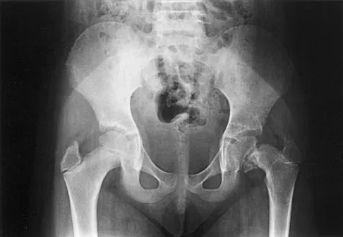
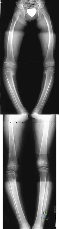
An obese 4-year-old boy has infantile Blount's disease. Radiographs reveal a metaphyseal-diaphyseal angle of 18 degrees and a depression of the medial proximal tibial physis. Management should consist of

Pediatrics 2004 Practice Questions: Set 1 (Solved) - Figure 3


Explanation

The deformity is too severe for observation, and at age 4 years, the child is too old for orthotic treatment. To prevent recurrence, surgery should be performed before irreversible changes occur in the medial physis. A proximal tibial osteotomy should overcorrect the mechanical axis to 10 degrees of valgus. Bar resection has not been shown to be as effective in this severe deformity, especially without a concomitant osteotomy. Raney EM, Topoleski TA, Yaghoubian R, Guidera KJ, Marshall JG: Orthotic treatment of infantile tibia vara. J Pediatr Orthop 1998;18:670-674.

Question 4

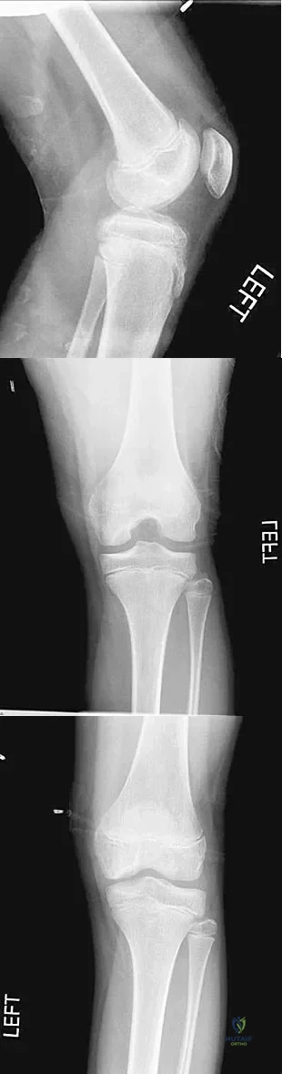
A 10-year-old boy has activity-related knee pain that is poorly localized. He denies locking, swelling, or giving way. Examination shows mild tenderness at the medial femoral condyle and painless full range of motion without ligamentous instability. Radiographs are shown in Figures 2a through 2c. What is the best course of action?


Explanation

The radiographs show an osteochondritis dissecans (OCD) lesion in the medial femoral condyle of a skeletally immature patient. The lesion is not displaced from its bed. Nonsurgical management of a stable OCD lesion in a patient with open physes consists of a period of activity limitation and occasional immobilization. Unstable lesions, loose bodies, and patients with closed physes require more aggressive treatment. Most of the surgical procedures can be done arthroscopically. Because the radiographic appearance is typical, biopsy is unnecessary. The radiographs do not show an osteocartilaginous loose body, and the patient reports no catching or locking; therefore, removal of the loose body is not indicated. Linden B: Osteochondritis dissecans of the femoral condyles: A long term follow-up study. J Bone Joint Surg Am 1977;59:769-776. Cahill BR: Osteochondritis dissecans of the knee: Treatment of juvenile and adult forms. J Am Acad Orthop Surg 1995;3:237-247.

Question 5

Figure 3a shows the preoperative radiograph of a 5-year-old girl who achieved complete correction with valgus osteotomies. Figure 3b shows a radiograph obtained 2 years later. What is the cause of the recurrent deformity on the right side?


Explanation

Although inadequate correction, obesity, patient age of older than 5 years and an increased metaphyseal-diaphyseal angle are all associated with a poorer outcome, the radiographs show a growth arrest of the medial tibial physis. If not recognized and treated with early surgery, progressive genu varum will occur with continued growth of the lateral physis. In addition to repeat osteotomy, options for treating the arrest include physeal bar resection or, as necessary, completion of the growth arrest by epiphyseodesis of the lateral physes, followed by a limb equalization procedure at a later date. Brooks WC, Gross RH: Genu varum in children: Diagnosis and treatment. J Am Acad Orthop Surg 1995;3:326-335. Herring JA: Tachdjian's Pediatric Orthopedics, ed 4. Philadelphia, PA, WB Saunders, 2002, pp 840-950.

Question 6

An 8-year-old boy reports ankle pain after striking the ground with the medial aspect of his foot while attempting to kick a soccer ball. Radiographs reveal slight distal tibial physeal widening but no other abnormalities. In treating this injury, which of the following associated conditions is most likely present but may be missed without careful evaluation?

Pediatrics 2004 Practice Questions: Set 1 (Solved) - Figure 9


Explanation

Malrotation of the foot is frequently overlooked in this clinical setting. This can be judged by evaluating and comparing the transmalleolar axes of the affected and unaffected legs. The rotation occurs through the physis and frequently is not recognized until the patient has been walking for a few months. The other conditions are not expected to occur in the clinical setting described. Phan VC, Wroten E, Yngve DA: Foot progression angle after distal tibial physeal fractures. J Pediatr Orthop 2002;22:31-35.

Question 7

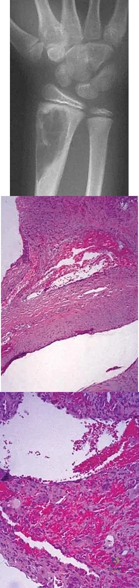
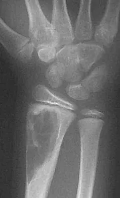
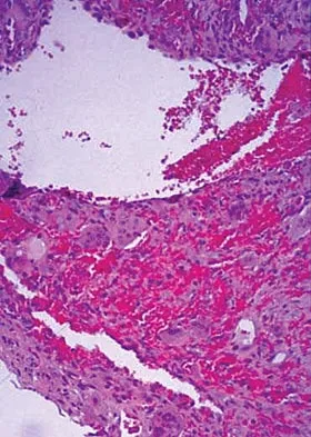
An 11-year-old girl has wrist pain. Figure 4a shows the radiograph, and Figures 4b and 4c show the low- and medium-power photomicrographs of a lesion in the distal radius. What is the most likely diagnosis?


Explanation

The radiograph shows an osteolytic eccentric lesion in the metaphyseal-diaphyseal region of the bone, and the photomicrographs show an aneurysmal bone cyst. The low-power photomicrograph shows large empty spaces with fibrous stroma and multinucleated giant cells. The red area in the center is hemorrhage in the stroma. The large empty spaces are cysts, which would be filled with blood in vivo. The medium-power photomicrograph shows a large cyst-like space and hemorrhage in the surrounding stoma. Giant cell tumors have "sheets" of giant cells. A nonossifying fibroma would have spindle cells, and a unicameral bone cyst may have a few giant cells, but blood is rare. Springfield DS, Gebhardt MC: Bone and soft tissue tumors, in Morrissy RT, Weinstein SL (eds): Lovell and Winter's Pediatric Orthopaedics, ed 5. Philadelphia, PA, Lippincott Williams and Wilkins, 2001, pp 540-542.

Question 8

In a patient with vertebral tuberculosis, which of the following characteristics is most predictive of progression of the kyphosis?

Pediatrics 2004 Practice Questions: Set 1 (Solved) - Figure 13


Explanation

In patients with vertebral tuberculosis, involvement of the anterior and posterior elements creates an instability and severe kyphotic collapse can occur. This characteristic has been shown to have a stronger association than level of involvement, age, or pretreatment degree of deformity. In the absence of instability, anterior growth can resume after treatment, leading to a decrease in the deformity. Rajasekaran S: The natural history of post-tubercular kyphosis in children: Radiological signs which predict late increase in deformity. J Bone Joint Surg Br 2001;83:954-962.

Question 9

When planning scoliosis surgery for a patient with a 50-degree thoracolumbar curve and spinal muscular atrophy, it is most important to include

Pediatrics 2004 Practice Questions: Set 1 (Solved) - Figure 14


Explanation

Typically, posterior spinal fusion to the pelvis is recommended for patients with spinal muscular atrophy and advanced scoliosis. Examination for lower extremity muscle contractures is important because the contractures may interfere with good sitting balance. Anterior release and fusion usually are not advised. Diaphragmatic pacing is not indicated because diaphragm function usually is not affected. Patients with spinal muscular atrophy usually are not ambulatory or only marginally ambulatory at the time of scoliosis surgery; therefore, gait analysis usually is not relevant. While a muscle biopsy may have a role in the diagnosis of this disorder, it plays no subsequent role in determining life expectancy or the value of spinal surgery. Daher YH, Lonstein JE, Winter RB, Bradford DS: Spinal surgery in spinal muscular atrophy. J Pediatr Orthop 1985;5:391-395.

Question 10

An 8-year-old boy sustains injuries to his head, abdomen, and left lower extremity after being struck by a truck. In the emergency department, his mental status deteriorates and he is intubated after assessment reveals a Glasgow Coma Scale score of 3; the score subsequently improves to 10. A CT scan reveals a right parietal intracranial hemorrhage, and an abdominal ultrasound reveals free fluid. Prior to an emergency laparotomy, the swollen left thigh is evaluated. Radiographs reveal a transverse fracture of the mid-diaphysis. Management of the fracture should consist of

Pediatrics 2004 Practice Questions: Set 1 (Solved) - Figure 15


Explanation

The prognosis for a young patient with a head injury is more favorable compared to that for adults. Full neurologic recovery generally occurs. Spasticity may occur within a few days after injury, which can lead to fracture displacement if immediate spica casting or traction is used. Early surgical stabilization will reduce problems with shortening and malunion and will facilitate transportation of the child for diagnostic tests. Surgery may be performed when it is best for the patient, either on the day of injury or later if time is needed for stabilization. In this patient, the fracture is ideally suited to stabilization using flexible intramedullary nails. Heinrich and associates' report of 78 diaphyseal femur fractures stabilized with flexible intramedullary nails included 14 patients with an associated closed head injury. All fractures healed, and there were no major complications. Tolo VT: Management of the multiply injured child, in Rockwood CA, Wilkins KE, Beaty JH (eds): Fractures in Children, ed 4. Philadelphia, PA, Lippincott-Raven, 1996, pp 83-95.

Question 11

A 3-year-old boy has a rigid 40-degree lumbar scoliosis that is the result of a fully segmented L5 hemivertebra. All other examination findings are normal. Management should consist of

Pediatrics 2004 Practice Questions: Set 1 (Solved) - Figure 16


Explanation

Near complete correction and rebalancing of the spine can be achieved by hemivertebral resection that may be done as either a simultaneous or a staged procedure in the young patient. This eliminates the problem of future progression and possible development of compensatory curves. Nonsurgical management is not indicated in congenital scoliosis. Convex hemiepiphyseodesis is best suited for patients younger than age 5 years who have a short curve caused by fully segmented hemivertebrae that correct to less than 40 degrees with the patient supine. Hemiepiphyseodesis and isolated posterior fusion are not indicated. Bradford DS, Boachie-Adjei O: One-stage anterior and posterior hemivertibral resection and arthrodesis for congenital scoliosis. J Bone Joint Surg Am 1990;72:536-540.

Question 12

A newborn with bilateral talipes equinovarus undergoes serial manipulation and casting. What is the primary goal of manipulation?

Pediatrics 2004 Practice Questions: Set 1 (Solved) - Figure 17


Explanation

Manipulative treatment and casting of talipes equinovarus has become popular because of disappointing surgical results and enthusiasm for the Ponseti method of manipulation. In this technique, the primary goal is to rotate the foot laterally around a talus that is held fixed by the manipulating surgeon's hands. While the navicular may be rotated anterolaterally with this technique, the primary focus is on the calcaneus. The calcaneus is rotated laterally and superiorly, not translated. Some dorsiflexion of the calcaneus can be obtained by manipulation, but the primary focus is on the rotational relationship of the talus and calcaneus, not the degree of calcaneal dorsiflexion. Ponseti IV: Common errors in the treatment of congenital clubfoot. Int Orthop 1997;21:137-141.

Question 13

Figure 5 shows the radiograph of a 10-year-old girl who reports chronic shoulder pain after her gymnastics classes. Examination reveals pain on internal and external rotation but no instability. What is the most likely diagnosis?

Pediatrics 2004 Practice Questions: Set 1 (Solved) - Figure 18


Explanation

The patient has a very wide humeral growth plate, indicating the presence of a proximal humeral stress fracture, an uncommon diagnosis in gymnasts. Gymnasts are prone to stress fractures of the scaphoid, distal radius, elbow, and clavicle. Proximal humeral stress fractures are more commonly seen in those participating in racket or throwing sports. Stress fractures can lead to growth arrest or inhibition, particularly in the distal radius. The radiograph shows normal findings for the acromion, acromioclavicular joint, scapula, and triceps origin. Fallon KE, Fricker PA: Stress fracture of the clavicle in a young female gymnast. Br J Sports Med 2001;35:448-449. Sinha AK, Kaeding CC, Wadley GM: Upper extremity stress fractures in athletes: Clinical features of 44 cases. Clin J Sports Med 1999;9:199-202. Caine D, Howe W, Ross W, Bergman G: Does repetitive physical loading inhibit radial growth in female gymnasts? Clin J Sports Med 1997;7:302-308.

Question 14

Figure 6 shows the clinical photographs of a newborn who underwent a colostomy for an imperforate anus. Examination shows extended knees, flexed hips, and equinovarus feet. Dimpling is noted over the buttocks. Patients with these findings differ from patients with myelodysplasia in that they

Pediatrics 2004 Practice Questions: Set 1 (Solved) - Figure 19


Explanation

The patient has sacral agenesis. Clinical signs include the classic dimpling over the buttocks and the characteristic lower extremity deformities. Imperforate anus is often associated with this disorder. Although motor function correlates with the level of vertebral defect, sensation is usually intact. This is important therapeutically, because patients are not as prone to pressure sores as are those with myelodysplasia. Kyphosis may develop in many patients with lumbosacral agenesis, but lordosis is unusual. Latex allergy and progressive neural deterioration may occur in patients with either myelodysplasia or sacral agenesis but is more common in the former.

Question 15

Which of the following patients is considered the most appropriate candidate for selective dorsal rhizotomy?

Pediatrics 2004 Practice Questions: Set 1 (Solved) - Figure 20


Explanation

While other surgical and nonsurgical options exist for management of spasticity, the criteria originally laid out by Peacock and associates describe the most appropriate candidate for rhizotomy as a patient with spastic diplegia who is between the ages of 4 to 8 years and has a stable gait pattern that is limited by lower extremity spasticity. Rhizotomy is not recommended in patients with athetosis because of unpredictable results. In addition, rhizotomy should be avoided in nonambulatory patients with spastic quadriplegia because it is associated with significant spinal deformities. Peacock WJ, Arens LJ, Berman B: Cerebral palsy spasticity: Selective posterior rhizotomy. Pediatr Neurosci 1987;13:61-66. Oppenheim WL: Selective posterior rhizotomy for spastic cerebral palsy: A review. Clin Orthop 1990;253:20-29.

Question 16

A 2-day-old infant has the hyperextended knee deformity shown in Figure 7. No other deformities are found on examination. A radiograph shows that the ossified portion of the proximal tibia is slightly anterior to that of the distal femur. Management should consist of

Pediatrics 2004 Practice Questions: Set 1 (Solved) - Figure 21


Explanation

Congenital dislocation of the knee is an uncommon deformity that varies in presentation from simple hyperextension to complete anterior dislocation of the tibia on the femur. Treatment varies with the age at presentation and the severity of the deformity. Most authors recommend early nonsurgical management. A recent study of 24 congenital knee dislocations in 17 patients found that satisfactory results were obtained in most instances using closed treatment. Based on their findings, the authors concluded that immediate reduction or serial casting should be performed when the patient is seen early after birth. If the patient is seen late and correction cannot be achieved by serial casting, traction followed by closed or open reduction may be necessary. Early percutaneous quadriceps recession has been described for complex congenital knee dislocations associated with underlying disorders, such as arthrogryposis and Ehlers-Danlos syndrome. Ko JY, Shih CH, Wenger DR: Congenital dislocation of the knee. J Pediatr Orthop 1999;19:252-259. Johnson E, Audell R, Oppenheim WL: Congenital dislocation of the knee. J Pediatr Orthop 1987;7:194-200.

Question 17

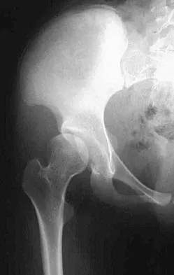
Figures 8a and 8b show the current radiographs of a 10-year-old boy with a hip disorder who was treated with an abduction orthosis 3 years ago. If no further remodeling occurs, what is the most likely prognosis?
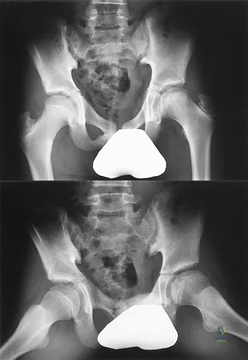


Explanation

The radiographs show a child with Legg-Calve-Perthes disease (LCPD) that has healed. Deformity (asphericity) of the femoral head is evident, but the femoral head and acetabulum are congruous. Stulberg and associates found that hips with aspherical congruity at skeletal maturity functioned well until the fifth or sixth decade of life. Similarly, another study found that degenerative arthritis caused deteriorating hip function after age 40 years in patients with this degree of residual deformity. Repeated episodes of ischemic necrosis are unlikely. Although some studies suggested coagulation abnormalities such as protein C and S deficiencies in children with LCPD, other studies failed to show any evidence of inherited thrombophila in most children with this disorder. There are no studies to suggest growth acceleration occurs following LCPD. Stulberg SD, Cooperman DR, Wallenstein R: The natural history of Legg-Calve-Perthes disease. J Bone Joint Surg Am 1984;66:479-489.

Question 18

In girls with idiopathic scoliosis, peak height velocity (PHV) typically occurs at what point?

Pediatrics 2004 Practice Questions: Set 1 (Solved) - Figure 24


Explanation

PHV generally occurs while girls are still Risser 0; menarche typically occurs before Risser 1, which has a wide variation in its timing. The curve magnitude at the PHV is the best prognostic indicator available. Most untreated patients with curves greater than 30 degrees at PHV require surgery, while patients with smaller curves at that stage typically do not require surgery. Little DG, Song KM, Katz D, Herring JA: Relationship of peak height velocity to other maturity indicators in idiopathic scoliosis in girls. J Bone Joint Surg Am 2000;82:685-693.

Question 19

Examination of a 6-year-old boy who sustained a displaced Salter-Harris type II fracture of the distal radius reveals 35 degrees of volar angulation. A satisfactory reduction is obtained with the aid of a hematoma block. At the 10-day follow-up examination, radiographs show loss of reduction and 35 degrees of volar angulation. Management should now consist of

Pediatrics 2004 Practice Questions: Set 1 (Solved) - Figure 25


Explanation

In a 6-year-old child with a physeal fracture, the healing response 10 days after injury is so advanced that manipulation would have to be very forceful to be successful. A forceful manipulation in a patient this age increases the risk of early growth arrest and a significant disability because 80% of the growth of the radius comes from the distal physis. Because of the large contribution of growth from the distal radial physis and the angulation being in the plane of wrist motion, the potential for remodeling of this fracture is great. It is highly probable that this fracture will completely remodel in 1 to 2 years of growth. In this patient, even a "gentle" open reduction would probably require enough force that the physis would be damaged. Dimeglio A: Growth in pediatric orthopaedics, in Morrissy RT, Weinstein SL (eds): Lovell and Winter's Pediatric Orthopaedics, ed 5. Philadelphia, PA, Lippincott Williams and Wilkins, 2001, pp 33-62.

Question 20

Figures 9a and 9b show the radiographs of a 12-year-old girl who has had right hip pain for the past 4 months. She reports that the pain is so severe that she is unable to walk and is now using a wheelchair. Examination reveals pain with any attempted range of motion. Management should include
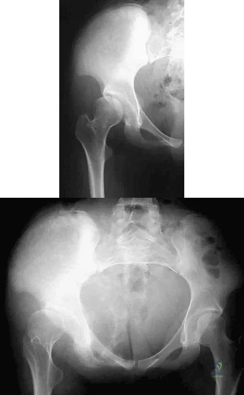


Explanation

In addition to mild hip dysplasia, the radiograph shows an osteoblastic lesion of the right ilium. The patient's symptoms are much more severe than is typical for late hip dysplasia. MRI can determine the extent of the lesion in the bone and soft tissues. Following work-up and biopsy, the patient was diagnosed with Ewing's sarcoma. Springfield DS, Gebhardt MC: Bone and soft tissue tumors, in Morrissy RT, Weinstein SL (eds): Lovell and Winter's Pediatric Orthopaedics, ed 5. Philadelphia, PA, Lippincott Williams and Wilkins, 2001, pp 507-518, 542-544.

Question 21

An 18-month-old boy has 45 degrees of kyphosis in the thoracolumbar spine secondary to type I congenital kyphosis. Examination reveals that he is neurologically intact, and an MRI scan shows no evidence of intraspinal pathology. Management should consist of

Pediatrics 2004 Practice Questions: Set 1 (Solved) - Figure 28


Explanation

Surgery is indicated for congenital kyphosis once the deformity reaches a certain size or if significant progression is documented. In a young patient with a relatively small deformity, the treatment of choice is isolated in situ posterior fusion and postoperative immobilization. If an adequate posterior fusion can be obtained, an epiphyseodesis effect can be generated, allowing the remaining anterior growth to cause some correction. Because there is no evidence of neurologic compression and the deformity is less than 50 degrees, anterior surgery is not indicated. There is no role for bracing in the management of congenital kyphosis. Winter RB: Congenital Deformities of the Spine. New York, NY, Thieme-Stratton, 1983, pp 229-261.

Question 22

Following an acute dislocation of the patella, the risk of a recurrent dislocation is greater if the patient has which of the following findings?

Pediatrics 2004 Practice Questions: Set 1 (Solved) - Figure 29


Explanation

Recurrent dislocations may follow an earlier dislocation. One study found that in patients who had a patellar dislocation between the ages of 11 to 14 years, 60% had a recurrent dislocation. The incidence of recurrent dislocation dropped to 33% in patients who had a patellar dislocation between the ages of 15 to 18 years. The authors also found that the incidence of recurrence was greater in patients who demonstrated a predisposition to dislocation as determined by evaluation of the unaffected knee. Predisposing signs included passive lateral hypermobility of the patella, a dysplastic distal third of the vastus medialis obliquis muscle, and a high and/or lateral position of the patella. A second study found that the risk of redislocation was considerably higher in patients who were in their teens at the first episode of dislocation compared to older patients. There are no studies linking either a patella baja or a bipartite patella to an increased risk of redislocation. Cash JD, Hughston JC: Treatment of acute patellar dislocation. Am J Sports Med 1988;16:244-249.

Question 23

Which of the following findings can cause a dorsal bunion in a patient with neuromuscular disease?

Pediatrics 2004 Practice Questions: Set 1 (Solved) - Figure 30


Explanation

Unopposed action of the tibialis anterior with weakness of the peroneus longus will lead to a dorsal bunion and supination deformity. Overpull of the gastrocnemius-soleus complex and posterior tibialis with weakness of the peroneus brevis will cause equinovarus deformity. A strong posterior tibialis with weakness of the peroneals will cause varus of the hindfoot. Unopposed peroneus brevis and incompetence of the posterior tibialis will lead to a flatfoot deformity. The etiology of a cavus foot is complex, but findings usually include a contracted plantar fascia and weakness of the tibialis anterior. Drennan JC (ed): The Child's Foot and Ankle. New York, NY, Raven Press, 1992, pp 289-302.

Question 24

Which of the following studies is considered most sensitive in monitoring a therapeutic response in acute hematogenous osteomyelitis?

Pediatrics 2004 Practice Questions: Set 1 (Solved) - Figure 31


Explanation

C-reactive protein declines rapidly as the clinical picture improves. Failure of the C-reactive protein to decline after 48 to 72 hours of treatment should indicate that treatment may need to be altered. Blood culture is positive only 50% of the time and will be negative soon after antibiotics are administered, even if treatment is not progressing satisfactorily. WBC count is highly variable and poorly correlated with treatment. The ESR rises rapidly but declines too slowly to guide treatment. Radiographic findings may not change but can take up to 2 weeks to show changes.

Question 25

Figure 10 shows the radiograph of a 7-year-old patient who has a bilateral Trendelenburg limp and limited range of hip motion but no pain. His work-up should include

Pediatrics 2004 Practice Questions: Set 1 (Solved) - Figure 32


Explanation

The radiograph shows bilateral flattening of the femoral heads with mottling and "fragmentation" suggestive of Legg-Calve-Perthes disease. However, when these changes occur bilaterally and are symmetric, multiple epiphyseal dysplasia or spondyloepiphyseal dysplasia should be suspected. Skeletal survey will show irregularity of the secondary ossification centers. With these conditions, there is no true osteonecrosis and no evidence that orthotic or surgical "containment" will alter the outcome of progressive degenerative arthritis. Cardiac anomalies and coagulopathies are not associated with the epiphyseal dysplasias. Crossan JF, Wynne-Davies R, Fulford GE: Bilateral failure of the capital femoral epiphysis: Bilateral Perthes disease, multiple epiphyseal dysplasia, pseudoachondroplasia, and spondyloepiphyseal dysplasia congenita and tarda. J Pediatr Orthop 1983;3:297-301.

Question 26

A 3-month-old girl with Developmental Dysplasia of the Hip (DDH) is treated with a Pavlik harness. At her two-week follow-up, the mother reports that the infant is no longer kicking her right leg as much. On examination, the infant demonstrates decreased active knee extension on the right side. What is the most likely cause of this finding?


Explanation

Hyperflexion of the hips in a Pavlik harness can cause compression of the femoral nerve against the inguinal ligament, leading to a femoral nerve palsy. This presents as decreased active extension of the knee (quadriceps weakness). The treatment is to decrease the amount of flexion or temporarily discontinue the harness until nerve function returns. Avascular necrosis is typically caused by excessive abduction.

Question 27

A 7-year-old boy with spastic quadriplegic cerebral palsy has progressive lateral subluxation of the right hip. Radiographs reveal a migration percentage of 50%, a neck-shaft angle of 155 degrees, and an acetabular index of 35 degrees. What is the most appropriate definitive management?


Explanation

In older children (typically >4 years) with spastic cerebral palsy and significant hip subluxation (migration percentage >40%), both femoral dysplasia (coxa valga and excessive anteversion) and acetabular dysplasia are usually present. Soft tissue releases alone are inadequate. Comprehensive bony reconstruction with a proximal femoral varus derotational osteotomy (VDRO) and a concomitant pelvic osteotomy (e.g., Dega or San Diego) is required to achieve and maintain concentric reduction.

Question 28

A 13-year-old obese boy undergoes in situ single screw fixation for a stable mild slipped capital femoral epiphysis (SCFE) of the left hip. Nine months later, he returns complaining of progressive left hip pain, severe stiffness, and an inability to participate in sports. Radiographs reveal diffuse joint space narrowing of the left hip and subchondral irregularities, with the screw threads completely within the femoral head. What is the most likely diagnosis?


Explanation

Chondrolysis is a devastating complication of SCFE characterized by acute or insidious onset of pain, marked stiffness, and diffuse joint space narrowing on radiographs. Although hardware penetration into the joint is a known risk factor, chondrolysis can occur even with properly placed implants or unoperated cases. Avascular necrosis typically presents with subchondral collapse, sclerosis, or segmental changes rather than global joint space narrowing.

Question 29

A 5-year-old boy presents with a lateral condyle fracture of the distal humerus that was treated with a long-arm cast at an outside facility 4 weeks ago. Radiographs now demonstrate 4 mm of displacement of the fracture fragment and no evidence of callus formation. What is the most appropriate next step in management?


Explanation

Lateral condyle fractures with displacement greater than 2 mm or those that displace secondarily in a cast require operative intervention. Because this injury involves the articular surface and the physis, and has poor healing potential due to the pull of the extensor origin and bathing in synovial fluid, delayed presentation with >2 mm of displacement should be treated with open reduction and internal fixation (ORIF) to prevent nonunion, progressive cubitus valgus, and subsequent tardy ulnar nerve palsy.

Question 30

A 12-year-old premenarcheal girl presents for evaluation of a spinal deformity. She is Risser 0. Standing posteroanterior and lateral radiographs of the spine reveal a right thoracic curve of 36 degrees and normal sagittal alignment. Which of the following is the most appropriate treatment recommendation?


Explanation

Indications for bracing in adolescent idiopathic scoliosis (AIS) include a curve magnitude between 25 and 45 degrees in a skeletally immature patient (Risser 0-2, premenarcheal or less than 1 year postmenarcheal). A full-time TLSO has been shown in multicenter randomized trials (e.g., BrAIST) to significantly decrease the progression of curves to the surgical threshold.

Question 31

A 4-week-old infant is undergoing treatment for an idiopathic right clubfoot using the Ponseti method. After four sequential casts, the forefoot is abducted to 60 degrees, and the heel is in valgus. However, the ankle can only be dorsiflexed to neutral. What is the most appropriate next step in management?


Explanation

In the Ponseti method, serial casting corrects cavus, adductus, and varus deformities first. Once the forefoot is abducted to roughly 60 degrees and the hindfoot varus is corrected to valgus, the equinus is addressed. If there is less than 15 degrees of ankle dorsiflexion at this stage (which occurs in about 80% of patients), a percutaneous Achilles tenotomy is indicated, followed by a final cast in hyperdorsiflexion and abduction for 3 weeks.

Question 32

A 6-year-old boy sustains a supracondylar fracture of the humerus. Radiographs demonstrate a Gartland Type III fracture with posteromedial displacement of the distal fragment. Which of the following neurologic deficits is most likely to be present?


Explanation

In a supracondylar humerus fracture with posteromedial displacement of the distal fragment, the proximal fragment displaces anterolaterally. This places the radial nerve at risk of tenting or laceration as it passes through the lateral intermuscular septum. Radial nerve palsy presents with wrist drop and inability to extend the fingers and thumb at the MCP joints. Conversely, posterolateral displacement puts the anterior interosseous nerve (AIN) at risk.

Question 33

A 4-year-old boy with blue sclerae and a history of four extremity fractures after minimal trauma is diagnosed with severe Osteogenesis Imperfecta (OI). Which of the following pharmacological therapies is currently the standard of care to decrease fracture incidence and increase bone mineral density in this patient?


Explanation

Intravenous bisphosphonates (such as pamidronate or zoledronic acid) are the standard of care for children with moderate to severe Osteogenesis Imperfecta. They inhibit osteoclast-mediated bone resorption, which leads to increased bone mineral density, decreased bone pain, and a significant reduction in the incidence of fractures.

Question 34

An 11-year-old boy presents with a 6-month history of bilateral foot pain and recurrent ankle sprains. Examination reveals rigid flatfeet with absent subtalar motion and peroneal spasticity. Computed tomography (CT) confirms bilateral talocalcaneal coalitions involving approximately 25% of the posterior facet, with no degenerative changes. Nonoperative management with casting and orthotics has failed to relieve his symptoms. What is the most appropriate surgical intervention?


Explanation

Talocalcaneal coalitions commonly present in late childhood or early adolescence as a rigid, painful flatfoot. If conservative management fails, surgical resection with interposition (e.g., fat graft or extensor digitorum brevis) is indicated, provided the coalition involves less than 50% of the posterior facet and there are no significant degenerative changes in the subtalar or surrounding joints.

Question 35

A 13-year-old boy presents to the emergency department with severe left hip pain after tripping over a curb. He is completely unable to bear weight on the left leg, even with the assistance of crutches. Radiographs demonstrate a severe slipped capital femoral epiphysis (SCFE). Which of the following factors is the most significant predictor for the development of avascular necrosis in this patient?


Explanation

According to the Loder classification, SCFE is categorized as stable or unstable based entirely on the clinical ability of the patient to bear weight (with or without crutches). Unstable SCFE (inability to bear weight) carries a high risk of avascular necrosis (ranging up to nearly 50%), compared to a minimal risk (<5%) in stable SCFE.

Question 36

A 13-year-old boy weighing 95 kg presents with left groin pain and an obligatory external rotation of the hip with flexion. Radiographs confirm a left slipped capital femoral epiphysis (SCFE). Which of the following factors is the strongest indication for prophylactic in situ pinning of the contralateral asymptomatic hip?


Explanation

The risk of a contralateral slip in patients with SCFE is approximately 25% to 60%. Absolute or strong relative indications for prophylactic pinning of the contralateral asymptomatic hip include underlying endocrine disorders (such as hypothyroidism, panhypopituitarism, or renal osteodystrophy), radiation therapy to the pelvis, open triradiate cartilage (young age, e.g., females <10 and males <12), and patients for whom reliable follow-up is unlikely. Age >12, male sex, and obesity alone are not universal indications for prophylactic pinning.

Question 37

A 4-month-old infant is being treated with a Pavlik harness for developmental dysplasia of the hip (DDH). During a routine follow-up, the parents report that the child has stopped kicking the affected leg. On examination, there is decreased active extension of the knee, but the child moves the foot and toes spontaneously. What is the most likely cause, and what is the appropriate initial management?


Explanation

Femoral nerve palsy is the most common nerve palsy associated with the Pavlik harness and is caused by excessive hyperflexion of the hip. Clinically, it presents as a loss of active knee extension. The appropriate management is to temporarily discontinue the harness or loosen the anterior straps to reduce the degree of hip flexion, allowing the nerve to recover. Sciatic nerve palsy is rare and would typically present with distal deficits. AVN and septic arthritis would present with pain and systemic signs, not isolated knee extension weakness.

Question 38

A 6-year-old boy sustains a completely displaced, extension-type supracondylar fracture of the humerus. During the neurologic examination, he is unable to flex the interphalangeal joint of his thumb and the distal interphalangeal joint of his index finger. Which nerve is most likely injured?


Explanation

The anterior interosseous nerve (AIN), a motor branch of the median nerve, is the most commonly injured nerve in extension-type supracondylar humerus fractures. It innervates the flexor pollicis longus (FPL), the flexor digitorum profundus (FDP) to the index and middle fingers, and the pronator quadratus. Injury results in the inability to flex the IP joint of the thumb and the DIP joint of the index finger (the 'OK' sign). Radial nerve injuries are more common in posteromedial displacement, while ulnar nerve injuries are more common in flexion-type fractures or as an iatrogenic injury during medial pinning.

Question 39

An 8-year-old boy with spastic quadriplegic cerebral palsy is found to have a migration percentage of 45% on his anteroposterior pelvis radiograph. He has pain with diaper changes and limited hip abduction. What is the most appropriate management?


Explanation

In children with cerebral palsy, hip subluxation is a common problem related to muscle imbalance. A migration percentage of >40-50% with progressive deformity or pain indicates a high risk of complete dislocation and joint degeneration. Soft tissue releases alone (adductor/iliopsoas tenotomies) are generally ineffective once the migration percentage exceeds 40% and bony changes are present. The gold standard treatment involves a bony reconstruction: a proximal femoral varus derotational osteotomy (VDRO) combined with a pelvic osteotomy (e.g., Dega or San Diego) to restore joint congruity and contain the femoral head.

Question 40

A 9-year-old boy presents with a painless limp and restricted hip internal rotation. Radiographs demonstrate fragmentation of the capital femoral epiphysis consistent with Legg-Calvé-Perthes disease. Which of the following factors carries the worst prognosis for this patient?


Explanation

Age at the onset of symptoms is the most significant prognostic factor in Legg-Calvé-Perthes disease. Children who develop the disease after age 8 have a worse prognosis because they have less time for the femoral head to remodel before skeletal maturity, often leading to a residual aspherical femoral head and early osteoarthritis. The extent of lateral pillar involvement (Herring classification) is also a critical radiographic prognostic factor. Male sex is a risk factor for developing the disease, but older age carries the worst outcome.

Question 41

When treating a rigid idiopathic clubfoot using the Ponseti method of serial casting, what is the correct order of deformity correction?


Explanation

The Ponseti method follows a strict sequence of correcting the deformities associated with clubfoot, remembered by the acronym CAVE: Cavus, Adductus, Varus, and Equinus. The cavus is corrected first by supinating the forefoot and elevating the first ray to align it with the hindfoot. Following this, the foot is abducted to correct the adductus and varus simultaneously, pivoting around the head of the talus. Finally, the equinus is corrected, often requiring a percutaneous Achilles tenotomy.

Question 42

A 10-year-old boy presents to the emergency department after sustaining a minor twisting injury to his right arm while throwing a baseball. Radiographs reveal a minimally displaced pathologic fracture through a centrally located, completely radiolucent lesion in the proximal humerus metaphysis. The lesion demonstrates a 'fallen leaf' sign. What is the most appropriate initial management?


Explanation

The clinical presentation and radiographic finding of a 'fallen leaf' sign (a fragment of cortical bone sitting at the dependent portion of a cystic lesion) are pathognomonic for a unicameral bone cyst (UBC). When a UBC presents with a pathologic fracture, the initial management is conservative immobilization (e.g., a sling) to allow the fracture to heal. In many cases, the cyst may undergo partial or complete spontaneous healing during fracture consolidation. If the cyst persists and is large enough to pose a risk of recurrent fracture, elective interventions such as injections or curettage/grafting can be considered later.

Question 43

A 3-year-old girl is diagnosed with congenital scoliosis secondary to a fully segmented hemivertebra at T8. Before proceeding with any surgical intervention, which of the following screening evaluations is most critical to perform?


Explanation

Congenital scoliosis occurs due to a failure of formation (hemivertebra) or segmentation during early embryogenesis. This critical period coincides with the development of the genitourinary, cardiovascular, and central nervous systems. As a result, up to 60% of patients with congenital scoliosis have associated anomalies (VACTERL association). Renal anomalies are present in 20-30%, requiring a renal ultrasound. Cardiac defects are found in 10-15%, necessitating an echocardiogram. Additionally, intraspinal anomalies (such as tethered cord, diastematomyelia, or syringomyelia) occur in 20-40% of cases, making an MRI of the entire neuraxis mandatory before surgical intervention.

Question 44

A 4-year-old boy sustains a minimally displaced (<2 mm) lateral condyle fracture of the humerus that is treated in a long-arm cast. He is lost to follow-up and returns 15 years later. Radiographs demonstrate an established nonunion of the lateral condyle. Which of the following clinical findings is most likely to be present on physical examination?


Explanation

Lateral condyle fractures of the humerus have a high propensity for nonunion if not perfectly immobilized or surgically fixed when displaced, because fracture fluid bathes the site and muscle pull from the extensor origin prevents apposition. An established nonunion of the lateral condyle typically leads to progressive cubitus valgus due to the continued growth of the intact medial physis while the lateral side is deficient. This progressive valgus deformity stretches the ulnar nerve behind the medial epicondyle, classically resulting in a tardy ulnar nerve palsy years or decades after the initial injury.

Question 45

An 11-year-old girl falls off her bicycle and injures her left knee. Radiographs reveal a Type III tibial eminence fracture (completely displaced). This injury is considered the pediatric equivalent of which of the following adult sports injuries?


Explanation

Tibial eminence (or tibial spine) fractures in children are avulsion fractures of the tibial insertion of the anterior cruciate ligament (ACL). Because the ligaments in a child are relatively stronger than the adjacent physeal/metaphyseal bone, the bone fails before the mid-substance of the ligament tears. Thus, a tibial eminence fracture is considered the pediatric equivalent of an adult ACL tear. A Type III fracture (completely displaced) typically requires surgical reduction and internal fixation to restore knee stability and prevent mechanical blocks to extension.

Question 46

A 4-month-old infant presents with a persistently dislocated left hip after a failed 6-week trial of Pavlik harness treatment. Ultrasound confirms the femoral head is dislocated but reducible with moderate force. The acetabulum appears dysplastic. What is the most appropriate next step in management?


Explanation

After a failed trial of a Pavlik harness in an infant under 6 months of age with developmental dysplasia of the hip (DDH), the next appropriate step is an examination under anesthesia, arthrogram, and closed reduction with spica casting. If closed reduction cannot be achieved or maintained within a safe zone, an open reduction is indicated. Continued observation or switching to another brace has a high failure rate once the Pavlik harness has definitively failed.

Question 47

A 12-year-old obese boy presents with acute on chronic left knee pain and an antalgic gait. Physical examination reveals obligate external rotation of the thigh during passive hip flexion. He is diagnosed with a stable slipped capital femoral epiphysis (SCFE) and undergoes in situ pinning with a single cannulated screw. Which of the following is the most significant risk factor for the development of chondrolysis in this patient?


Explanation

Chondrolysis is a devastating complication of SCFE characterized by rapid cartilage destruction and joint stiffness. The most significant recognized risk factor for chondrolysis is unrecognized intra-articular pin penetration during surgical fixation. Meticulous fluoroscopic evaluation (including the approach-withdraw technique) is essential to ensure the screw has not breached the articular surface.

Question 48

A 6-year-old girl sustains a severely displaced extension-type supracondylar fracture of the humerus. On examination, the hand is pink but the radial pulse is absent. Neurologic examination reveals an inability to flex the interphalangeal joint of the thumb and the distal interphalangeal joint of the index finger. Which nerve is most likely injured?


Explanation

The anterior interosseous nerve (AIN) is a branch of the median nerve and is the most commonly injured nerve in extension-type supracondylar humerus fractures, particularly those with posterolateral displacement. AIN palsy presents with weakness of the flexor pollicis longus and flexor digitorum profundus to the index and middle fingers, manifesting as an inability to make an 'OK' sign.

Question 49

A 13-year-old boy presents with a painful, rigid flatfoot and a history of recurrent ankle sprains. Examination shows a lack of subtalar motion and peroneal muscle spasm. Radiographs show a continuous C-shaped radiodense line formed by the medial outline of the talar dome and the posteroinferior aspect of the sustentaculum tali. Which of the following is the most likely diagnosis?


Explanation

The clinical presentation of a rigid flatfoot with peroneal spasticity in an adolescent is classic for a tarsal coalition. The 'C-sign' on a lateral radiograph is highly indicative of a talocalcaneal coalition (specifically involving the middle facet). Calcaneonavicular coalitions are typically seen on an oblique radiograph as an 'anteater nose' sign.

Question 50

An 8-year-old boy presents with a painless limp. Radiographs demonstrate fragmentation of the capital femoral epiphysis. The treating orthopedic surgeon notes multiple 'head-at-risk' signs on the radiograph. According to Catterall, which of the following is considered a 'head-at-risk' sign in Legg-Calve-Perthes disease?


Explanation

Catterall described five 'head-at-risk' clinical and radiographic signs indicating a poorer prognosis in Legg-Calve-Perthes disease. The radiographic signs include: Gage sign (a V-shaped radiolucent defect in the lateral portion of the epiphysis and adjacent metaphysis), calcification lateral to the epiphysis, lateral subluxation of the femoral head, a horizontal growth plate, and metaphyseal cysts.

Question 51

A 6-year-old nonambulatory child with spastic quadriplegic cerebral palsy presents for routine evaluation. An anteroposterior pelvis radiograph demonstrates a right hip migration percentage of 55%. The child is currently asymptomatic. What is the most appropriate management?


Explanation

In a child with cerebral palsy, a hip migration percentage greater than 40-50% indicates significant subluxation and a high risk of progression to dislocation. Soft tissue releases (like adductor tenotomy) alone are insufficient at this stage. Bony reconstruction with a proximal femoral varus derotational osteotomy (VDRO), often combined with a pelvic osteotomy, is the most appropriate treatment to achieve joint congruity and prevent painful dislocation.

Question 52

A 14-year-old boy sustains a Salter-Harris type II fracture of the distal femur during a football game. He undergoes an anatomic closed reduction and casting. Which of the following complications is most commonly associated with this specific injury?


Explanation

Distal femur physeal fractures, particularly Salter-Harris II fractures, have a notoriously high rate of physeal growth arrest, reported to be up to 50% or more. This is due to the highly undulating nature of the distal femoral physis, which sustains significant crush injury (Salter-Harris V type damage) even during a shear or bending mechanism. Close radiographic follow-up is required to monitor for leg length discrepancies and angular deformities.

Question 53

A 2-year-old boy presents with an anterolateral bowing of the tibia and a pseudoarthrosis. Physical examination reveals multiple café-au-lait spots on his trunk and axillary freckling. Which of the following conditions is most strongly associated with this orthopedic presentation?


Explanation

Congenital pseudarthrosis of the tibia (CPT) with anterolateral bowing is highly associated with Neurofibromatosis type 1 (NF1). Approximately 50% of patients with CPT have NF1. The presence of multiple café-au-lait spots and axillary freckling further supports this diagnosis. Management is surgical and notoriously difficult, often requiring excision of the pseudarthrosis, bone grafting, and intramedullary stabilization.

Question 54

During the Ponseti casting technique for the treatment of idiopathic clubfoot, the foot is sequentially manipulated to correct the complex deformity. Which of the following describes the correct sequential order of deformity correction?


Explanation

The Ponseti method utilizes a specific sequence to correct clubfoot deformity, remembered by the acronym CAVE: Cavus, Adductus, Varus, and Equinus. The cavus is corrected first by supinating the forefoot and elevating the first ray to align it with the hindfoot. Then, the adductus and varus are corrected simultaneously by abducting the foot around the head of the talus. Finally, the equinus is corrected, often requiring a percutaneous Achilles tenotomy.

Question 55

A 14-year-old female gymnast presents with progressive low back pain. Radiographs demonstrate a grade II isthmic spondylolisthesis at L5-S1. She has failed 6 months of conservative management (rest, bracing, physical therapy), and her pain significantly limits her activities of daily living. Neurologic examination is entirely normal. What is the most appropriate surgical treatment?


Explanation

For symptomatic low-grade (Meyerding Grade I or II) isthmic spondylolisthesis in children and adolescents that fails conservative management, the gold standard surgical treatment is a posterior in situ posterolateral fusion. Neurologic decompression is generally not required if there are no neurologic deficits. Reduction of low-grade slips does not significantly improve outcomes and increases the risk of L5 nerve root injury. Pars repair is typically reserved for select patients with early pars stress fractures or very low-grade slips without significant disc degeneration.

Question 56

A 3-year-old boy presents to the emergency department after falling off a slide. He has a closed, isolated midshaft femur fracture with 1.5 cm of shortening. He is normally developing and has no other injuries. What is the most appropriate definitive management for this patient?


Explanation

For children aged 6 months to 5 years with isolated, closed femur fractures with acceptable shortening (typically <2 cm), early hip spica casting is the standard of care. Flexible intramedullary nailing is typically reserved for children older than 5 years (or heavier than 50 lbs), while the Pavlik harness is indicated for infants under 6 months of age. Submuscular plating and external fixation are generally reserved for older patients, polytrauma, open fractures, or unstable fracture patterns where length cannot be maintained.

Question 57

A 7-year-old girl with spastic quadriplegic cerebral palsy presents for routine surveillance. She is non-ambulatory (GMFCS Level V). Radiographs of the pelvis demonstrate a right hip migration percentage of 55%, coxa valga, and an intact Shenton's line on the left. She has mild pain with hip abduction. What is the most appropriate management?


Explanation

In children with cerebral palsy, hip surveillance is critical. Soft tissue releases (adductor tenotomies) are prophylactic and most effective when the migration percentage (MP) is less than 30-40% and the child is young (typically <4-5 years). Once the migration percentage exceeds 40-50% in a 7-year-old, soft tissue releases alone have a high failure rate. Bony reconstructive surgery, specifically a proximal femoral varus derotation osteotomy (VDRO) combined with a pelvic osteotomy (such as a Dega or San Diego), is required to safely contain the hip and halt progression of subluxation to dislocation.

Question 58

A 9-month-old girl presents for an initial orthopedic evaluation. She has asymmetric thigh folds and limited abduction of the left hip. Ultrasound at 6 weeks of age was reportedly abnormal but the parents did not follow up. Current radiographs demonstrate a dislocated left hip with a dysplastic acetabulum. What is the most appropriate next step in management?


Explanation

The patient has late-presenting developmental dysplasia of the hip (DDH). The Pavlik harness is generally indicated for infants under 6 months of age; its success rate drops significantly and the risk of complications rises in older infants. For children aged 6 to 18 months, the gold standard initial treatment is closed reduction and application of a hip spica cast, often preceded by or performed concurrently with an adductor tenotomy. Open reduction with or without pelvic/femoral osteotomies is typically reserved for failures of closed reduction or primary treatment in children older than 18 months.

Question 59

A 13-year-old girl sustains a twisting injury to her right ankle. Radiographs reveal a Salter-Harris III fracture of the anterolateral distal tibia.

This specific fracture pattern (Juvenile Tillaux fracture) occurs as a direct result of the asymmetrical closure of the distal tibial physis. Which of the following describes the LAST portion of the distal tibial physis to close?


Explanation

The distal tibial physis closes in a predictable sequence over an 18-month period, typically between ages 12 and 15. The sequence begins centrally, progresses medially, then posteromedially, and finally closes anterolaterally. Because the anterolateral physis is the last to fuse, the anterior inferior tibiofibular ligament (AITFL) can avulse this open anterolateral portion of the epiphysis during an external rotation injury, leading to the classic juvenile Tillaux fracture (Salter-Harris III).

Question 60

A 6-year-old boy falls onto an outstretched hand and sustains a widely displaced, extension-type supracondylar fracture of the humerus. On physical examination in the emergency department, his hand is pink and well-perfused with a palpable radial pulse. However, he is unable to flex the interphalangeal joint of his thumb and the distal interphalangeal joint of his index finger. Which nerve is most likely injured?


Explanation

The anterior interosseous nerve (AIN), a branch of the median nerve, is the most commonly injured nerve in extension-type supracondylar humerus fractures. Injury to the AIN manifests clinically as an inability to flex the interphalangeal joint of the thumb (flexor pollicis longus) and the distal interphalangeal joint of the index finger (flexor digitorum profundus), resulting in the loss of the 'OK' sign. These neurapraxias typically resolve spontaneously over several months with conservative management.

Question 61

An 11-year-old boy with a BMI in the 99th percentile presents with left knee pain and a limp for 4 weeks. Radiographs show a mild, stable slipped capital femoral epiphysis (SCFE) of the left hip. The parents ask about the risk to the other hip. Which of the following is the strongest indication for prophylactic pinning of the contralateral, asymptomatic right hip?


Explanation

The decision to perform prophylactic pinning of the contralateral hip in a unilateral SCFE patient requires balancing the risks of surgery against the risk of a subsequent slip. Patients with underlying endocrine disorders (such as hypothyroidism, growth hormone deficiency, or panhypopituitarism) or renal osteodystrophy have an extremely high risk (up to 100%) of bilateral involvement, making an underlying endocrine disorder a strong, widely accepted indication for prophylactic pinning. Other relative indications may include young age (<10 for girls, <11 for boys) or inability to follow up.

Question 62

An 8-year-old boy presents with a 2-month history of a painless limp. Radiographs demonstrate sclerosis and fragmentation of the proximal femoral epiphysis consistent with Legg-Calve-Perthes disease. Which of the following is considered the most important radiographic prognostic factor for determining the final outcome of the hip?


Explanation

The Herring lateral pillar classification system evaluates the height of the lateral portion of the capital femoral epiphysis on an AP pelvis radiograph during the fragmentation stage of Legg-Calve-Perthes disease. It is widely recognized as the most reliable radiographic prognostic indicator for final hip outcome. Hips in Group A (no lateral pillar involvement) have the best prognosis, while Group C (<50% lateral pillar height maintained) have the poorest outcome. Age of onset (especially >8 years) is the most significant clinical prognostic factor.

Question 63

A 14-year-old boy complains of recurrent right ankle sprains and deep midfoot pain that worsens with activity. Examination reveals a rigid flat foot, prominent peroneal tendons, and limited subtalar inversion and eversion. Lateral radiographs show an elongated anterior process of the calcaneus (the 'anteater nose' sign). What is the most likely diagnosis?


Explanation

The clinical presentation of a rigid flatfoot with peroneal spasticity in an adolescent is classic for a tarsal coalition. The 'anteater nose' sign on the lateral foot radiograph is the pathognomonic finding of a calcaneonavicular coalition, representing a tubular elongation of the anterior process of the calcaneus attempting to bridge to the navicular. Talocalcaneal coalitions are typically best seen on a Harris axial view or CT scan and may show a 'C-sign' on the lateral radiograph.

Question 64

A 12-year-old girl presents with a destructive diaphyseal lesion of the femur with a permeative pattern and an 'onion skin' periosteal reaction.

A core needle biopsy is performed, revealing sheets of uniform small, round, blue cells. Immunohistochemistry is strongly positive for CD99. Which of the following chromosomal translocations is most characteristic of this tumor?


Explanation

The clinical, radiographic ('onion skin' periosteal reaction, permeative diaphyseal lesion), and histologic (small round blue cells, CD99+) findings are highly characteristic of Ewing sarcoma. Ewing sarcoma is classically associated with the t(11;22)(q24;q12) chromosomal translocation, which creates the EWS-FLI1 fusion gene. t(9;22) is associated with CML (Philadelphia chromosome) and myxoid chondrosarcoma; t(12;16) is seen in myxoid liposarcoma; t(X;18) in synovial sarcoma; and t(2;13) in alveolar rhabdomyosarcoma.

Question 65

A newborn male is evaluated for bilateral idiopathic clubfeet (talipes equinovarus). The treating orthopedic surgeon initiates the Ponseti method of serial casting. What is the correct physiological sequence in which the components of the deformity are systematically corrected?


Explanation

The Ponseti method addresses the components of the clubfoot deformity in a specific sequence, easily remembered by the mnemonic CAVE: Cavus, Adductus, Varus, Equinus. The first cast corrects the cavus by supinating the forefoot to align it with the hindfoot. Subsequent casts correct the adductus and varus by gradually abducting the foot around the head of the talus. Finally, the equinus is corrected, which often requires a percutaneous Achilles tenotomy before the final cast is applied.

Question 66

A 12-year-old boy with a BMI of 35 presents with an acute on chronic slipped capital femoral epiphysis (SCFE) of the left hip. He undergoes uneventful in situ pinning. When considering prophylactic pinning of the contralateral right hip, which of the following is the most important risk factor for developing a subsequent SCFE?


Explanation

Prophylactic pinning of the contralateral hip in patients with a unilateral SCFE is indicated in patients with endocrine disorders (e.g., hypothyroidism, growth hormone supplementation, renal osteodystrophy), prior radiation therapy, and an age of presentation less than 10 years in boys or 11 years in girls. Male sex and family history alone do not mandate prophylactic pinning. Slip angle determines severity but not automatically contralateral risk in the absence of other specific factors.

Question 67

A 6-year-old boy presents with a completely displaced posteromedial supracondylar humerus fracture. Based on the direction of displacement, which nerve is at highest risk of injury?


Explanation

Supracondylar humerus fractures are the most common elbow fractures in children. Posteromedial displacement is the most common pattern of an extension-type supracondylar humerus fracture. In this pattern, the distal fragment goes posteromedially, causing the proximal fragment to displace anterolaterally. The radial nerve is located anterolaterally and is tethered at the lateral intermuscular septum, making it the most vulnerable structure to injury by the proximal fragment spike. The anterior interosseous nerve (AIN) is most commonly injured in posterolateral displacement.

Question 68

An infant is placed in a Pavlik harness for treatment of developmental dysplasia of the hip (DDH). During a follow-up visit, the parents report that the child has stopped kicking the leg. Examination reveals an absence of active knee extension on the affected side, but intact ankle dorsiflexion and plantar flexion. What is the most appropriate next step in management?


Explanation

Femoral nerve palsy is a known complication of Pavlik harness treatment for DDH. It is caused by excessive hip flexion, which can compress the femoral nerve against the rim of the pelvis. The diagnosis is made clinically by a loss of active knee extension (decreased quadriceps function) while ankle motion remains intact. Management consists of either adjusting the anterior straps to reduce hip flexion or removing the harness entirely until nerve function returns. Continuing the harness without adjustment risks permanent nerve injury.

Question 69

When treating idiopathic clubfoot using the Ponseti method, what is the proper sequence of deformity correction?


Explanation

The Ponseti method for the treatment of idiopathic clubfoot relies on a specific sequence of deformity correction summarized by the acronym CAVE: Cavus, Adduction, Varus, and Equinus. First, the cavus is corrected by elevating the first ray to align the forefoot with the hindfoot. Then, the forefoot is abducted to correct the adduction and varus simultaneously while counter-pressure is applied to the lateral aspect of the talar head. Finally, the equinus is corrected, most often requiring a percutaneous Achilles tenotomy.

Question 70

Which of the following is the most important prognostic factor for long-term outcome in a child with Legg-Calvé-Perthes disease?


Explanation

In Legg-Calvé-Perthes disease, the age at the onset of symptoms is the most consistently reliable prognostic factor for final hip outcome. Children who present at less than 6 years of age have a better prognosis because they have greater potential for remodeling of the femoral head. Children presenting after 8 years of age have a worse prognosis. The other most important prognostic factor is the degree of lateral (not medial) pillar involvement according to the Herring classification.

Question 71

A 14-year-old boy presents with knee pain. Radiographs reveal a mixed sclerotic and lytic lesion in the distal femoral metaphysis with a sunburst periosteal reaction and a Codman triangle. A biopsy shows malignant spindle cells producing osteoid. What is the most significant prognostic factor for survival in this patient?


Explanation

Osteosarcoma is the most common primary pediatric malignant bone tumor. Standard treatment involves neoadjuvant chemotherapy followed by wide surgical resection and adjuvant chemotherapy. The most significant prognostic factor for long-term survival is the histologic response of the tumor to the neoadjuvant chemotherapy, specifically the percentage of tumor necrosis. A necrosis rate of greater than 90% is considered a favorable response and correlates strongly with improved survival.

Question 72

A 5-year-old child with spastic quadriplegic cerebral palsy is evaluated in the clinic. The patient has a Gross Motor Function Classification System (GMFCS) level of V. What is the most appropriate radiographic screening protocol for hip displacement in this patient?


Explanation

Children with cerebral palsy (CP) are at a high risk for progressive hip displacement (subluxation and dislocation) due to muscle spasticity and imbalance. The risk of hip displacement is directly correlated with the Gross Motor Function Classification System (GMFCS) level. A child with GMFCS level V is non-ambulatory and has the highest risk (over 90%) of hip displacement. Therefore, rigorous surveillance is required. The recommended screening protocol for a GMFCS level V child includes an anteroposterior (AP) pelvis radiograph every 6 to 12 months.

Question 73

A 12-year-old boy presents with a history of recurrent ankle sprains and rigid flatfeet. On examination, he has decreased subtalar motion and peroneal spasticity. Radiographs demonstrate a C-sign on the lateral view of the foot. Which of the following is the most appropriate initial management?


Explanation

Tarsal coalition is a fusion between two or more tarsal bones, most commonly talocalcaneal or calcaneonavicular. The C-sign on a lateral foot radiograph is a radiographic indicator of a talocalcaneal coalition, formed by the continuous outline of the medial outline of the talar dome and the posterior outline of the sustentaculum tali. The initial management for a symptomatic tarsal coalition is non-operative, focusing on resting the foot and breaking the pain cycle with immobilization, typically in a short leg walking cast or a controlled ankle motion (CAM) boot for 4 to 6 weeks.

Question 74

A 3-year-old child sustains a closed, isolated midshaft femur fracture with 2 cm of shortening after falling from a playground slide. Which of the following is the most appropriate definitive management?


Explanation

The standard of care for an isolated, closed midshaft femur fracture in a 3-year-old child with acceptable shortening (less than 2 cm) is early spica casting. Surgical management, such as flexible intramedullary nailing, is typically reserved for children aged 5 to 11 years or in cases of polytrauma, open fractures, or inability to achieve or maintain acceptable alignment in a cast. Immediate spica cast application in young children yields excellent clinical and radiographic outcomes with minimal complications.

Question 75

A 14-year-old female gymnast complains of lower back pain that worsens with extension activities. Neurological exam is normal. AP and lateral radiographs of the lumbar spine are normal. What is the most appropriate next step in imaging to diagnose an acute pars interarticularis stress reaction?


Explanation

In a pediatric athlete presenting with extension-related low back pain and normal radiographs, an acute pars interarticularis stress reaction (spondylolysis) must be suspected. While CT scans offer excellent bony detail, they involve significant ionizing radiation. A bone scan combined with SPECT is sensitive but also entails high radiation exposure. Magnetic resonance imaging (MRI) of the lumbar spine, specifically utilizing fluid-sensitive (STIR or T2 fat-suppressed) sequences, is now considered the gold standard for early diagnosis as it can detect bone marrow edema in the pars interarticularis without exposing the pediatric patient to ionizing radiation.

Question 76

An 11-year-old girl with a history of renal osteodystrophy presents with a 3-week history of left groin and knee pain. She is diagnosed with a moderate slipped capital femoral epiphysis (SCFE) on the left side. Her right hip is completely asymptomatic, and radiographs of the right hip are normal. What is the most appropriate management regarding the contralateral right hip?


Explanation

Prophylactic in situ pinning of the contralateral hip is highly recommended in patients with SCFE associated with an underlying endocrinopathy or metabolic bone disease (such as renal osteodystrophy, hypothyroidism, or growth hormone deficiency). These patients have an extremely high rate of bilateral involvement (up to 100% in some metabolic conditions) compared to idiopathic cases. Observation is generally reserved for idiopathic cases with a reliable follow-up.

Question 77

A 6-year-old boy falls from the monkey bars and sustains a displaced extension-type supracondylar humerus fracture. Radiographs demonstrate posterolateral displacement of the distal fragment.

Based on the direction of displacement, which nerve is most at risk for injury, and what is its characteristic motor deficit?


Explanation

In extension-type supracondylar humerus fractures, the direction of distal fragment displacement predicts the nerve at risk due to the sharp spike of the proximal fragment. Posterolateral displacement of the distal fragment causes the proximal fragment to displace anteromedially, placing the median nerve and particularly the anterior interosseous nerve (AIN) at greatest risk. The classic clinical sign of an AIN palsy is the inability to form an 'OK' sign, due to weakness of the flexor pollicis longus and the flexor digitorum profundus to the index finger.

Question 78

A 3-month-old infant with developmental dysplasia of the hip is being treated with a Pavlik harness. At a routine 2-week follow-up, the mother reports that the child is no longer kicking her right leg. On examination, there is an absence of active knee extension on the right side, but foot and ankle motion are preserved. What is the most likely cause of this finding?


Explanation

Femoral nerve palsy is a known complication of Pavlik harness treatment and is typically caused by excessive flexion of the hips, which compresses the femoral nerve against the inguinal ligament. It manifests as a loss of active knee extension. The appropriate management is to temporarily loosen the anterior straps to decrease hip flexion, which usually allows for complete recovery of nerve function. Excessive abduction places the hip at risk for avascular necrosis (AVN).

Question 79

A 7-year-old child with spastic quadriplegic cerebral palsy is evaluated for progressive left hip dysplasia. Current anteroposterior pelvis radiographs show a migration percentage of 65%, a neck-shaft angle of 155 degrees, and an intact triradiate cartilage. The articular cartilage appears well-preserved on MRI. What is the most appropriate surgical intervention?


Explanation

In children with cerebral palsy and a subluxated but reconstructable hip (migration percentage > 50% and preserved cartilage), the gold standard surgical treatment is a combined one-stage reconstruction. This typically involves a varus derotational osteotomy (VDRO) of the proximal femur to correct coxa valga and excessive anteversion, combined with a pelvic osteotomy (such as a Dega or San Diego osteotomy) to correct acetabular dysplasia. Soft tissue releases are also performed concurrently. Salvage procedures like the Castle procedure are reserved for painful, chronically dislocated, and non-reconstructable hips.

Question 80

A 7-year-old boy is diagnosed with Legg-Calvé-Perthes disease. According to the Herring classification system, which of the following radiographic parameters evaluated during the fragmentation stage is the most reliable predictor of long-term outcome?


Explanation

The Herring Lateral Pillar Classification is based on AP radiographs during the fragmentation stage of Legg-Calvé-Perthes disease. It divides the femoral head into three pillars (medial, central, and lateral). The height of the lateral pillar is the most reliable prognostic indicator. Group A (>100% height maintained) has the best outcome, Group B (>50% maintained) has an intermediate outcome, and Group C (<50% maintained) has the worst outcome regarding future joint congruency and arthritis risk.

Question 81

When correcting an infant's idiopathic clubfoot deformity using the Ponseti casting technique, the deformities must be sequentially addressed. What is the correct chronological order of correction?


Explanation

The Ponseti method dictates a specific sequence for the correction of the clubfoot deformity, easily remembered by the acronym CAVE: Cavus, Adductus, Varus, Equinus. The cavus is corrected first by elevating the first ray to align the forefoot with the hindfoot. Then, the adductus and varus are corrected simultaneously by abducting the midfoot around the fixed head of the talus. Finally, the equinus is corrected, often requiring a percutaneous Achilles tenotomy.

Question 82

A 9-year-old boy presents to the emergency department after a minor fall with right arm pain. Radiographs reveal a centrally located, lucent metaphyseal lesion in the proximal humerus with a nondisplaced pathologic fracture. A 'fallen leaf' sign is noted within the lesion.

What is the most appropriate initial management?


Explanation

The clinical and radiographic presentation is classic for a unicameral bone cyst (UBC) complicated by a pathologic fracture. The 'fallen leaf' (or 'fallen fragment') sign is pathognomonic for a UBC. When a UBC presents with a nondisplaced fracture, the initial treatment is immobilization to allow the fracture to heal. Up to 15-20% of UBCs may go on to spontaneous resolution after a fracture. Surgical intervention or injections are typically reserved for cysts that fail to resolve and pose a persistent risk of re-fracture after the initial fracture has healed.

Question 83

A 5-year-old girl falls onto an outstretched hand and complains of elbow pain. Radiographs demonstrate a lateral condyle fracture of the distal humerus.

The fracture fragment is displaced by 4 millimeters. What is the most appropriate definitive management?


Explanation

Lateral condyle fractures of the humerus are intra-articular injuries. Displacement of greater than 2 mm is an indication for open reduction and internal fixation (ORIF) to anatomically restore the joint surface and prevent complications such as nonunion, malunion, cubitus valgus, and tardy ulnar nerve palsy. Closed reduction is often inadequate because the fracture fragment is subjected to the deforming pull of the common extensor origin.

Question 84

A 13-year-old boy sustains a twisting injury to his ankle while playing soccer. Radiographs demonstrate a Salter-Harris type III fracture of the anterolateral aspect of the distal tibia (Tillaux fracture).

This specific fracture pattern is governed by the normal physiological closure pattern of the distal tibial physis. In what order does the distal tibial physis close?


Explanation

The juvenile Tillaux fracture occurs due to the asymmetric closure of the distal tibial physis, which typically occurs over an 18-month period. The physis closes first in the central portion, then proceeds medially, and finally closes laterally. Because the anterolateral physis is the last to close, it remains vulnerable to the avulsion force of the anterior inferior tibiofibular ligament (AITFL) during external rotation injuries in adolescents.

Question 85

A 4-year-old child with a history of recurrent fractures, blue sclerae, and dentinogenesis imperfecta is diagnosed with osteogenesis imperfecta (OI) type III. In order to decrease the fracture burden and correct progressive long-bone deformities, what is the current gold standard combined medical and surgical management strategy?


Explanation

The medical management of severe osteogenesis imperfecta heavily relies on bisphosphonates (often administered intravenously, like pamidronate), which inhibit osteoclast resorption and increase bone density, significantly reducing fracture rates. Surgically, the use of telescopic (growing) intramedullary rods, such as the Fassier-Duval rod, is the gold standard for long-bone deformities in growing children with OI. Telescopic rods accommodate growth, providing internal splinting that decreases the likelihood of recurrent fractures and rod migration compared to static solid nails or plates.

Question 86

An obese 12-year-old boy presents with acute-on-chronic left thigh pain and is unable to bear weight on the left leg, even with crutches. Radiographs demonstrate a posterior and medial displacement of the proximal femoral epiphysis. According to the Loder classification, what is the most significant complication directly associated with his specific presentation type?


Explanation

The Loder classification divides Slipped Capital Femoral Epiphysis (SCFE) into stable and unstable slips based on the patient's ability to bear weight (with or without crutches). This patient cannot bear weight, indicating an unstable SCFE. Unstable SCFE has a notoriously high rate of avascular necrosis (AVN), reported to be as high as 47-50%, compared to nearly 0% in stable SCFE. Chondrolysis is more commonly associated with unrecognized pin penetration into the joint space.

Question 87

A 13-year-old boy complains of frequent ankle sprains and lateral foot pain. Examination reveals a rigid flatfoot with peroneal spasticity. Oblique radiographs of the foot demonstrate an 'anteater sign.'

If a 6-month trial of short leg casting and orthotics fails to relieve his symptoms, and no degenerative changes are noted on CT scan, what is the most appropriate definitive surgical management?


Explanation

The patient's clinical presentation and the 'anteater sign' (an elongated anterior process of the calcaneus) on oblique radiographs are classic for a calcaneonavicular coalition. Initial treatment consists of conservative measures (cast immobilization, orthotics). If symptoms persist in the absence of degenerative arthritic changes, the gold standard surgical treatment is resection of the calcaneonavicular bar with interposition of a tissue spacer, most commonly the extensor digitorum brevis (EDB) muscle belly or fat, to prevent recurrence.

Question 88

A 6-year-old boy falls from monkey bars and sustains a completely displaced, extension-type supracondylar humerus fracture. On arrival at the emergency department, his hand is pink and warm with brisk capillary refill, but no radial pulse is palpable. What is the next best step in management?


Explanation

In the setting of a displaced supracondylar humerus fracture with a 'pulseless but pink' (well-perfused) hand, the initial management is urgent closed reduction and percutaneous pinning (CRPP). The pulse often returns following anatomical alignment of the fracture. If the hand remains pink and well-perfused after CRPP, observation is the appropriate management. Open vascular exploration is indicated if the hand is dysvascular (pulseless, white, and cold) before or after reduction.

Question 89

A 6-year-old child with spastic quadriplegic cerebral palsy is evaluated during routine hip surveillance. Radiographs demonstrate a Reimer's migration percentage of 45% in the right hip. There are no advanced degenerative changes. What is the most appropriate management?


Explanation

In children with cerebral palsy, hip subluxation is a common complication. A Reimer's migration index greater than 40% to 50% in an older child (e.g., >4-5 years old) indicates significant hip displacement that typically requires bony reconstruction to prevent complete dislocation and painful arthritis. The standard of care is a proximal femoral varus derotational osteotomy (VDRO) combined with a pelvic osteotomy (e.g., Dega or San Diego) to improve acetabular coverage. Soft tissue release alone (adductor tenotomy) is insufficient for this degree of subluxation at this age.

Question 90

A 4-month-old infant with developmental dysplasia of the hip (DDH) has been managed with a Pavlik harness for 4 weeks. Repeat dynamic ultrasound demonstrates that the affected hip remains completely dislocated and cannot be reduced in the harness. What is the most appropriate next step in management?


Explanation

If a dislocated hip fails to reduce after 3 to 4 weeks of Pavlik harness treatment, the harness must be discontinued. Prolonged use of the harness in an unreduced hip can lead to 'Pavlik harness disease' (erosion and damage to the posterior acetabular cartilage) and increases the risk of avascular necrosis. The next step is a transition to an alternative rigid abduction orthosis or, more definitively, a closed reduction with spica casting under general anesthesia (with or without an arthrogram and adductor tenotomy).

Question 91

An 8-year-old boy presents with a painless limp. Radiographs demonstrate fragmentation of the capital femoral epiphysis consistent with Legg-Calvé-Perthes disease.

The lateral pillar maintains 60% of its normal height. According to the prospective multicenter study by Herring et al., what is the most appropriate management for this specific presentation?


Explanation

The patient has Legg-Calvé-Perthes disease. Maintaining 60% of the lateral pillar height places him in Herring Lateral Pillar Group B. The multicenter prospective study by Herring et al. demonstrated that children aged 8 years or older at the time of disease onset who have Group B (or B/C border) lateral pillar involvement have significantly better long-term radiographic and clinical outcomes when treated with surgical containment (such as a proximal femoral varus osteotomy or pelvic osteotomy) compared to non-operative management.

Question 92

A 2-week-old infant is diagnosed with congenital idiopathic clubfoot and is scheduled to begin serial casting using the Ponseti method. What is the correct chronological sequence of deformity correction in this technique?


Explanation

The Ponseti method addresses the components of a clubfoot deformity in a specific sequential order, best remembered by the acronym CAVE: Cavus, Adductus, Varus, and Equinus. The cavus is corrected first by supinating the forefoot to elevate the first ray. The adductus and varus are then corrected simultaneously by abducting the foot around the fixed head of the talus. Finally, the equinus is addressed, which frequently requires a percutaneous Achilles tenotomy.

Question 93

An 8-year-old boy weighing 35 kg sustains a closed, length-stable, transverse midshaft fracture of the femur. His neurovascular examination is intact. What is the most appropriate definitive management?


Explanation

For pediatric length-stable midshaft femur fractures in children aged 5 to 11 years (typically weighing less than 50 kg), flexible intramedullary nailing (e.g., Titanium Elastic Nails, or TENs) is the standard of care. It provides excellent alignment, allows early mobilization, and avoids the complications of prolonged immobilization. Early spica casting is generally reserved for children under 5 years of age. Rigid nailing through the piriformis fossa is contraindicated in this age group due to the high risk of iatrogenic avascular necrosis of the femoral head.

Question 94

A 12-year-old boy sustains a Salter-Harris type II fracture of the distal femur. The fracture is managed with a closed reduction and percutaneous pinning. Post-operative radiographs show perfect anatomical alignment. Despite an optimal reduction, the parents should be thoroughly counseled that the child remains at significant risk for which of the following complications?


Explanation

Distal femoral physeal fractures are notorious for having a very high rate of premature physeal closure and subsequent growth arrest, reported to occur in 40% to 90% of cases. This complication can happen regardless of the exact Salter-Harris classification or the quality of the anatomical reduction. The vulnerability is largely due to the undulating, wave-like anatomy of the distal femoral physis, which sustains significant microscopic crushing and shearing forces at the time of injury.

Question 95

A 3-year-old girl with blue sclerae and dentinogenesis imperfecta presents with her third low-energy long bone fracture. Genetic testing confirms a mutation affecting type I collagen. Which of the following systemic pharmacological treatments is most commonly indicated to decrease fracture incidence and improve bone mass in this condition?


Explanation

The patient's clinical presentation and genetic defect are consistent with Osteogenesis Imperfecta (OI). Intravenous bisphosphonates, such as pamidronate or zoledronic acid, are the current gold standard medical therapy for moderate to severe pediatric OI. By inhibiting osteoclast-mediated bone resorption, bisphosphonates increase bone mineral density, decrease the frequency of fractures, and often alleviate chronic bone pain. Teriparatide is generally contraindicated in children with open physes.

Question 96

A 2-week-old infant is brought to the clinic for evaluation of a bilateral congenital foot deformity. Examination reveals rigid equinus, varus of the hindfoot, adductus of the forefoot, and a cavus midfoot. The decision is made to initiate the Ponseti method of serial casting. According to the principles of this technique, what is the correct sequence of deformity correction?


Explanation

The Ponseti method is the gold standard for the treatment of idiopathic clubfoot. The correction follows a specific sequence remembered by the acronym CAVE: Cavus, Adductus, Varus, and Equinus. The cavus is corrected first by elevating the first ray, which aligns the forefoot with the hindfoot. Subsequent casts correct the adductus and varus by abducting the foot around the talar head. Finally, the equinus is corrected, which often requires a percutaneous Achilles tenotomy in the majority of patients.

Question 97

A 6-year-old boy falls from the monkey bars and sustains a completely displaced, extension-type supracondylar fracture of the humerus. On presentation to the emergency department, his hand is pink and well-perfused. However, neurologic examination reveals that he is unable to flex the interphalangeal joint of his thumb or the distal interphalangeal joint of his index finger. Which of the following nerve structures is most likely injured?


Explanation

The anterior interosseous nerve (AIN), a motor branch of the median nerve, is the most commonly injured nerve in extension-type pediatric supracondylar humerus fractures. It innervates the flexor pollicis longus (FPL), the flexor digitorum profundus (FDP) to the index and middle fingers, and the pronator quadratus. The classic physical exam finding for AIN palsy is the inability to make an 'OK' sign, presenting as a 'pinch' posture instead due to the inability to flex the IP joint of the thumb and the DIP joint of the index finger. Sensory examination remains intact.

Question 98

A 7-month-old girl presents for evaluation of a persistent limp and asymmetric thigh folds. She has had no prior orthopedic treatment. Ultrasound and plain radiographs confirm a completely dislocated left hip consistent with developmental dysplasia of the hip (DDH). Clinical examination demonstrates limited abduction of the left hip and a positive Galeazzi sign. What is the most appropriate initial management for this patient?


Explanation

For children 6 to 18 months of age with previously untreated developmental dysplasia of the hip (DDH), closed reduction with hip spica casting is generally the initial treatment of choice. The Pavlik harness is highly successful in infants under 6 months of age but has a significantly higher failure rate in older, larger, and more active infants. Open reduction is indicated if closed reduction fails to achieve a stable, concentric reduction within a safe zone, or as a primary option in children presenting over 18 months of age. Pelvic or femoral osteotomies are typically reserved for children older than 18 months or those with residual dysplasia following successful reduction.

Question 99

A 12-year-old obese boy with a history of end-stage renal disease presents with a 3-week history of left thigh pain and a noticeable limp. He denies trauma. Examination reveals obligate external rotation of the left hip upon passive flexion. Radiographs demonstrate a mild left slipped capital femoral epiphysis (SCFE). Radiographs of the right hip appear normal. What is the most appropriate definitive management?


Explanation

This patient presents with a stable slipped capital femoral epiphysis (SCFE) and underlying chronic renal failure (renal osteodystrophy). The standard treatment for a stable SCFE is in situ single-screw fixation. Prophylactic pinning of the contralateral asymptomatic hip remains controversial in purely idiopathic SCFE but is highly recommended in patients with endocrine or metabolic disorders (e.g., hypothyroidism, chronic renal failure, prior radiation, growth hormone therapy). These patients have an exceptionally high risk (up to 50-100%) of developing a bilateral slip. Closed reduction of a SCFE is contraindicated as it significantly increases the risk of avascular necrosis (AVN).

Question 100

A 5-year-old girl with spastic quadriplegic cerebral palsy is evaluated in the clinic. She is non-ambulatory (GMFCS Level V) and requires full assistance for transfers. Pelvic radiographs reveal an anteroposterior view with a bilateral migration percentage (Reimers' index) of 45%. Clinical examination shows bilateral hip abduction is limited to 20 degrees with the hips in extension. She has an established coxa valga deformity. What is the most appropriate management to prevent painful hip dislocation?


Explanation

Hip displacement is a common and severe complication in patients with spastic quadriplegic cerebral palsy (GMFCS IV and V). A migration percentage (Reimers' index) greater than 30-40% indicates significant subluxation requiring surgical intervention. While isolated soft tissue releases (adductor tenotomies) may be effective early on (migration <30%), once the migration percentage exceeds 40% and structural bony changes (coxa valga, excessive femoral anteversion) are present, soft tissue releases alone have an unacceptably high failure rate. Bony reconstruction, specifically bilateral proximal femoral varus derotational osteotomies (often combined with a pelvic osteotomy), is the standard of care to achieve concentric reduction and prevent progression to a painful dislocated hip.

None

Clinic OS
Medically Verified Content by
Prof. Clinic OS
Consultant Orthopedic & Spine Surgeon
Chapter Index