العربية
Part of the Master Guide

AAOS Pediatric Orthopedic MCQs (Set 2): DDH, SCFE & Spinal Deformities | Board Review

Orthopedic Pediatrics 2026 MCQs: Board Review Questions & Answers (Part 3)

23 Apr 2026 60 min read 83 Views
Figure for Pediatrics 2004 MCQs - Part 3 - Question 51

Key Takeaway

Learn more about Orthopedic Pediatrics 2026 MCQs: Board Review Questions & Answers (Part 3) and how to manage it. Top-rated Orthopedic Pediatrics 2026 MCQs bank. Practice with clinical case questions, orthopedic surgery board review, and evidence-based answers updated for 2026.

Orthopedic Pediatrics 2026 MCQs: Board Review Questions & Answers (Part 3)

Comprehensive 100-Question Exam


00:00

Start Quiz

Question 1

A 10-year-old boy with an L1 myelomeningocele has a low-grade fever and a swollen thigh that is warm to touch and erythematous. AP and lateral radiographs are shown in Figures 24a and 24b. Management should consist of


Explanation

24b Fractures of the long bones are common in patients with myelodysplasia, and the frequency of fracture increases with higher level defects. Fractures also occur following surgery and immobilization secondary to disuse osteoporosis. The response to the fracture (swelling, fever, warmth, erythema) is often confused with infection, osteomyelitis, or cellulitis. Management should consist of a short period of immobilization in a well-padded splint. Long-term casting results in further osteopenia and repeated fractures. Lock TR, Aronson DD: Fractures in patients who have myelomeningocele. J Bone Joint Surg Am 1989;71:1153-1157.

Question 2

A 6-year-old African-American boy with sickle cell disease has had pain and limited use of his right arm for the past 3 days. History reveals that he sustained a humeral fracture approximately 3 years ago. A lateral radiograph is shown in Figure 25. Based on these findings, a presumptive diagnosis of chronic osteomyelitis is made. What are the two most likely organisms?


Explanation

The risk of Salmonella osteomyelitis is much greater in patients with sickle cell disease than the general population. The exact reason for this increased risk is still unclear, but it appears to be associated with an increased incidence of gastrointestinal microinfarcts and abscesses. Both Staphylococcus aureus and Salmonella have been mentioned as the most prevalent causative organisms. Piehl FC, David RJ, Prugh SI: Osteomyelitis in sickle cell disease. J Pediatr Orthop 1993;13:225-227. Givner LB, Luddy RE, Schwartz AD: Etiology of osteomyelitis in patients with major sickle hemoglobinopathies. J Pediatr 1981;99:411-413. Epps CH Jr, Bryant DD III, Coles MJ, Castro O: Osteomyelitis in patients who have sickle-cell disease: Diagnosis and management. J Bone Joint Surg Am 1991;73:1281-1294.

Question 3

A 7-year-old child is unresponsive, tachycardic, and has a systolic blood pressure of 50 mm Hg after being struck by a car. The patient is intubated and venous access is obtained. The secondary survey reveals an unstable pelvis. Despite adequate resuscitation, the patient continues to be hemodynamically unstable. What is the best course of action?


Explanation

The patient is hemodynamically unstable, so any treatment should be aimed at stabilization. Airway, breathing, and circulation are the most important areas to control initially; the patient has been intubated and has adequate venous access. Despite fluid resuscitation, the child remains hypotensive, indicating continued blood loss. With an unstable pelvic fracture there can be significant hemorrhage. Decreasing the pelvic volume can decrease blood loss related to the pelvic fracture. This can be done in the emergency department by applying a pelvic sling. Other means of decreasing pelvic volume include a pelvic clamp, a simple anterior frame pelvic external fixator, or a simple sheet tied around the pelvis. These maneuvers may stabilize the patient so that further evaluation and treatment can be undertaken. All of the other choices will delay stabilization and should be postponed until the patient is stabilized. Torode I, Zieg D: Pelvic fractures in children. J Pediatr Orthop 1985;5:76-84. Eichelberger MR, Randolph JG: Pediatric trauma-initial resuscitation, in Moore EE, Eisman B, Van Way CE (eds): Critical Decisions in Trauma. St Louis, MO, CV Mosby, 1984, p 344.

Question 4

A 3-year-old boy with severe cerebral palsy is unable to sit independently and does not crawl. Examination reveals a 40-degree hip flexion contracture by the Thomas test and 25 degrees of passive abduction. A radiograph of the pelvis shows subluxation of both hips, with a migration index of 30%. Management should consist of


Explanation

Progressive hip subluxation occurs in up to 50% of children with spastic quadriparesis. The subluxation is the result of chronic muscle hypertonicity, especially in the adductor muscle group. In time, the constant muscle tension will lead to dislocation, dysplastic changes in the acetabulum, and erosive changes in the cartilage of the femoral head. Many of these children will experience pain. Two recent studies have shown that early soft-tissue releases can successfully prevent progressive subluxation in children who are younger than age 4 years and who have a Reimers index (migration index) of less than 40%. Botulinum toxin A injections may reduce tone in the adductors for 4 to 6 months, but it is difficult to inject into the iliopsoas. Additionally, there are no long-term studies documenting the efficacy of botulinum toxin A to treat progressive hip subluxation in patients who have spastic quadriparesis. In general, proximal femoral osteotomy, combined with soft-tissue release as necessary, is indicated in older children (older than age 4 years) with progressive subluxation. Although selective dorsal rhizotomy has been used in nonambulatory patients, outcomes are less well documented than in ambulatory patients. There are no studies documenting the effect of selective dorsal rhizotomy on progressive hip subluxation in nonambulatory children. Miller F, Cardoso Dias R, Dabney KW, et al: Soft-tissue release for spastic hip subluxation in cerebral palsy. J Pediatr Orthop 1997;17:571-584.

Question 5

The parents of a 3-year-old girl who has had pain and swelling in the right ankle for the past 3 months now report that she has a limp and that the right knee and both ankles are painful and swollen. The limp and difficulty walking are most severe in the morning when the child first gets out of bed and are also more severe after extended walking. The parents deny fever, chills, weight loss, or night pain. Examination shows mild swelling and slightly restricted motion of the right knee and both ankles but is otherwise normal. In addition to initiation of treatment, the child should be referred to which of the following specialists?


Explanation

Pauciarticular juvenile rheumatoid arthritis (JRA) is the most common subgroup of JRA. It typically presents between the ages of 2 to 4 years with a mild swelling of one to four joints. Girls are affected four times more often than boys. The ankle and knee are commonly involved, and limping is typically worse in the morning and after extended activity. The diagnosis of pauciarticular JRA is typically one of exclusion because laboratory studies, including erythrocyte sedimentation rate and rheumatoid factor, are usually within normal limits. Pauciarticular JRA has the highest incidence of chronic uveitis, and in the subgroup with elevated antinuclear antibody (ANA) titers, the incidence is 75%. In JRA, uveitis usually occurs after the onset of synovitis but may precede the joint symptoms. At the early stage of uveitis, the patient is asymptomatic. If the eye condition is not detected and treated, progressive loss of vision may occur. Orthopaedic surgeons may be instrumental in making the diagnosis of pauciarticular JRA. Pauciarticular JRA is not associated with conditions that require input from the other specialists. Carey TP: Inflammatory arthritides: Juvenile rheumatoid arthritis, seronegative spondyloarthropathies, transient synovitis, hemophilic arthropathy, in Fitzgerald RH, Kaufer H, Malkani AL (eds): Orthopaedics. St Louis, MO, Mosby, 2002, pp 1315-1321.

Question 6

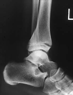
A 9-year-old boy has pain over the midfoot medially with activity. Based on the findings shown in Figures 26a and 26b, which of the following is considered the most effective short-term management?


Explanation

26b While Kohler's disease has a benign course, temporary cast immobilization has been shown to result in a shortened duration of symptoms. Core decompression or other surgery is not warranted because of the benign nature. Shoe inserts may be beneficial, but there are no studies to support their use. Borges JL, Guile JT, Bowen JR: Kohler's bone disease of the tarsal navicular. J Pediatr Orthop 1995;15:596-598.

Question 7

During the first 2 years of life, which of the following actions is most responsible for increasing structural stability of the physis?


Explanation

The zone of Ranvier provides the earliest increase in strength of the physis. During the first year of life, the zone spreads over the adjacent metaphysis to form a fibrous circumferential ring bridging from the epiphysis to the diaphysis. This ring increases the mechanical strength of the physis. The zone also helps the physis grow latitudinally. In turn, the increased width of the physis helps the physis further resist mechanical forces. The change in shape of the physis to its progressively more undulating form is also a factor in increasing physeal strength, but this occurs over a longer period of time, as the child's activity level increases. The undulations of the physis seen in some growth plates also add to stability but to a lesser extent. The other changes contribute little toward increasing physeal strength. Burkus J, Ogden J: Development of the distal femoral epiphysis: A microscopic morphological investigation of the zone of Ranvier. J Pediatr Orthop 1984;4:661-668.

Question 8

Because the patient shown in Figure 27 can no longer fit in shoes, treatment of the deformity should consist of


Explanation

In local gigantism, a ray resection allows proper fitting of shoes. The ray resection narrows the foot and shortens the length. The foot may require further surgery with growth. Debulking, physeal arrest, and distal phalanx amputation are unlikely to be effective. Turra S, Santini S, Cagnoni G, Jacopetti T: Gigantism of the foot: Our experience in seven cases. J Pediatr Orthop 1998;18:337-345.

Question 9

Examination of a 9-year-old girl who injured her left elbow in a fall reveals tenderness and swelling localized to the medial aspect of the elbow. Motor and sensory examinations of the hand are normal, and circulation is intact. A radiograph is seen in Figure 28. Management should consist of


Explanation

Avulsion fractures of the medial epicondyle are caused by a valgus stress applied to the immature elbow and usually occur in children between the ages of 9 and 14 years. Long-term studies have shown that isolated fractures of the medial epicondyle with between 5 to 15 mm of displacement heal well. Brief immobilization (1 to 2 weeks) in a long arm cast or splint yields results similar to open reduction and internal fixation. Fibrous union of the fragment is not associated with significant symptoms or diminished function. Surgical excision of the fragment yielded the worst results in one study and should be avoided. Open reduction is best reserved for those injuries in which the medial epicondylar fragment becomes entrapped in the elbow joint during reduction and cannot be extracted by closed manipulation. Farsetti P, Potenza V, Caterini R, Ippolito E: Long-term results of treatment of fractures of the medial humeral epicondyle in children. J Bone Joint Surg Am 2001;83:1299-1305.

Question 10

A 2-year-old child has refused to bear weight on his leg for the past 2 days. His parents report that he will crawl, has no fever, and has painless full range of motion of his hip and knee. Examination reveals no deformity or bruising, but there is mild swelling and tenderness over the anterior tibia. C-reactive protein, WBC count, and erythrocyte sedimentation rate studies are normal. Radiographs are negative. What is the best course of action?


Explanation

Despite the negative radiographic findings, the child's age and presentation are most consistent with a toddler's fracture. There is often not a witnessed injury. The differential diagnosis of infection is unlikely given that the child is afebrile and shows no signs of illness. Immobilization will make the child more comfortable and will often allow weight bearing. Repeat radiographs at the end of treatment will show a healing fracture and confirm the diagnosis. Aspiration of the tibial metaphysis would be indicated to obtain material for culture. The bone scan and MRI would show abnormalities, but these studies are nonspecific, costly, and time-consuming. Occasionally, oblique radiographs will show the fracture. Halsey MF, Finzel KC, Carrion WV, Haralabatos SS, et al: Toddler's fracture: Presumptive diagnosis and treatment. J Pediatr Orthop 2001;21:152-156.

Question 11

A 7-year-old girl sustains the fracture shown in Figure 29a. Casting results in uneventful healing. Ten months later, the patient has a progressive valgus deformity of the right lower extremity. A radiograph is shown in Figure 29b. Management should now consist of


Explanation

29b Although fractures of the proximal tibial metaphysis in young children appear innocuous, development of a progressive valgus deformity is possible despite adequate and appropriate treatment. When treating a child with this injury, it is prudent to warn the parents that a valgus deformity of the tibia may develop. The most likely cause is asymmetric growth of the proximal tibial physis. Because spontaneous angular improvement can be expected in most patients, surgery to correct these deformities should be delayed at least 2 to 3 years and should be limited to patients who have symptoms. There are no studies that document the efficacy of bracing for this deformity. Tuten HR, Keeler KA, Gabos PG, et al: Posttraumatic tibia valga in children: A long-term follow-up note. J Bone Joint Surg Am 1999;81:799-810.

Question 12

An obese 10-year-old boy has had left groin pain and a limp for the past 2 months. Examination reveals decreased abduction and internal rotation. Laboratory studies show normal renal function and an elevated thyroid-stimulating hormone (TSH) level. AP and frog lateral radiographs of the pelvis are shown in Figures 30a and 30b. What is the best course of action?


Explanation

30b The radiographs show a grade I slipped capital femoral epiphysis (SCFE) that is classified as stable because the child is able to bear weight. The elevated TSH level indicates possible hypothyroidism. SCFE usually occurs in boys age 12 to 14 years. Because of the patient's young age and hypothyroidism, he is at increased risk for slippage of the contralateral hip; therefore, prophylactic pinning of the uninvolved side also should be considered. Because of the risk of slip progression, crutch treatment and nonsteroidal anti-inflammatory drugs are not indicated. Realignment osteotomy is not indicated for grade I SCFE. Traction to reduce the slip, followed by pinning, has been advocated for unstable slips but is not indicated here. Loder RT, Wittenberg B, DeSilva G: Slipped capital femoral epiphysis associated with endocrine disorders. J Pediatr Orthop 1995;15:349-356.

Question 13

A 7-year-old boy has had chronic left leg pain that is worse at night but is not activity related. Use of nonsteroidal anti-inflammatory drugs for the past 6 months has failed to provide relief. A CBC count with differential, erythrocyte sedimentation rate, and C-reactive protein are within normal limits. Radiographs and a CT scan are shown in Figures 31a through 31c. Management should consist of


Explanation

31b 31c Osteoid osteomas are painful bone lesions, with radiographs revealing a dense sclerotic cortex surrounding a small radiolucency or nidus. Symptoms often are worse at night but usually are not activity related. While treatment in the past has consisted of open en bloc excision, current means of removal include percutaneous drilling under CT guidance and percutaneous radiofrequency coagulation. Success rates of percutaneous treatment are comparable to those seen following open procedures. The characteristic radiographic appearance of this lesion usually obviates the need for biopsy. Because the lesion is not caused by pyogenic organisms, antibiotics are not indicated. Donahue F, Ahmad A, Mnaymneh W, Pevsner NH: Osteoid osteoma: Computed tomography guided percutaneous excision. Clin Orthop 1999;366:191-196.

Question 14

In patients with neurofibromatosis, what is the most important sign of impending rapid progression of a spinal deformity?


Explanation

Neurofibromatosis can progress very rapidly. Rib penciling is the only singular prognostic factor. Significant progression has been observed in 87% of the curves with three or more penciled ribs. The other factors are often present but do not have a high correlation with rapid, severe progression. Crawford AH, Schorry EK: Neurofibromatosis in children: The role of the orthopaedist. J Am Acad Orthop Surg 1999;7:217-230.

Question 15

The fracture shown in Figure 32 is strongly indicative of what diagnosis?


Explanation

Fractures that occur through the primary spongiosa at the subphyseal region of the metaphysis are highly specific for child abuse. On radiographic studies, the metaphyseal lucency in these injuries may appear as either the so-called "bucket-handle" or "metaphyseal corner" fracture. These fractures are not typical features of osteogenesis imperfecta or vitamin D-resistant rickets. The ingestion of lead may lead to thick, transverse bands of increased density at the distal metaphysis. Fractures in the subphyseal region of the metaphysis are not typically seen in children who have osteomyelitis. Kocher MS, Kasser JR: Orthopaedic aspects of child abuse. J Am Acad Orthop Surg 2000;8:10-20.

Question 16

Figures 33a and 33b show the radiographs of a 10-year-old girl who reports a 4-month history of medial foot pain after she was kicked while playing soccer. The pain is worse with activity and partially relieved by rest. Examination reveals tenderness directly over a prominent navicular tuberosity. Management should consist of


Explanation

33b An accessory tarsal navicular is located at the medial tuberosity of the navicular bone. Nearly all children and adolescents who have a symptomatic accessory tarsal navicular bone become asymptomatic when they reach skeletal maturity. Initial management should include activity restrictions, shoe modification to avoid pressure over the prominent navicular, and non-narcotic analgesics. Although anecdotal, the use of arch supports may be helpful. When pain is refractory to these methods, a short period of cast immobilization may be useful. Surgery should be reserved for patients who have disabling symptoms despite a prolonged period of nonsurgical management. When surgery is indicated, simple excision of the accessory navicular is recommended. Sella EJ, Lawson JP, Ogden JA: The accessory navicular synchondrosis. Clin Orthop 1986;209:280-285.

Question 17

An 18-month-old child with obstetrical palsy has a maximum external rotation as shown in Figure 34. The parents should be advised that without surgical treatment the likelihood that glenoid dysplasia will develop is approximately what percent?


Explanation

Based on the available literature, the probability of development of glenoid dysplasia in the setting of a significant limitation of external rotation is close to 70%. Humeral dysplasia is also likely and can be managed surgically. Efforts are being made to identify procedures that will prevent glenoid dysplasia and help maintain function. Pearl ML, Edgerton BW: Glenoid deformity secondary to brachial plexus birth palsy. J Bone Joint Surg Am 1998;80:659-667. Waters PM, Smith GR, Jaramillo D: Glenohumeral deformity secondary to brachial plexus birth palsy. J Bone Joint Surg Am 1998;80:668-677.

Question 18

A 10-year-old boy has a painful, swollen knee after falling off his bicycle. Examination reveals no other injuries. Radiographs are shown in Figures 35a and 35b. Initial management of this fracture should consist of


Explanation

35b The radiographs show a minimally displaced fracture of the tibial eminence, which is classified as a McKeever type II injury. In a number of studies, it has been found that most of these fractures will reduce with extension of the knee. This is often made easier with evacuation of the hemarthrosis. The position of knee immobilization is controversial, with some authors preferring full extension and others preferring 20 degrees of flexion. Flexion to 90 degrees will further displace the fragment. If the fragment does not reduce or if the patient has a McKeever type III or IV injury, reduction and internal fixation are required. This can be done with either an open or an arthroscopic procedure. Excision of the fragment is not indicated. Meyers MH, McKeever FM: Fractures of the intercondylar eminence of the tibia. J Bone Joint Surg Am 1970;52:1677-1684. Wiley JJ, Baxter MP: Tibial spine fractures in children. Clin Orthop 1990;255:54-60 Janarv PM, Westblad P, Johansson C, Hirsch G: Long-term follow-up of anterior tibial spine fractures in children. J Pediatr Orthop 1995;15:63-68.

Question 19

Figures 36a and 36b show the radiographs of a 3-year old child who has a congenital upper extremity deformity. Which of the following features would be a major contraindication to a centralization procedure?


Explanation

36b The patient has bilateral absent radii or radial clubhand. Patients who lack elbow flexion take advantage of the hand position to reach their mouths, and a centralization procedure would take away that ability. This procedure can be performed on patients with partial to complete absence of the radius. A hypoplastic thumb can be addressed at a staged procedure; it does not represent a contraindication to centralization. Complete thumb absence can be addressed by pollicizing the index ray. Green DP: Operative Hand Surgery, ed 2. New York, NY, Churchill Livingstone, 1988, pp 269-271.

Question 20

Examination of a 4-year old child with obstetrical palsy reveals weak deltoids, pectoralis major strength of 4-5, and normal hand function. External rotation of the shoulder is limited. What is the most appropriate surgical procedure to restore external rotation?


Explanation

Transfer of the latissimus dorsi and teres major to the posterior rotator cuff will restore external rotation and some abduction. The procedure should be performed in children who are approximately age 4 years, following spontaneous recovery and prior to significant stiffness. External rotation osteotomy is more appropriate for an older child. Fusion should not be performed until skeletal maturity. Distal biceps rerouting restores pronation for a supination deformity. Latissimus dorsi and teres major transfer to the subscapularis would accentuate the internal rotation. In younger patients without significant bony deformity, a subscapularis slide or lengthening can restore external rotation. Strecker WB, McAllister JW, Manske PR, Schoenecker PL, Dailey LA: Sever-L'Episcopo transfers in obstetrical palsy: A retrospective review of twenty cases. J Pediatr Orthop 1990;10:442-444.

Question 21

A 7-month-old girl has had a severe flatfoot deformity since birth. The talar head is prominent in the medial plantar arch of the foot. No other deformities of the spine or extremities are present. Motor and sensory examinations of the extremities are normal. Figures 37a through 37c show simulated weight-bearing AP and lateral radiographs and a planter flexion lateral view. What is the most likely diagnosis?


Explanation

37b 37c Congenital vertical talus is a fixed dorsal dislocation of the talonavicular joint with equinus of the ankle joint. The AP radiograph shows valgus of the midfoot and an increased talocalcaneal angle; the lateral radiograph shows a vertically positioned talus and equinus of the ankle joint, and the plantar flexion lateral view shows that the talonavicular joint does not reduce. A line drawn through the long axis of the talus passes below the long axis of the first metatarsal. Initial management should consist of serial casting to stretch the dorsal soft-tissue structures; surgery eventually will be required to reduce the talonavicular joint. The differential diagnosis of congenital vertical talus includes pes calcaneovalgus, flexible pes planus, and peroneal spastic flatfoot. Pes calcaneovalgus, flexible pes planus, congenital short Achilles tendon, and peroneal spastic flatfoot would not show resistent dorsal dislocation of the navicular on the plantar flexion view. Kodros SA, Dias LS: Single-stage surgical correction of congenital vertical talus. J Pediatr Orthop 1999;19:42-48.

Question 22

A 12-year-old boy has severe left shoulder pain after being struck by an automobile. A chest radiograph, AP and lateral radiographs, and a CT scan with three-dimensional reconstruction of the scapula are shown in Figures 38a through 38d. Management should consist of


Explanation

38b 38c 38d Scapular body fractures in children are rare and are often associated with other injuries of the chest and thorax. Management is generally nonsurgical, unless the injury is open, and usually consists of support with a sling and gentle range-of-motion exercises to minimize shoulder stiffness. Green N, Swiontkowski M: Skeletal Trauma in Children, ed 2. Philadelphia, PA, WB Saunders, 1998, vol 3, pp 319-341.

Question 23

Figure 39 shows the radiograph of a 4-month old infant who has been undergoing weekly casting since birth for a congenital equinovarus deformity. Management should now consist of


Explanation

The radiograph shows the development of a rocker-bottom foot deformity. A rocker-bottom foot occurs in the treatment of clubfoot when casting is continued in the presence of a very tight gastrocnemius-soleus complex and an uncorrected hindfoot. While there are some preliminary reports on using Botox injection and continued casting for the equinus deformity, most authors recommend posterior or posterior medial release. Percutaneous tenotomy has been recently recommended with the resurgence of the Ponsetti technique. Lehman WB, Atar D: Complications in the management of talipes equinovarus, in Drennan JC (ed): The Child's Foot and Ankle. New York, NY, Raven Press, 1992, pp 135-136. Herring JA: Tachdjian's Pediatric Orthopedics, ed 4. Philadelphia, PA, WB Saunders, 2002, pp 927-935.

Question 24

A 1-year-old infant has the hand deformities shown in Figure 40. What pathologic process is the most likely cause of these deformities?


Explanation

Streeter's dysplasia is clearly related to rupture of the amnion in utero and is now most commonly referred to as premature amnion rupture sequence. The deformities arise from amniotic bands that wrap about protruding parts and from uterine packing because of the accompanying oligohydramnios. Clubfoot can develop as a result of the latter mechanism. Three limb involvement is most commonly seen, along with syndactyly. Treatment involves resection of bands and Z-plasty of skin. The disease is not genetic and has not been related to teratogen exposure or to iatrogenic influences such as amniocentesis. Developmental field disruption is not seen in this disease, and the growth potential of the involved parts is normal unless neurovascular disruption has arisen from band formation.

Question 25

A 13-year-old girl with hallux valgus reports pain after playing basketball. Radiographs show a hallux valgus angle of 20 degrees, an intermetatarsal angle of 11 degrees, a distal metatarsal articular angle of 10 degrees, and a congruent joint. Management should consist of


Explanation

Shoe wear modification is the most appropriate management based on the patient's age, high activity level, and relatively minor symptoms. She also has a mild hallux valgus. Normal radiographic measurements are an intermetatarsal angle of less than 9 degrees, a hallux valgus angle of less than 15 degrees, and a distal metatarsal articular angle of less than 9 degrees. Surgical procedures should be reserved for patients with more severe or progressive deformities. Stephens HM: Bunions, in Fitzgerald RH, Kaufer H, Malkani AL (eds): Orthopaedics. St Louis, MO, Mosby, 2002, pp 1510-1519.

Question 26

A 12-year-old boy with chronic kidney disease presents with a stable slipped capital femoral epiphysis (SCFE) on the left. Which of the following is the strongest indication for prophylactic pinning of the contralateral right hip?


Explanation

Patients with endocrine disorders or renal osteodystrophy are at a highly elevated risk of bilateral SCFE. Prophylactic pinning of the contralateral hip is strongly recommended in these patients due to the high incidence of subsequent slip.

Question 27

A 6-year-old boy sustains a completely displaced supracondylar humerus fracture. After closed reduction and percutaneous pinning, the hand remains pink but the radial pulse is absent on Doppler. What is the most appropriate next step in management?


Explanation

A "pink, pulseless" hand after reduction of a supracondylar humerus fracture indicates adequate collateral perfusion. The standard of care is close observation and admission, as the pulse often returns within 24 to 48 hours without surgical exploration.

Question 28

A 4-year-old girl presents with acute onset of right hip pain and a limp. Which of the following parameters is NOT included in the original Kocher criteria for differentiating septic arthritis from transient synovitis?


Explanation

The classic Kocher criteria include history of non-weight-bearing, ESR > 40, temperature > 38.5 C, and WBC > 12,000. Although CRP is highly predictive and used in modified criteria, it was not part of the original four-part Kocher criteria.

Question 29

A 4-year-old boy who was successfully treated for idiopathic clubfoot as an infant using the Ponseti method presents with a dynamic supination deformity of the foot during the swing phase of gait. His ankles have 15 degrees of passive dorsiflexion. What is the treatment of choice?


Explanation

Dynamic supination during gait in a previously corrected clubfoot is a classic sign of relapse caused by an overpowering tibialis anterior. Provided there is adequate ankle dorsiflexion, a full tibialis anterior tendon transfer to the lateral cuneiform is the treatment of choice.

Question 30

A 13-year-old girl sustains a Salter-Harris III fracture of the anterolateral distal tibia. Which of the following ligaments is responsible for the avulsion of this specific fracture fragment?


Explanation

A juvenile Tillaux fracture is a Salter-Harris III fracture of the anterolateral distal tibia. It occurs due to avulsion by the anterior inferior tibiofibular ligament (AITFL) during an external rotation injury as the medial physis closes before the lateral physis.

Question 31

An infant with achondroplasia presents with hypotonia, apnea, and hyperreflexia. MRI of the cervicomedullary junction confirms severe stenosis at the foramen magnum. Which underlying genetic mutation is responsible for this patient's syndrome?


Explanation

Achondroplasia is caused by a gain-of-function mutation in the FGFR3 gene. Severe foramen magnum stenosis can lead to cervicomedullary compression, causing central apnea and sudden death, which necessitates urgent surgical decompression.

Question 32

A 6-year-old child with spastic quadriplegic cerebral palsy is found on routine surveillance screening to have a hip migration percentage of 45%. The hip is painful and abduction is limited to 15 degrees. What is the most appropriate management?


Explanation

In a child with cerebral palsy, a hip migration percentage greater than 30-40% with clinical symptoms (pain, limited abduction) indicates progressive subluxation. Bony reconstruction with a VDRO, and often a concomitant pelvic osteotomy, is required to stabilize the hip.

Question 33

A 4-year-old girl is diagnosed with infantile Blount disease. Radiographs show a Langenskiöld stage IV lesion with a distinct osseous bar forming across the medial physis. What is the most appropriate treatment?


Explanation

Langenskiöld stage IV infantile Blount disease is characterized by an osseous bridge across the medial proximal tibial physis. Treatment requires excision of the physeal bar to allow growth, combined with a proximal tibial valgus osteotomy to correct the mechanical axis.

Question 34

An 8-year-old boy weighing 30 kg sustains a midshaft, transverse femur fracture. He is treated with titanium elastic nails (TENs). To optimize biomechanical stability, how should the diameter of the nails be selected?


Explanation

For maximum stability using titanium elastic nails in pediatric femur fractures, two nails of identical diameter should be used. Each nail should represent approximately 40% (totaling 80%) of the narrowest diameter of the medullary canal.

Question 35

A 14-year-old boy presents with a rigid flatfoot and a history of recurrent ankle sprains. Radiographs show a "C sign" on the lateral view. Which of the following is the best imaging modality to confirm the suspected diagnosis and assist in preoperative planning?


Explanation

The "C sign" on a lateral radiograph is indicative of a talocalcaneal coalition. A CT scan is the gold standard for defining the location, extent, and joint involvement of tarsal coalitions, which is critical for preoperative planning.

Question 36

A 2-year-old boy presents with anterolateral bowing of the tibia and an impending fracture. Which of the following systemic conditions is most commonly associated with this presentation?


Explanation

Anterolateral bowing of the tibia is highly associated with congenital pseudarthrosis of the tibia (CPT). Approximately 50% of patients with CPT have a concurrent diagnosis of Neurofibromatosis type 1 (NF1).

Question 37

A 7-year-old boy presents with a painful, snapping right knee. MRI confirms a Wrisberg-variant discoid lateral meniscus. What is the primary anatomical deficiency in this specific variant?


Explanation

The Wrisberg-variant discoid meniscus lacks the normal posterior meniscotibial (coronary) ligament attachments, making it hypermobile. Its only posterior attachment is the meniscofemoral ligament of Wrisberg, leading to subluxation and the classic "snapping" knee.

Question 38

A 14-year-old elite baseball pitcher presents with vague anterior shoulder pain that worsens with throwing. Radiographs demonstrate widening and irregularity of the proximal humeral physis. What is the first-line treatment?


Explanation

Little Leaguer's shoulder is an overuse injury resulting in epiphysiolysis of the proximal humerus. Treatment consists of complete rest from throwing (typically 3 months) to allow physeal healing, followed by a gradual return-to-throwing program.

Question 39

A 4-year-old girl is brought to the emergency department after a minor fall. Her lateral cervical spine radiograph shows 3 mm of anterior displacement of C2 on C3. Swischuk's line is drawn from the anterior aspect of the C1 spinous process to the anterior aspect of the C3 spinous process. The anterior aspect of the C2 spinous process touches this line. What is the most appropriate management?


Explanation

Pseudosubluxation of C2 on C3 is a normal physiologic variant in young children due to ligamentous laxity. A normal Swischuk line (the C2 spinous process is within 1-2 mm of the line) confirms this is not a true traumatic subluxation.

Question 40

In a patient diagnosed with Legg-Calvé-Perthes disease, which of the following is considered the most significant prognostic factor for long-term hip joint congruency and function?


Explanation

The age at the onset of Legg-Calvé-Perthes disease is the most reliable prognostic indicator. Children who develop the disease before age 6 to 8 generally have a much better capacity for remodeling and superior long-term outcomes than older children.

Question 41

A 13-year-old male presents with acute thigh pain and inability to bear weight after a minor fall. He had mild groin pain for 3 months prior. Radiographs confirm a displaced slipped capital femoral epiphysis (SCFE). He undergoes urgent in-situ pinning. Which of the following factors is most predictive of developing avascular necrosis (AVN) in this patient?


Explanation

Instability, defined by Loder as the inability to bear weight even with crutches, is the most significant risk factor for AVN in SCFE, with rates up to 47% compared to near 0% in stable slips.

Question 42

A 5-month-old female has been treated in a Pavlik harness for developmental dysplasia of the hip (DDH) for 4 weeks. A follow-up ultrasound demonstrates the femoral head remains dislocated out of the acetabulum. What is the most appropriate next step in management?


Explanation

Prolonged use of a Pavlik harness in a persistently dislocated hip increases the risk of 'Pavlik harness disease' (posterior acetabular wear). If reduction is not achieved within 3-4 weeks, the harness should be abandoned for closed reduction and spica casting.

Question 43

An infant is born with an idiopathic congenital talipes equinovarus (clubfoot). The treating orthopedic surgeon initiates the Ponseti method of serial casting. According to this method, which component of the deformity must be corrected first?


Explanation

In the Ponseti method, the first step is correcting the forefoot cavus. This is achieved by elevating the first ray to align the forefoot with the hindfoot, before abducting the foot to correct adduction and varus.

Question 44

A 6-year-old boy falls from monkey bars and sustains a completely displaced, extension-type supracondylar fracture of the humerus. On examination, the hand is pink and warm, but the radial pulse is non-palpable. What is the most appropriate initial management?


Explanation

For a pulseless but well-perfused (pink) hand following a supracondylar humerus fracture, the standard of care is urgent closed reduction and percutaneous pinning. The pulse frequently returns after the fracture is anatomically reduced.

Question 45

A 3-year-old obese male presents with progressive bowing of the left leg. Standing radiographs reveal a sharp varus deformity at the proximal tibial metaphysis. The metaphyseal-diaphyseal angle (MDA) is measured at 18 degrees. What is the most appropriate management?


Explanation

An MDA greater than 16 degrees in a child under 4 years old indicates infantile Blount disease with a high risk of progression. The initial treatment for stage I/II infantile Blount disease in a 3-year-old is bracing with a KAFO.

Question 46

An 8-year-old boy presents with a 2-day history of right knee pain, limp, and a fever of 38.8 C. Labs show a WBC of 16,000/mm3, ESR of 65 mm/hr, and CRP of 5.2 mg/dL. Knee aspiration yields turbid fluid with 85,000 WBCs/mm3 (>90% polymorphonuclear cells). Which of the following is the most appropriate definitive management?


Explanation

The clinical presentation and aspirate findings (>50,000 WBCs with >75% PMNs) are highly diagnostic of septic arthritis. This is a surgical emergency requiring urgent joint irrigation and debridement to prevent irreversible cartilage destruction.

Question 47

A 7-year-old boy is diagnosed with Legg-Calve-Perthes disease. Radiographs show fragmentation of the femoral head. Which of the following radiographic findings is considered a 'head-at-risk' sign indicating a poorer prognosis?


Explanation

Catterall's 'head-at-risk' signs include lateral subluxation of the femoral head, Gage sign, calcification lateral to the epiphysis, metaphyseal cysts, and a horizontal physis. These signs portend a poorer prognosis and hinge abduction.

Question 48

A 12-year-old girl is diagnosed with a unilateral slipped capital femoral epiphysis (SCFE). Which of the following underlying conditions represents the strongest indication for prophylactic in-situ pinning of the contralateral, asymptomatic hip?


Explanation

Prophylactic pinning of the contralateral hip is strongly indicated in patients with underlying endocrinopathies or metabolic disorders, such as renal osteodystrophy or hypothyroidism, due to the high risk (up to 100%) of bilateral involvement.

Question 49

A 6-year-old boy weighing 22 kg sustains an isolated, closed, length-stable midshaft femur fracture after falling off a trampoline. What is the most appropriate definitive treatment?


Explanation

Flexible intramedullary nailing is the standard of care for length-stable midshaft femur fractures in children aged 5-11 years weighing less than 50 kg (100 lbs). Spica casting is typically reserved for children under 5 years of age.

Question 50

A 7-year-old non-ambulatory child with spastic quadriplegic cerebral palsy presents for routine surveillance. AP pelvis radiographs reveal a Reimers migration percentage of 55% on the right hip with associated acetabular dysplasia. The hip is reducible on exam. What is the most appropriate surgical management?


Explanation

In a child with CP and a hip migration percentage >50% with acetabular dysplasia, soft tissue release alone is insufficient. Bony reconstruction with a proximal femoral VDRO and a volume-reducing pelvic osteotomy is required.

Question 51

A 14-year-old male sustains an ankle injury while playing basketball. Radiographs reveal a Triplane fracture of the distal tibia. Which of the following accurately describes the Salter-Harris (SH) fracture patterns typically seen on standard anteroposterior (AP) and lateral ankle radiographs?


Explanation

A Triplane fracture is a multiplanar injury that typically appears as a Salter-Harris III fracture on the AP radiograph (vertical fracture through the epiphysis) and a Salter-Harris II fracture on the lateral radiograph (posterior metaphyseal fragment).

Question 52

A 12-year-old boy presents with a 2-day history of severe left hip pain and is unable to bear weight. Radiographs demonstrate a left slipped capital femoral epiphysis (SCFE). He undergoes urgent in situ pinning. Which of the following factors most strongly correlates with the development of avascular necrosis in this patient?


Explanation

Unstable SCFE, defined clinically by the inability to bear weight even with crutches, is the most significant risk factor for AVN, with rates up to 47%.

Question 53

A 4-month-old female is undergoing treatment with a Pavlik harness for developmental dysplasia of the left hip. During a follow-up visit, the mother reports the infant has stopped moving the left leg. Examination reveals decreased active extension of the left knee, but sensation and perfusion are intact. What is the most appropriate next step in management?


Explanation

The clinical picture describes femoral nerve palsy, a known complication of hyperflexion in a Pavlik harness. Management requires loosening the anterior straps or discontinuing the harness temporarily to allow nerve recovery.

Question 54

A 13-year-old obese boy presents with acute-on-chronic left hip pain and an inability to bear weight. Radiographs demonstrate a slipped capital femoral epiphysis (SCFE), and he is classified as having an unstable slip. What is the primary theoretical advantage of performing an urgent open reduction and capsulotomy (e.g., modified Dunn procedure) rather than in situ pinning?


Explanation

Unstable SCFE has a high risk of AVN. Open reduction with capsulotomy decompresses the joint hematoma and anatomically aligns the epiphysis without forceful manipulation, theoretically lowering AVN risk.

Question 55

A 6-year-old girl sustains a severely displaced extension-type supracondylar humerus fracture. After closed reduction and percutaneous pinning in the operating room, her hand is pink and well-perfused, but the radial pulse remains non-palpable. A biphasic Doppler signal is present at the wrist. What is the most appropriate management?


Explanation

A 'pink, pulseless' hand after reduction and pinning of a supracondylar fracture with good capillary refill and Doppler signals should be observed closely. Vascular exploration is indicated only if the hand becomes pale and poorly perfused (white and pulseless).

Question 56

A 14-year-old male presents after feeling a 'pop' in his anterior knee while jumping. Radiographs show a displaced Ogden type III tibial tubercle avulsion fracture. Which of the following complications requires the most vigilant monitoring in the acute postoperative period following internal fixation?


Explanation

Tibial tubercle avulsion fractures are highly associated with anterior compartment syndrome due to disruption of the anterior tibial recurrent artery. Vigilant postoperative monitoring is essential.

Question 57

A 4-year-old boy treated successfully for idiopathic clubfoot as an infant with the Ponseti method presents with a relapsed deformity. He walks with a dynamic supination of the foot during the swing phase of gait. His foot is completely passively correctable. What is the most appropriate surgical intervention?


Explanation

Dynamic supination in a relapsed clubfoot that is passively correctable is treated with transferring the entire tibialis anterior tendon to the lateral cuneiform. This rebalances the foot and prevents further relapse.

Question 58

A 4-year-old boy presents with a 2-day history of right hip pain. His temperature is 38.6 C (101.5 F), WBC count is 14,000/mm3, ESR is 45 mm/hr, and he refuses to bear weight. According to the Kocher criteria, what is the approximate probability that this child has septic arthritis of the hip?


Explanation

The Kocher criteria include history of fever (>38.5 C), non-weight bearing, ESR >40 mm/hr, and WBC >12,000/mm3. Having all four predictors yields a >99% probability of septic arthritis.

Question 59

A 2-year-old girl is noted to have anterolateral bowing of her left tibia. Radiographs reveal sclerosis and a narrowed medullary canal at the apex of the bow. Her history is significant for multiple cafe-au-lait spots. What is the most likely diagnosis?


Explanation

Anterolateral bowing of the tibia with a narrowed medullary canal is the classic presentation of congenital pseudarthrosis of the tibia (CPT). It is strongly associated with Neurofibromatosis type 1 (NF1).

Question 60

A 7-year-old boy presents with a painless limp. Radiographs demonstrate sclerosis and fragmentation of the left femoral head. According to the Herring lateral pillar classification for Legg-Calve-Perthes disease, a patient with >50% but <100% maintenance of lateral pillar height is classified as:


Explanation

In the Herring lateral pillar classification, Group B indicates >50% but <100% maintenance of the lateral pillar height. Group C is defined as <50% of the lateral pillar height maintained.

Question 61

A 3-year-old boy weighing 15 kg (33 lbs) sustains an isolated, closed, midshaft femur fracture after a fall from a playground structure. What is the treatment of choice?


Explanation

Early Spica casting is the standard of care for isolated, closed femur fractures in children aged 6 months to 5 years. Flexible nailing is generally reserved for children >5 years old or those weighing >20 kg.

Question 62

A 12-year-old boy complains of recurrent ankle sprains and lateral foot pain. On examination, he has restricted subtalar motion and peroneal spasticity. An oblique radiograph of the foot demonstrates the 'anteater nose' sign. What is the most likely diagnosis?


Explanation

The 'anteater nose' sign on an oblique radiograph of the foot is pathognomonic for a calcaneonavicular coalition. Talocalcaneal coalitions are best seen on Harris axial views and may show the 'C-sign' on a lateral radiograph.

Question 63

A 6-year-old girl with spastic quadriplegic cerebral palsy (GMFCS Level V) is evaluated for hip surveillance. An AP pelvis radiograph demonstrates a migration percentage (Reimer's index) of 45% bilaterally. What is the most appropriate management?


Explanation

In non-ambulatory children with CP and a hip migration percentage >40%, reconstructive surgery including VDRO and often a pelvic osteotomy is indicated to prevent painful dislocation. Soft tissue releases alone are insufficient at this stage.

Question 64

A 2-year-old boy is evaluated for bilateral genu varum. Clinical examination shows a lateral thrust during gait. Radiographs show prominent beaking of the medial metaphysis, and the metaphyseal-diaphyseal angle (Drennan angle) is 18 degrees. What is the most appropriate initial management?


Explanation

A metaphyseal-diaphyseal angle >16 degrees with metaphyseal beaking is highly predictive of infantile Blount disease. In children <3 years old, bracing with KAFOs is the appropriate initial management.

Question 65

A 13-year-old premenarchal female with Adolescent Idiopathic Scoliosis presents with a right thoracic curve of 32 degrees. Her Risser stage is 0, and her Sanders maturity scale is 2. What is the most appropriate treatment recommendation?


Explanation

Bracing is indicated for skeletally immature patients (Risser 0-2, premenarchal) with an AIS curve between 25 and 45 degrees. A TLSO brace worn 16-23 hours a day decreases the risk of curve progression to a surgical threshold.

Question 66

A 5-year-old child with severe osteogenesis imperfecta (Type III) has suffered multiple femur and tibia fractures resulting in progressive bowing deformities. Which surgical intervention is the gold standard for correcting the deformity and preventing further fractures in this patient?


Explanation

The Sofield-Millar procedure (multiple osteotomies) combined with extensible (telescoping) intramedullary rods, such as Fassier-Duval nails, is the surgical gold standard. This corrects long bone deformities and prevents fractures in growing children with severe OI.

Question 67

A 14-year-old gymnast presents with chronic lower back pain that radiates to her posterior thighs. Radiographs reveal a grade III L5-S1 isthmic spondylolisthesis. She has failed 6 months of conservative management. What is the most appropriate surgical treatment?


Explanation

High-grade (Grade III or IV) or symptomatic slipped dysplastic spondylolisthesis failing non-operative care requires surgical stabilization. L5-S1 posterolateral fusion (with or without instrumentation/reduction) is the standard treatment.

Question 68

An 11-year-old boy with obesity presents with 2 weeks of worsening left groin pain and an inability to bear weight that started acutely yesterday. Radiographs confirm an acute-on-chronic slipped capital femoral epiphysis. Which of the following is the most significant risk factor for developing avascular necrosis in this patient?


Explanation

According to the Loder classification, an unstable SCFE is defined by the inability to bear weight, even with crutches. Unstable slips carry a much higher risk of avascular necrosis (up to 47%) compared to stable slips.

Question 69

A 5-year-old girl sustains a severely displaced extension-type supracondylar humerus fracture. On presentation, her hand is warm and pink, but the radial pulse is not palpable. After closed reduction and percutaneous pinning, the hand remains pink and warm, but the pulse is still absent. What is the most appropriate next step in management?


Explanation

In a pulseless but pink and well-perfused hand following reduction and pinning of a supracondylar fracture, observation is recommended. The collateral circulation is adequate, and the radial pulse often returns within a few days due to the resolution of vasospasm.

Question 70

A 4-year-old boy who was successfully treated for idiopathic clubfoot with the Ponseti method presents with a relapse. Examination reveals dynamic supination during the swing phase of gait and fixed equinus. He has been compliant with bracing. What is the most appropriate surgical intervention?


Explanation

Relapse in a young child treated with the Ponseti method often presents with dynamic supination. The treatment of choice is a split anterior tibial tendon transfer (to the lateral cuneiform) combined with Achilles tendon lengthening for the equinus contracture.

Question 71

A 6-week-old female is being treated with a Pavlik harness for developmental dysplasia of the hip. During a follow-up visit, the parents note she has stopped kicking her left leg. On exam, there is an absent patellar reflex and lack of active knee extension on the left. What is the most appropriate management?


Explanation

The patient has developed a femoral nerve palsy, a known complication of hyperflexion in a Pavlik harness. The harness must be removed immediately to allow for neurologic recovery, which usually occurs within a few weeks, before resuming DDH treatment.

Question 72

A 14-year-old boy presents with rigid flatfeet and recurrent ankle sprains. Lateral radiographs demonstrate an elongated lateral process of the talus (the "anteater nose" sign). Which of the following is the best initial diagnostic modality to confirm the suspected diagnosis?


Explanation

The "anteater nose" sign is pathognomonic for a calcaneonavicular coalition. The best initial study to clearly visualize this specific coalition is a 45-degree internal rotation oblique radiograph of the foot.

Question 73

An 8-year-old boy is diagnosed with Legg-Calvé-Perthes disease. Radiographs reveal fragmentation with maintenance of more than 50% of the lateral pillar height. According to the Herring classification, which group does this represent, and what is the typical recommendation?


Explanation

This is a Herring Group B classification (lateral pillar height >50% but <100%). In children over the age of 8, surgical containment (e.g., femoral or pelvic osteotomy) for Group B hips yields better long-term outcomes than conservative management.

Question 74

An 11-year-old boy weighing 55 kg (121 lbs) sustains a midshaft transverse femur fracture. Which of the following stabilization methods is most appropriate and carries the lowest risk of complications for this specific patient?


Explanation

In children older than 11 or weighing more than 49 kg (100 lbs), flexible nails have an unacceptably high failure rate. Rigid locked nailing using a lateral entry (to avoid the piriformis fossa and risk of AVN) is the preferred treatment.

Question 75

A 6-year-old child with spastic quadriplegic cerebral palsy is unable to walk and sits in a wheelchair (GMFCS level V). Routine hip surveillance radiographs show a Reimers migration percentage of 45% bilaterally. What is the recommended management?


Explanation

A migration percentage greater than 40% in a child with spastic cerebral palsy typically necessitates bony reconstructive surgery (VDRO) to prevent painful dislocation. Soft tissue releases alone are insufficient at this degree of subluxation.

Question 76

A 13-year-old girl presents after an external rotation ankle injury. Radiographs and CT show a fracture line extending through the distal tibial epiphysis on the sagittal view and through the metaphysis on the coronal view. How is this fracture classified overall according to the Salter-Harris system?


Explanation

A triplane fracture is considered an overall Salter-Harris IV equivalent because the fracture lines cross the epiphysis, physis, and metaphysis. It typically occurs in early adolescence as the distal tibial physis closes asymmetrically.

Question 77

An 11-year-old boy with obesity presents with acute left groin pain and inability to bear weight after a minor fall. He has a 3-week history of mild limp. Radiographs confirm an unstable slipped capital femoral epiphysis (SCFE). In addition to urgent in situ pinning of the left hip, what is the most appropriate management regarding the contralateral hip?


Explanation

Prophylactic pinning of the contralateral hip is indicated in patients with a high risk of a second slip, such as those with endocrine disorders or significant remaining growth (e.g., modified Oxford bone age score <= 16). Routine prophylactic pinning without risk stratification is not universally recommended.

Question 78

A 6-year-old girl sustains a severely displaced extension-type supracondylar humerus fracture. After closed reduction and percutaneous pinning, the hand is pink but lacks a palpable radial pulse. Capillary refill is less than 2 seconds. What is the most appropriate next step in management?


Explanation

A "pink, pulseless" hand after reduction and pinning of a supracondylar humerus fracture with adequate perfusion (capillary refill < 2 seconds) should be observed closely. Vascular exploration is indicated if the hand becomes pale and dysvascular after an anatomic reduction.

Question 79

A 5-year-old boy with Gross Motor Function Classification System (GMFCS) level V cerebral palsy is undergoing routine surveillance. An anteroposterior pelvic radiograph shows a Reimers migration percentage of 45% bilaterally. He is pain-free. What is the most appropriate treatment?


Explanation

In a child with cerebral palsy, a migration percentage >40-50% indicates significant hip subluxation with a high risk of progression to dislocation. Reconstructive surgery, typically involving a varus derotational osteotomy (VDRO) and often a pelvic osteotomy, is the treatment of choice to prevent painful dislocation.

Question 80

A 3-year-old child with a history of idiopathic clubfoot treated successfully with the Ponseti method presents with a dynamic supination deformity during the swing phase of gait. Passive range of motion is full, and the foot is plantigrade. What is the most appropriate surgical intervention?


Explanation

Dynamic supination of the foot in a toddler with a treated clubfoot represents a relapse often driven by an overactive anterior tibial tendon. Transfer of the entire anterior tibial tendon to the lateral cuneiform balances the foot and corrects the dynamic deformity.

Question 81

An 18-month-old girl is brought in for a waddling gait. Radiographs reveal a completely dislocated left hip with a false acetabulum and an acetabular index of 42 degrees. What is the most appropriate management?


Explanation

Children presenting with DDH between 18 and 24 months of age typically require open reduction due to soft tissue contractures and acetabular dysplasia. Concomitant femoral shortening minimizes avascular necrosis risk, and a pelvic osteotomy corrects the severe acetabular dysplasia.

Question 82

A 13-year-old boy complains of recurrent right foot sprains and vague midfoot pain. Physical examination demonstrates rigid pes planovalgus and decreased subtalar motion. Oblique radiographs demonstrate an elongation of the anterior process of the calcaneus. What is the most appropriate initial management?


Explanation

The clinical and radiographic findings (anteater sign) indicate a calcaneonavicular coalition. Initial management of symptomatic tarsal coalition is conservative, utilizing immobilization in a cast or walking boot to reduce inflammation before considering surgical resection.

Question 83

A 12-year-old girl presents with adolescent idiopathic scoliosis. Her primary right thoracic curve measures 25 degrees. She has not reached menarche. Hand radiograph shows a Sanders bone age stage of 2 (early adolescent growth phase). What is her estimated risk of curve progression to >50 degrees, and what is the best treatment?


Explanation

A 25-degree curve in a premenarchal female at Sanders stage 2 has a high risk of progression (roughly 60-80%). Rigid thoracolumbosacral orthosis (TLSO) bracing is indicated for curves between 25-40 degrees in highly immature patients to prevent progression to surgical magnitude.

Question 84

A newborn male is noted to have a short webbed neck, low posterior hairline, and severely limited cervical range of motion. Radiographs demonstrate fusion of multiple cervical vertebrae. Which of the following conditions must be urgently evaluated in this patient?


Explanation

Klippel-Feil syndrome is characterized by congenital fusion of cervical vertebrae. The most critical and potentially life-threatening associated issue is cervical spine instability (often at adjacent unfused segments or the atlantoaxial junction), requiring careful evaluation to prevent spinal cord injury.

Question 85

A 4-year-old girl with obesity presents with progressive bowing of her left leg. Radiographs show varus deformity of the proximal tibia with a metaphyseal-diaphyseal angle of 18 degrees and profound depression of the medial tibial plateau (Langenskiöld stage IV). What is the recommended treatment?


Explanation

Infantile Blount's disease with severe medial plateau depression (Langenskiöld stage IV or higher) in a child older than 3-4 years requires surgical intervention. Proximal tibial osteotomy combined with elevation of the depressed medial tibial plateau is necessary to restore joint congruity and correct the mechanical axis.

Question 86

A 4-year-old boy sustains a completely displaced, midshaft spiral fracture of the right femur from a fall off a slide. He weighs 16 kg. What is the most appropriate definitive management?


Explanation

For children aged 6 months to 5 years with a femur fracture and appropriate weight (usually < 20 kg), early spica casting is the standard of care with excellent outcomes. Flexible nailing is typically reserved for older children aged 5 to 11 years.

Question 87

An 8-year-old boy presents with a 6-month history of right hip pain and a painless limp. AP and frog-leg lateral radiographs show sclerosis and fragmentation of the lateral pillar of the femoral head. Dynamic arthrogram demonstrates hinge abduction. What is the most appropriate management?


Explanation

In Legg-Calvé-Perthes disease, hinge abduction is a sign of severe impingement between the extruded, deformed femoral head and the lateral acetabular margin. Surgical containment via proximal femoral or pelvic osteotomy is required to redirect the head into the acetabulum and restore congruity.

Question 88

A 9-year-old girl presents with a painful, loud "clunk" in her left knee during extension. MRI demonstrates a thickened lateral meniscus covering the entire tibial plateau without tears, but lacking posterior meniscotibial attachments. What is the diagnosis and best treatment?


Explanation

The Wrisberg variant of a discoid meniscus lacks normal posterior meniscotibial attachments (coronary ligaments), leading to hypermobility and the classic snapping knee syndrome. Treatment requires saucerization (partial meniscectomy) along with surgical stabilization of the posterior horn to the capsule.

Question 89

A 14-year-old boy sustains an ankle injury while playing soccer. Radiographs and CT scan reveal a displaced Salter-Harris III fracture of the anterolateral aspect of the distal tibial epiphysis. Which ligament is primarily responsible for the avulsion of this fracture fragment?


Explanation

A juvenile Tillaux fracture is a Salter-Harris III fracture of the anterolateral distal tibial epiphysis. It occurs because the anterior inferior tibiofibular ligament (AITFL) avulses the fragment during an external rotation force, due to the asymmetric closure pattern of the distal tibial physis.

None

Clinic OS
Medically Verified Content by
Prof. Clinic OS
Consultant Orthopedic & Spine Surgeon
Chapter Index