العربية
Part of the Master Guide

AAOS Pediatric Orthopedic MCQs (Set 2): DDH, SCFE & Spinal Deformities | Board Review

Orthopedic Pediatrics 2026 MCQs: Board Review Questions & Answers (Part 1)

23 Apr 2026 62 min read 70 Views
Figure for Pediatrics 2004 MCQs - Part 1 - Question 1

Key Takeaway

This topic focuses on Orthopedic Pediatrics 2026 MCQs: Board Review Questions & Answers (Part 1), Top-rated Orthopedic Pediatrics 2026 MCQs bank. Practice with clinical case questions, orthopedic surgery board review, and evidence-based answers updated for 2026.

Orthopedic Pediatrics 2026 MCQs: Board Review Questions & Answers (Part 1)

Comprehensive 100-Question Exam


00:00

Start Quiz

Question 1

An 8-year-old boy sustains nondisplaced midshaft fractures of the tibia and fibula after being struck by a car while he was riding his bicycle. No other injuries are noted, but the patient reports pain with passive motion of his toes. His neurovascular examination is otherwise normal. What is the best course of action?


Explanation

Pain with passive motion of the toes is a recognized early sign of increased compartment pressures. At a minimum, a baseline evaluation of the leg compartment pressures should be obtained. While it is normal for the patient to have pain related to the associated muscle contusions, any significant concerns should be addressed immediately in light of the severe consequences likely when a compartment syndrome occurs. Mubarak SJ, Owen CA, Hargens AR, et al: Acute compartment syndromes: Diagnosis and treatment with the aid of the wick catheter. J Bone Joint Surg Am 1978;60:1091-1095.

Question 2

A 6-year-old girl has the bilateral foot deformity shown in Figure 1. There is no family history of disease. Examination reveals fixed hindfoot equinus, and muscle function testing shows strong posterior tibial function, fair plus anterior tibial function, poor peroneal function, and strong gastrocnemius function. A Coleman block test shows a correctable hindfoot. Nerve conduction velocity studies show diminished function in the peroneal and ulnar nerves on both sides. Pathologic changes found in a sural nerve biopsy include "onion bulb" formation, and DNA testing confirms the presence of a mutation in the MPZ gene, consistent with hereditary motor sensory neuropathy type III (HMSN-III). What is the best course of action?


Explanation

The patient has HMSN-III or Dejerine-Sottas syndrome. This form of HMSN progresses very rapidly and frequently results in severe foot deformity in early childhood. The changes are progressive and are the result of muscle imbalance during growth. Balancing of the foot musculature is essential, particularly during the phases of rapid growth of the foot. However, this cannot be accomplished using the anterior tibial muscle because it is already weak and the transfer will further weaken it. Bony procedures also may be required, and tendon transfers cannot be depended on to correct bony deformity. However, these procedures can be deferred until the foot is closer to adult size. Surgeries that lead to joint arthrodesis, such as triple arthrodesis and some midfoot osteotomies, are contraindicated because the feet may lose protective sensation as the disease progresses. Fusions in insensate feet are less successful than realignment procedures that maintain mobility. Wetmore RS, Drennan JC: Long-term results of triple arthrodesis in Charcot-Marie-Tooth disease. J Bone Joint Surg Am 1989;71:417-422. Roper BA, Tibrewal SB: Soft tissue surgery in Charcot-Marie-Tooth. J Bone Joint Surg Br 1989;71:17-20.

Question 3

An obese 4-year-old boy has infantile Blount's disease. Radiographs reveal a metaphyseal-diaphyseal angle of 18 degrees and a depression of the medial proximal tibial physis. Management should consist of


Explanation

The deformity is too severe for observation, and at age 4 years, the child is too old for orthotic treatment. To prevent recurrence, surgery should be performed before irreversible changes occur in the medial physis. A proximal tibial osteotomy should overcorrect the mechanical axis to 10 degrees of valgus. Bar resection has not been shown to be as effective in this severe deformity, especially without a concomitant osteotomy. Raney EM, Topoleski TA, Yaghoubian R, Guidera KJ, Marshall JG: Orthotic treatment of infantile tibia vara. J Pediatr Orthop 1998;18:670-674.

Question 4

A 10-year-old boy has activity-related knee pain that is poorly localized. He denies locking, swelling, or giving way. Examination shows mild tenderness at the medial femoral condyle and painless full range of motion without ligamentous instability. Radiographs are shown in Figures 2a through 2c. What is the best course of action?


Explanation

2b 2c The radiographs show an osteochondritis dissecans (OCD) lesion in the medial femoral condyle of a skeletally immature patient. The lesion is not displaced from its bed. Nonsurgical management of a stable OCD lesion in a patient with open physes consists of a period of activity limitation and occasional immobilization. Unstable lesions, loose bodies, and patients with closed physes require more aggressive treatment. Most of the surgical procedures can be done arthroscopically. Because the radiographic appearance is typical, biopsy is unnecessary. The radiographs do not show an osteocartilaginous loose body, and the patient reports no catching or locking; therefore, removal of the loose body is not indicated. Linden B: Osteochondritis dissecans of the femoral condyles: A long term follow-up study. J Bone Joint Surg Am 1977;59:769-776. Cahill BR: Osteochondritis dissecans of the knee: Treatment of juvenile and adult forms. J Am Acad Orthop Surg 1995;3:237-247.

Question 5

Figure 3a shows the preoperative radiograph of a 5-year-old girl who achieved complete correction with valgus osteotomies. Figure 3b shows a radiograph obtained 2 years later. What is the cause of the recurrent deformity on the right side?


Explanation

3b Although inadequate correction, obesity, patient age of older than 5 years and an increased metaphyseal-diaphyseal angle are all associated with a poorer outcome, the radiographs show a growth arrest of the medial tibial physis. If not recognized and treated with early surgery, progressive genu varum will occur with continued growth of the lateral physis. In addition to repeat osteotomy, options for treating the arrest include physeal bar resection or, as necessary, completion of the growth arrest by epiphyseodesis of the lateral physes, followed by a limb equalization procedure at a later date. Brooks WC, Gross RH: Genu varum in children: Diagnosis and treatment. J Am Acad Orthop Surg 1995;3:326-335. Herring JA: Tachdjian's Pediatric Orthopedics, ed 4. Philadelphia, PA, WB Saunders, 2002, pp 840-950.

Question 6

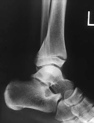
An 8-year-old boy reports ankle pain after striking the ground with the medial aspect of his foot while attempting to kick a soccer ball. Radiographs reveal slight distal tibial physeal widening but no other abnormalities. In treating this injury, which of the following associated conditions is most likely present but may be missed without careful evaluation?


Explanation

Malrotation of the foot is frequently overlooked in this clinical setting. This can be judged by evaluating and comparing the transmalleolar axes of the affected and unaffected legs. The rotation occurs through the physis and frequently is not recognized until the patient has been walking for a few months. The other conditions are not expected to occur in the clinical setting described. Phan VC, Wroten E, Yngve DA: Foot progression angle after distal tibial physeal fractures. J Pediatr Orthop 2002;22:31-35.

Question 7

An 11-year-old girl has wrist pain. Figure 4a shows the radiograph, and Figures 4b and 4c show the low- and medium-power photomicrographs of a lesion in the distal radius. What is the most likely diagnosis?


Explanation

4b 4c The radiograph shows an osteolytic eccentric lesion in the metaphyseal-diaphyseal region of the bone, and the photomicrographs show an aneurysmal bone cyst. The low-power photomicrograph shows large empty spaces with fibrous stroma and multinucleated giant cells. The red area in the center is hemorrhage in the stroma. The large empty spaces are cysts, which would be filled with blood in vivo. The medium-power photomicrograph shows a large cyst-like space and hemorrhage in the surrounding stoma. Giant cell tumors have "sheets" of giant cells. A nonossifying fibroma would have spindle cells, and a unicameral bone cyst may have a few giant cells, but blood is rare. Springfield DS, Gebhardt MC: Bone and soft tissue tumors, in Morrissy RT, Weinstein SL (eds): Lovell and Winter's Pediatric Orthopaedics, ed 5. Philadelphia, PA, Lippincott Williams and Wilkins, 2001, pp 540-542.

Question 8

In a patient with vertebral tuberculosis, which of the following characteristics is most predictive of progression of the kyphosis?


Explanation

In patients with vertebral tuberculosis, involvement of the anterior and posterior elements creates an instability and severe kyphotic collapse can occur. This characteristic has been shown to have a stronger association than level of involvement, age, or pretreatment degree of deformity. In the absence of instability, anterior growth can resume after treatment, leading to a decrease in the deformity. Rajasekaran S: The natural history of post-tubercular kyphosis in children: Radiological signs which predict late increase in deformity. J Bone Joint Surg Br 2001;83:954-962.

Question 9

When planning scoliosis surgery for a patient with a 50-degree thoracolumbar curve and spinal muscular atrophy, it is most important to include


Explanation

Typically, posterior spinal fusion to the pelvis is recommended for patients with spinal muscular atrophy and advanced scoliosis. Examination for lower extremity muscle contractures is important because the contractures may interfere with good sitting balance. Anterior release and fusion usually are not advised. Diaphragmatic pacing is not indicated because diaphragm function usually is not affected. Patients with spinal muscular atrophy usually are not ambulatory or only marginally ambulatory at the time of scoliosis surgery; therefore, gait analysis usually is not relevant. While a muscle biopsy may have a role in the diagnosis of this disorder, it plays no subsequent role in determining life expectancy or the value of spinal surgery. Daher YH, Lonstein JE, Winter RB, Bradford DS: Spinal surgery in spinal muscular atrophy. J Pediatr Orthop 1985;5:391-395.

Question 10

An 8-year-old boy sustains injuries to his head, abdomen, and left lower extremity after being struck by a truck. In the emergency department, his mental status deteriorates and he is intubated after assessment reveals a Glasgow Coma Scale score of 3; the score subsequently improves to 10. A CT scan reveals a right parietal intracranial hemorrhage, and an abdominal ultrasound reveals free fluid. Prior to an emergency laparotomy, the swollen left thigh is evaluated. Radiographs reveal a transverse fracture of the mid-diaphysis. Management of the fracture should consist of


Explanation

The prognosis for a young patient with a head injury is more favorable compared to that for adults. Full neurologic recovery generally occurs. Spasticity may occur within a few days after injury, which can lead to fracture displacement if immediate spica casting or traction is used. Early surgical stabilization will reduce problems with shortening and malunion and will facilitate transportation of the child for diagnostic tests. Surgery may be performed when it is best for the patient, either on the day of injury or later if time is needed for stabilization. In this patient, the fracture is ideally suited to stabilization using flexible intramedullary nails. Heinrich and associates' report of 78 diaphyseal femur fractures stabilized with flexible intramedullary nails included 14 patients with an associated closed head injury. All fractures healed, and there were no major complications. Tolo VT: Management of the multiply injured child, in Rockwood CA, Wilkins KE, Beaty JH (eds): Fractures in Children, ed 4. Philadelphia, PA, Lippincott-Raven, 1996, pp 83-95.

Question 11

A 3-year-old boy has a rigid 40-degree lumbar scoliosis that is the result of a fully segmented L5 hemivertebra. All other examination findings are normal. Management should consist of


Explanation

Near complete correction and rebalancing of the spine can be achieved by hemivertebral resection that may be done as either a simultaneous or a staged procedure in the young patient. This eliminates the problem of future progression and possible development of compensatory curves. Nonsurgical management is not indicated in congenital scoliosis. Convex hemiepiphyseodesis is best suited for patients younger than age 5 years who have a short curve caused by fully segmented hemivertebrae that correct to less than 40 degrees with the patient supine. Hemiepiphyseodesis and isolated posterior fusion are not indicated. Bradford DS, Boachie-Adjei O: One-stage anterior and posterior hemivertibral resection and arthrodesis for congenital scoliosis. J Bone Joint Surg Am 1990;72:536-540.

Question 12

A newborn with bilateral talipes equinovarus undergoes serial manipulation and casting. What is the primary goal of manipulation?


Explanation

Manipulative treatment and casting of talipes equinovarus has become popular because of disappointing surgical results and enthusiasm for the Ponseti method of manipulation. In this technique, the primary goal is to rotate the foot laterally around a talus that is held fixed by the manipulating surgeon's hands. While the navicular may be rotated anterolaterally with this technique, the primary focus is on the calcaneus. The calcaneus is rotated laterally and superiorly, not translated. Some dorsiflexion of the calcaneus can be obtained by manipulation, but the primary focus is on the rotational relationship of the talus and calcaneus, not the degree of calcaneal dorsiflexion. Ponseti IV: Common errors in the treatment of congenital clubfoot. Int Orthop 1997;21:137-141.

Question 13

Figure 5 shows the radiograph of a 10-year-old girl who reports chronic shoulder pain after her gymnastics classes. Examination reveals pain on internal and external rotation but no instability. What is the most likely diagnosis?


Explanation

The patient has a very wide humeral growth plate, indicating the presence of a proximal humeral stress fracture, an uncommon diagnosis in gymnasts. Gymnasts are prone to stress fractures of the scaphoid, distal radius, elbow, and clavicle. Proximal humeral stress fractures are more commonly seen in those participating in racket or throwing sports. Stress fractures can lead to growth arrest or inhibition, particularly in the distal radius. The radiograph shows normal findings for the acromion, acromioclavicular joint, scapula, and triceps origin. Fallon KE, Fricker PA: Stress fracture of the clavicle in a young female gymnast. Br J Sports Med 2001;35:448-449. Sinha AK, Kaeding CC, Wadley GM: Upper extremity stress fractures in athletes: Clinical features of 44 cases. Clin J Sports Med 1999;9:199-202. Caine D, Howe W, Ross W, Bergman G: Does repetitive physical loading inhibit radial growth in female gymnasts? Clin J Sports Med 1997;7:302-308.

Question 14

Figure 6 shows the clinical photographs of a newborn who underwent a colostomy for an imperforate anus. Examination shows extended knees, flexed hips, and equinovarus feet. Dimpling is noted over the buttocks. Patients with these findings differ from patients with myelodysplasia in that they


Explanation

The patient has sacral agenesis. Clinical signs include the classic dimpling over the buttocks and the characteristic lower extremity deformities. Imperforate anus is often associated with this disorder. Although motor function correlates with the level of vertebral defect, sensation is usually intact. This is important therapeutically, because patients are not as prone to pressure sores as are those with myelodysplasia. Kyphosis may develop in many patients with lumbosacral agenesis, but lordosis is unusual. Latex allergy and progressive neural deterioration may occur in patients with either myelodysplasia or sacral agenesis but is more common in the former.

Question 15

Which of the following patients is considered the most appropriate candidate for selective dorsal rhizotomy?


Explanation

While other surgical and nonsurgical options exist for management of spasticity, the criteria originally laid out by Peacock and associates describe the most appropriate candidate for rhizotomy as a patient with spastic diplegia who is between the ages of 4 to 8 years and has a stable gait pattern that is limited by lower extremity spasticity. Rhizotomy is not recommended in patients with athetosis because of unpredictable results. In addition, rhizotomy should be avoided in nonambulatory patients with spastic quadriplegia because it is associated with significant spinal deformities. Peacock WJ, Arens LJ, Berman B: Cerebral palsy spasticity: Selective posterior rhizotomy. Pediatr Neurosci 1987;13:61-66. Oppenheim WL: Selective posterior rhizotomy for spastic cerebral palsy: A review. Clin Orthop 1990;253:20-29.

Question 16

A 2-day-old infant has the hyperextended knee deformity shown in Figure 7. No other deformities are found on examination. A radiograph shows that the ossified portion of the proximal tibia is slightly anterior to that of the distal femur. Management should consist of


Explanation

Congenital dislocation of the knee is an uncommon deformity that varies in presentation from simple hyperextension to complete anterior dislocation of the tibia on the femur. Treatment varies with the age at presentation and the severity of the deformity. Most authors recommend early nonsurgical management. A recent study of 24 congenital knee dislocations in 17 patients found that satisfactory results were obtained in most instances using closed treatment. Based on their findings, the authors concluded that immediate reduction or serial casting should be performed when the patient is seen early after birth. If the patient is seen late and correction cannot be achieved by serial casting, traction followed by closed or open reduction may be necessary. Early percutaneous quadriceps recession has been described for complex congenital knee dislocations associated with underlying disorders, such as arthrogryposis and Ehlers-Danlos syndrome. Ko JY, Shih CH, Wenger DR: Congenital dislocation of the knee. J Pediatr Orthop 1999;19:252-259. Johnson E, Audell R, Oppenheim WL: Congenital dislocation of the knee. J Pediatr Orthop 1987;7:194-200.

Question 17

Figures 8a and 8b show the current radiographs of a 10-year-old boy with a hip disorder who was treated with an abduction orthosis 3 years ago. If no further remodeling occurs, what is the most likely prognosis?


Explanation

8b The radiographs show a child with Legg-Calve-Perthes disease (LCPD) that has healed. Deformity (asphericity) of the femoral head is evident, but the femoral head and acetabulum are congruous. Stulberg and associates found that hips with aspherical congruity at skeletal maturity functioned well until the fifth or sixth decade of life. Similarly, another study found that degenerative arthritis caused deteriorating hip function after age 40 years in patients with this degree of residual deformity. Repeated episodes of ischemic necrosis are unlikely. Although some studies suggested coagulation abnormalities such as protein C and S deficiencies in children with LCPD, other studies failed to show any evidence of inherited thrombophila in most children with this disorder. There are no studies to suggest growth acceleration occurs following LCPD. Stulberg SD, Cooperman DR, Wallenstein R: The natural history of Legg-Calve-Perthes disease. J Bone Joint Surg Am 1984;66:479-489.

Question 18

In girls with idiopathic scoliosis, peak height velocity (PHV) typically occurs at what point?


Explanation

PHV generally occurs while girls are still Risser 0; menarche typically occurs before Risser 1, which has a wide variation in its timing. The curve magnitude at the PHV is the best prognostic indicator available. Most untreated patients with curves greater than 30 degrees at PHV require surgery, while patients with smaller curves at that stage typically do not require surgery. Little DG, Song KM, Katz D, Herring JA: Relationship of peak height velocity to other maturity indicators in idiopathic scoliosis in girls. J Bone Joint Surg Am 2000;82:685-693.

Question 19

Examination of a 6-year-old boy who sustained a displaced Salter-Harris type II fracture of the distal radius reveals 35 degrees of volar angulation. A satisfactory reduction is obtained with the aid of a hematoma block. At the 10-day follow-up examination, radiographs show loss of reduction and 35 degrees of volar angulation. Management should now consist of


Explanation

In a 6-year-old child with a physeal fracture, the healing response 10 days after injury is so advanced that manipulation would have to be very forceful to be successful. A forceful manipulation in a patient this age increases the risk of early growth arrest and a significant disability because 80% of the growth of the radius comes from the distal physis. Because of the large contribution of growth from the distal radial physis and the angulation being in the plane of wrist motion, the potential for remodeling of this fracture is great. It is highly probable that this fracture will completely remodel in 1 to 2 years of growth. In this patient, even a "gentle" open reduction would probably require enough force that the physis would be damaged. Dimeglio A: Growth in pediatric orthopaedics, in Morrissy RT, Weinstein SL (eds): Lovell and Winter's Pediatric Orthopaedics, ed 5. Philadelphia, PA, Lippincott Williams and Wilkins, 2001, pp 33-62.

Question 20

Figures 9a and 9b show the radiographs of a 12-year-old girl who has had right hip pain for the past 4 months. She reports that the pain is so severe that she is unable to walk and is now using a wheelchair. Examination reveals pain with any attempted range of motion. Management should include


Explanation

9b In addition to mild hip dysplasia, the radiograph shows an osteoblastic lesion of the right ilium. The patient's symptoms are much more severe than is typical for late hip dysplasia. MRI can determine the extent of the lesion in the bone and soft tissues. Following work-up and biopsy, the patient was diagnosed with Ewing's sarcoma. Springfield DS, Gebhardt MC: Bone and soft tissue tumors, in Morrissy RT, Weinstein SL (eds): Lovell and Winter's Pediatric Orthopaedics, ed 5. Philadelphia, PA, Lippincott Williams and Wilkins, 2001, pp 507-518, 542-544.

Question 21

An 18-month-old boy has 45 degrees of kyphosis in the thoracolumbar spine secondary to type I congenital kyphosis. Examination reveals that he is neurologically intact, and an MRI scan shows no evidence of intraspinal pathology. Management should consist of


Explanation

Surgery is indicated for congenital kyphosis once the deformity reaches a certain size or if significant progression is documented. In a young patient with a relatively small deformity, the treatment of choice is isolated in situ posterior fusion and postoperative immobilization. If an adequate posterior fusion can be obtained, an epiphyseodesis effect can be generated, allowing the remaining anterior growth to cause some correction. Because there is no evidence of neurologic compression and the deformity is less than 50 degrees, anterior surgery is not indicated. There is no role for bracing in the management of congenital kyphosis. Winter RB: Congenital Deformities of the Spine. New York, NY, Thieme-Stratton, 1983, pp 229-261.

Question 22

Following an acute dislocation of the patella, the risk of a recurrent dislocation is greater if the patient has which of the following findings?


Explanation

Recurrent dislocations may follow an earlier dislocation. One study found that in patients who had a patellar dislocation between the ages of 11 to 14 years, 60% had a recurrent dislocation. The incidence of recurrent dislocation dropped to 33% in patients who had a patellar dislocation between the ages of 15 to 18 years. The authors also found that the incidence of recurrence was greater in patients who demonstrated a predisposition to dislocation as determined by evaluation of the unaffected knee. Predisposing signs included passive lateral hypermobility of the patella, a dysplastic distal third of the vastus medialis obliquis muscle, and a high and/or lateral position of the patella. A second study found that the risk of redislocation was considerably higher in patients who were in their teens at the first episode of dislocation compared to older patients. There are no studies linking either a patella baja or a bipartite patella to an increased risk of redislocation. Cash JD, Hughston JC: Treatment of acute patellar dislocation. Am J Sports Med 1988;16:244-249.

Question 23

Which of the following findings can cause a dorsal bunion in a patient with neuromuscular disease?


Explanation

Unopposed action of the tibialis anterior with weakness of the peroneus longus will lead to a dorsal bunion and supination deformity. Overpull of the gastrocnemius-soleus complex and posterior tibialis with weakness of the peroneus brevis will cause equinovarus deformity. A strong posterior tibialis with weakness of the peroneals will cause varus of the hindfoot. Unopposed peroneus brevis and incompetence of the posterior tibialis will lead to a flatfoot deformity. The etiology of a cavus foot is complex, but findings usually include a contracted plantar fascia and weakness of the tibialis anterior. Drennan JC (ed): The Child's Foot and Ankle. New York, NY, Raven Press, 1992, pp 289-302.

Question 24

Which of the following studies is considered most sensitive in monitoring a therapeutic response in acute hematogenous osteomyelitis?


Explanation

C-reactive protein declines rapidly as the clinical picture improves. Failure of the C-reactive protein to decline after 48 to 72 hours of treatment should indicate that treatment may need to be altered. Blood culture is positive only 50% of the time and will be negative soon after antibiotics are administered, even if treatment is not progressing satisfactorily. WBC count is highly variable and poorly correlated with treatment. The ESR rises rapidly but declines too slowly to guide treatment. Radiographic findings may not change but can take up to 2 weeks to show changes.

Question 25

Figure 10 shows the radiograph of a 7-year-old patient who has a bilateral Trendelenburg limp and limited range of hip motion but no pain. His work-up should include


Explanation

The radiograph shows bilateral flattening of the femoral heads with mottling and "fragmentation" suggestive of Legg-Calve-Perthes disease. However, when these changes occur bilaterally and are symmetric, multiple epiphyseal dysplasia or spondyloepiphyseal dysplasia should be suspected. Skeletal survey will show irregularity of the secondary ossification centers. With these conditions, there is no true osteonecrosis and no evidence that orthotic or surgical "containment" will alter the outcome of progressive degenerative arthritis. Cardiac anomalies and coagulopathies are not associated with the epiphyseal dysplasias. Crossan JF, Wynne-Davies R, Fulford GE: Bilateral failure of the capital femoral epiphysis: Bilateral Perthes disease, multiple epiphyseal dysplasia, pseudoachondroplasia, and spondyloepiphyseal dysplasia congenita and tarda. J Pediatr Orthop 1983;3:297-301.

Question 26

A 13-year-old obese male presents with acute-on-chronic left groin pain after a minor fall. He is completely unable to bear weight on the affected extremity. Radiographs reveal a slipped capital femoral epiphysis (SCFE). Compared to a patient who is able to bear weight, this patient is at the highest risk for developing which of the following complications?


Explanation

This patient has an unstable SCFE, clinically defined by the inability to bear weight even with crutches. Unstable SCFE has a substantially higher risk of avascular necrosis (up to 47%) compared to stable SCFE.

Question 27

A 4-month-old female infant is undergoing treatment with a Pavlik harness for developmental dysplasia of the hip (DDH). After 3 weeks of strict full-time wear, an ultrasound reveals that the hip remains dislocated. What is the most appropriate next step in management?


Explanation

Failure to achieve reduction in a Pavlik harness after 3 to 4 weeks necessitates abandoning the harness to prevent 'Pavlik disease' (posterior acetabular wear). The next appropriate step is typically a trial of a rigid abduction orthosis or proceeding to closed reduction with spica casting.

Question 28

A 5-year-old girl sustains a Gartland type III supracondylar humerus fracture. Following closed reduction and percutaneous pinning, the radial pulse is not palpable, but her hand is warm and pink with a capillary refill time of 2 seconds. What is the most appropriate next step?


Explanation

A 'pink, pulseless' hand after reduction and pinning of a supracondylar fracture indicates adequate collateral perfusion. Current AAOS guidelines recommend close observation for 24-48 hours rather than routine surgical exploration.

Question 29

An infant with idiopathic clubfoot is undergoing serial casting using the Ponseti method. After sequential correction of the cavus, adductus, and varus deformities, the foot remains in 15 degrees of equinus. What is the most appropriate next step?


Explanation

The Ponseti method addresses deformities in the CAVE sequence: Cavus, Adductus, Varus, and Equinus. Once the midfoot score is 1 or less and only equinus remains, a percutaneous Achilles tenotomy is indicated to safely correct the equinus.

Question 30

A 7-year-old child with spastic quadriplegic cerebral palsy (GMFCS Level V) has bilateral hip pain. Anteroposterior pelvic radiographs demonstrate a Reimers migration percentage of 65% bilaterally with early acetabular dysplasia. What is the most appropriate treatment?


Explanation

In a child with CP, a migration percentage greater than 50% indicates hip dislocation with significant dysplasia. Soft tissue releases alone are insufficient; osseous reconstruction with VDRO and pelvic osteotomy is required.

Question 31

A 2-year-old boy with achondroplasia presents with a history of recurrent apneic episodes, delayed motor milestones, and hyperreflexia in the bilateral lower extremities. Which of the following is the most likely etiology?


Explanation

Infants and young children with achondroplasia are at risk for foramen magnum stenosis, which can cause cervicomedullary compression. Symptoms include central apnea, hyperreflexia, and hypotonia, warranting emergent MRI and potential surgical decompression.

Question 32

A 13-year-old premenarchal girl presents with adolescent idiopathic scoliosis. Radiographs demonstrate a right thoracic curve of 32 degrees. Her Risser stage is 0. What is the most appropriate management?


Explanation

Bracing is indicated for skeletally immature patients (Risser 0-2, premenarchal) with an idiopathic scoliotic curve between 25 and 45 degrees. A TLSO worn for at least 18 hours per day has been shown to significantly decrease the risk of progression to surgery.

Question 33

A 4-year-old boy presents with profound lower extremity bowing. Laboratory testing reveals hypophosphatemia, elevated alkaline phosphatase, and normal serum calcium. Genetic testing confirms a mutation in the PHEX gene. The pathophysiology of his bone disease is primarily driven by an excess of which of the following?


Explanation

The patient has X-linked hypophosphatemic (XLH) rickets due to a PHEX mutation. This mutation leads to an overproduction of FGF23, which impairs renal phosphate reabsorption, causing hypophosphatemia and rickets.

Question 34

A newborn is diagnosed with severe unilateral fibular hemimelia. Which of the following knee anomalies is most commonly associated with this condition?


Explanation

Fibular hemimelia is a longitudinal deficiency characterized by partial or complete absence of the fibula. It is highly associated with anterior cruciate ligament (ACL) deficiency, absent lateral rays, and tarsal coalitions.

Question 35

An 8-year-old boy is diagnosed with Legg-Calve-Perthes disease. Radiographs show greater than 50% collapse of the lateral pillar. According to the Herring lateral pillar classification, what is his prognosis and recommended treatment?


Explanation

This patient has Lateral Pillar C disease (>50% collapse). Children older than 8 years at the time of onset with Pillar B/C or C involvement have a poorer prognosis and generally benefit from surgical containment (femoral or pelvic osteotomy).

Question 36

A 3-year-old boy presents with an inability to bear weight on his right leg, a temperature of 38.8 C, a WBC count of 14,000/mm3, and an ESR of 55 mm/hr. According to the Kocher criteria, what is the probability that this child has septic arthritis of the hip?


Explanation

The patient meets all four Kocher criteria: non-weight-bearing, temperature >38.5 C, WBC >12,000, and ESR >40. The presence of all four criteria carries a 99% probability of septic arthritis, necessitating urgent joint aspiration.

Question 37

A 14-year-old boy sustains an ankle injury while playing soccer. Radiographs show a Salter-Harris type III fracture of the anterolateral aspect of the distal tibial epiphysis. What is the mechanism of injury for this specific fracture pattern?


Explanation

This is a Tillaux fracture, caused by an avulsion of the anterolateral tibial epiphysis by the anterior inferior tibiofibular ligament (AITFL). The mechanism is external rotation in an adolescent whose distal tibial physis is closing centrally and medially but remains open laterally.

Question 38

A 12-year-old boy presents with recurrent ankle sprains and a painful, rigid flatfoot. Physical examination reveals restricted subtalar motion. To identify the most common etiology of this condition, which imaging view is most helpful?


Explanation

The presentation is classic for a tarsal coalition, with calcaneonavicular being the most common type. An oblique radiograph of the foot is the best initial view to visualize the 'anteater sign' indicative of a calcaneonavicular coalition.

Question 39

A 6-year-old boy presents with a painless "clunking" in his knee that occurs when transitioning from flexion to extension. MRI reveals a discoid meniscus. The Wrisberg variant of a discoid lateral meniscus is uniquely characterized by the absence of which of the following?


Explanation

The Wrisberg variant of a discoid meniscus lacks the normal posterior meniscotibial (coronary ligament) capsular attachment. This hypermobility causes the meniscus to snap into the intercondylar notch during extension, resulting in the classic 'snapping knee' syndrome.

Question 40

An 8-year-old boy who weighs 35 kg (77 lbs) sustains an isolated, closed, length-stable midshaft femur fracture. What is the most appropriate definitive treatment?


Explanation

For a child aged 5 to 11 years weighing less than 50 kg (110 lbs) with a length-stable femur fracture, flexible intramedullary nailing is the gold standard treatment. It provides excellent outcomes with a rapid return to function and low risk of AVN.

Question 41

A 5-year-old boy is brought in after sustaining a lateral condyle fracture of the humerus. It is displaced by 4 mm on initial radiographs but the parents refuse surgery. Six months later, the fracture goes on to nonunion. If left untreated, what is the most likely long-term neurologic complication?


Explanation

Nonunion of a lateral condyle fracture often leads to progressive cubitus valgus deformity. This chronic stretching of the ulnar nerve behind the medial epicondyle can cause tardy (delayed) ulnar nerve palsy years later.

Question 42

A 3-year-old girl is evaluated for multiple long bone fractures following minimal trauma. Clinical exam reveals blue sclerae and mild dentinogenesis imperfecta. Genetic testing is most likely to reveal a mutation affecting the synthesis of which of the following proteins?


Explanation

The clinical picture describes Osteogenesis Imperfecta (OI), which is predominantly caused by autosomal dominant mutations in the COL1A1 or COL1A2 genes. These mutations result in quantitative or qualitative defects in Type I collagen.

Question 43

A 14-year-old gymnast presents with severe back pain and radiculopathy. Radiographs show an L5-S1 isthmic spondylolisthesis with a 65% slip (Meyerding Grade 3). If a surgical reduction of the slip is attempted, which nerve root is at the highest risk for iatrogenic injury?


Explanation

In high-grade spondylolisthesis at L5-S1, attempting to surgically reduce the L5 vertebra back onto the S1 sacral dome places the L5 nerve root under significant tension, posing a high risk for stretch injury or palsy.

Question 44

An 18-month-old girl presents with a painless limp and a positive Trendelenburg sign. Pelvic radiographs show a unilaterally dislocated left hip with an acetabular index of 40 degrees. What is the most appropriate management?


Explanation

In a child older than 18 months presenting with DDH and significant dysplasia (acetabular index >30-35), closed reduction has a high failure and AVN rate. Open reduction, often combined with a pelvic osteotomy (e.g., Salter or Pemberton) and capsulorrhaphy, is typically required.

Question 45

A 2-year-old boy is brought to the clinic for anterolateral bowing of the right tibia. Radiographs demonstrate diaphyseal narrowing and sclerosis. The parents are counseled regarding the high risk of fracture and congenital pseudarthrosis. This condition is most strongly associated with which of the following underlying disorders?


Explanation

Anterolateral bowing of the tibia in a young child is a classic presentation of impending congenital pseudarthrosis of the tibia (CPT). Approximately 50% to 90% of children with CPT have an underlying diagnosis of Neurofibromatosis type 1 (NF-1).

Question 46

A 5-month-old girl has been treated with a Pavlik harness for an initially irreducible, dislocated right hip for 4 weeks. Repeat ultrasound shows the hip remains persistently dislocated. What is the next best step in management?


Explanation

Continued use of a Pavlik harness for a dislocated hip after 3-4 weeks without successful reduction leads to 'Pavlik harness disease', which causes posterior wear of the acetabulum. Transitioning to a rigid orthosis is indicated for residual dysplasia but not for a dislocated hip, which requires closed (or open) reduction and spica casting.

Question 47

A 6-year-old boy falls from monkey bars and sustains a significantly displaced extension-type supracondylar humerus fracture. On presentation, his hand is pink but the radial pulse is absent. The fracture is closed reduced and pinned, and the hand remains pink, but the radial pulse remains absent. Capillary refill is brisk. What is the most appropriate next step?


Explanation

A 'pink, pulseless hand' following closed reduction and pinning of a supracondylar humerus fracture with good capillary refill indicates adequate collateral perfusion. Observation is the standard of care, as most pulses return within a few days and ischemic complications in this setting are exceedingly rare.

Question 48

A 12-year-old obese boy presents with right thigh pain and inability to bear weight after a minor trip 2 days ago. Radiographs reveal a severe, unstable right slipped capital femoral epiphysis (SCFE). During management, what is the most significant clinical risk associated with this specific type of injury?


Explanation

Unstable SCFE is defined by the inability to bear weight even with crutches and carries a high risk of avascular necrosis (AVN), ranging from 10-50%. Prompt, gentle reduction and stabilization are crucial to minimize capsular tension and preserve the tenuous blood supply.

Question 49

A 7-year-old boy presents with a painless limp of 3 months duration. Radiographs show fragmentation of the capital femoral epiphysis consistent with Legg-Calvé-Perthes disease. According to the Herring lateral pillar classification, which radiographic feature is most predictive of the final functional outcome?


Explanation

The Herring lateral pillar classification divides the femoral head into three pillars. The height of the lateral pillar on an AP radiograph during the fragmentation stage is the most reliable prognostic indicator for long-term outcome in Legg-Calvé-Perthes disease.

Question 50

A 3-week-old boy is undergoing Ponseti casting for bilateral idiopathic clubfoot. After 5 weekly casts, the cavus, adductus, and varus deformities have been corrected, but 15 degrees of rigid hindfoot equinus remains. What is the next recommended step in management?


Explanation

In the Ponseti method, once the forefoot and midfoot deformities (cavus, adductus, varus) are fully corrected, persistent hindfoot equinus (seen in >80% of cases) is treated with a percutaneous Achilles tendon tenotomy. A final cast is then applied for 3 weeks before transitioning to a foot abduction orthosis.

Question 51

A 4-year-old boy with spastic diplegic cerebral palsy presents for routine hip surveillance. His Reimer's migration percentage is 25% bilaterally. Physical exam shows limited hip abduction to 30 degrees bilaterally. What is the recommended prophylactic surgical treatment to prevent progressive hip subluxation?


Explanation

In children with cerebral palsy, soft-tissue releases (adductor and psoas lengthening) are indicated when the hip migration percentage is between 25% and 30% with decreasing abduction. Once the migration index exceeds 40%, bony reconstruction (VDRO) is typically required.

Question 52

An 11-year-old girl presents with a painful, swollen mass on her distal thigh. A biopsy reveals uniform small round blue cells. Cytogenetic analysis of the tumor tissue is most likely to show which of the following chromosomal translocations?


Explanation

Ewing sarcoma is a classic small round blue cell tumor strongly associated with the chromosomal translocation t(11;22)(q24;q12). This specific translocation results in the EWS-FLI1 fusion protein, which is diagnostic in approximately 85% of cases.

Question 53

A 14-year-old obese boy presents with progressive bowing of his left leg. Radiographs reveal a severe varus deformity centered at the proximal tibia, with medial physeal widening and an open proximal tibial physis. What is the most appropriate initial management?


Explanation

In adolescent Blount disease with open physes and sufficient remaining growth potential, guided growth (hemiepiphysiodesis) using lateral tension band plating is the preferred initial minimally invasive treatment to gradually correct the varus deformity.

Question 54

A 3-year-old boy presents with a completely displaced, spiral fracture of the femoral shaft following a reported fall from a 2-foot bed. He has no other injuries, and his neurovascular status is intact. What is the most appropriate definitive management?


Explanation

For children aged 6 months to 5 years with an isolated diaphyseal femur fracture and <2 cm of shortening, immediate hip spica casting is the gold standard of treatment. However, the reported low-energy mechanism warrants a high index of suspicion for child abuse.

Question 55

A 12-year-old boy complains of recurrent ankle sprains and midfoot pain. Examination reveals a rigid flatfoot with absent subtalar motion. Oblique radiographs demonstrate a "calcaneonavicular" coalition. He has failed 6 months of conservative management with custom orthotics and a short leg cast. What is the most appropriate surgical treatment?


Explanation

Symptomatic calcaneonavicular coalitions that fail extensive conservative management are best treated with surgical resection. Interposition of autologous fat or the extensor digitorum brevis muscle belly is performed to prevent recurrence of the coalition.

Question 56

A 10-year-old girl falls while skiing and presents with a swollen, painful knee. Radiographs reveal a type III (completely displaced) avulsion fracture of the anterior tibial spine. What is the most appropriate management?


Explanation

Meyers and McKeever type III tibial spine fractures are completely displaced and often have interposed meniscal tissue. They require anatomical reduction and internal fixation (using screws or sutures), usually performed arthroscopically, to restore ACL competency and prevent an extension block.

Question 57

A 4-year-old boy with multiple recurrent fractures, blue sclerae, and dentinogenesis imperfecta is diagnosed with Osteogenesis Imperfecta (OI) type I. He is started on intravenous pamidronate. What is the primary mechanism of action of this medication in the treatment of OI?


Explanation

Bisphosphonates, such as pamidronate, are the mainstay of medical therapy for moderate to severe Osteogenesis Imperfecta. They function by inhibiting osteoclast activity and inducing osteoclast apoptosis, thereby decreasing bone turnover and increasing bone mineral density.

Question 58

A 13-year-old boy undergoes in-situ percutaneous pinning of a stable slipped capital femoral epiphysis (SCFE) on the left side. Which of the following is considered an absolute indication for prophylactic pinning of the asymptomatic contralateral right hip?


Explanation

Prophylactic pinning of the contralateral hip in SCFE is strongly recommended for patients with an underlying endocrine disorder (such as hypothyroidism or renal osteodystrophy) or a history of radiation therapy, as their risk of eventual bilateral involvement approaches 100%.

Question 59

A 13-year-old girl twists her ankle while playing soccer and sustains a Salter-Harris III fracture of the anterolateral distal tibia. Which ligamentous structure transmits the avulsion force responsible for this specific fracture pattern?


Explanation

A Tillaux fracture is an avulsion of the anterolateral epiphysis of the distal tibia (Salter-Harris III). It is caused by tension from the anterior inferior tibiofibular ligament (AITFL) during an external rotation injury, typically occurring as the medial physis has already closed.

Question 60

A 3-month-old female is being treated with a Pavlik harness for Developmental Dysplasia of the Hip (DDH). At her 2-week follow-up, she is noted to have decreased active knee extension on the affected side. What is the most appropriate next step in management?


Explanation

Decreased active knee extension in a Pavlik harness indicates femoral nerve palsy due to excessive hyperflexion. The harness must be discontinued temporarily until neurologic function returns to prevent permanent damage.

Question 61

A 12-year-old obese boy presents with left thigh pain and a limp. Radiographs confirm a stable left slipped capital femoral epiphysis (SCFE). Which of the following is an absolute indication for prophylactic pinning of the contralateral right hip?


Explanation

Prophylactic pinning of the contralateral hip is strongly recommended in patients with endocrine or metabolic disorders, as well as in patients presenting prior to age 10 or those undergoing radiation therapy. Obesity is a risk factor but not an absolute indication.

Question 62

In the evaluation of a 7-year-old boy with Legg-Calvé-Perthes disease, which of the following is the most significant prognostic radiographic factor according to the Herring classification?


Explanation

The Herring (lateral pillar) classification assesses the height of the lateral aspect of the capital femoral epiphysis during the fragmentation phase. It is widely considered the most reliable prognostic indicator for long-term hip outcome in Legg-Calvé-Perthes disease.

Question 63

A 4-year-old boy with a history of idiopathic clubfoot treated successfully with the Ponseti method presents with a relapsed deformity. He walks with dynamic supination of the foot during the swing phase. Passive range of motion is full, and the foot is completely correctable. What is the most appropriate surgical management?


Explanation

Dynamic supination in a relapsed clubfoot that is passively correctable is best treated with a full transfer of the tibialis anterior tendon to the lateral cuneiform. This procedure balances the muscular forces of the foot and prevents further relapse.

Question 64

A 6-year-old boy sustains a completely displaced extension-type supracondylar humerus fracture. After closed reduction and percutaneous pinning, the hand is pink but the radial pulse is not palpable. Capillary refill is less than 2 seconds. What is the most appropriate next step?


Explanation

A "pink, pulseless" hand after reduction and pinning of a supracondylar humerus fracture with good perfusion (capillary refill <2 seconds) should be closely observed. Vascular exploration is indicated only if the hand becomes pale and poorly perfused.

Question 65

A 4-year-old girl sustains a lateral condyle fracture of the humerus. Radiographs show 3 mm of displacement. Which of the following complications is most likely if this fracture is treated non-operatively with a cast?


Explanation

Lateral condyle fractures with >2 mm of displacement are prone to nonunion if treated non-operatively due to poor vascularity and the presence of synovial fluid bathing the fracture. This typically leads to progressive cubitus valgus and tardy ulnar nerve palsy.

Question 66

A 3-year-old boy is brought to the emergency department after sustaining an isolated midshaft femur fracture from a low-energy fall. Examination reveals no other injuries. What is the gold standard treatment for this patient?


Explanation

For children aged 6 months to 5 years with isolated femur fractures and less than 2 cm of shortening, early spica casting is the gold standard treatment. Flexible nailing is generally reserved for older children (aged 5 to 11 years).

Question 67

A 14-year-old female presents with severe back pain and radiating bilateral leg pain. Radiographs reveal an L5-S1 isthmic spondylolisthesis with an 80% slip (Meyerding Grade IV) and a high slip angle. She has failed conservative management. What is the recommended surgical approach?


Explanation

High-grade spondylolisthesis (>50% slip) with radicular symptoms and failure of conservative management is best treated with posterior decompression and instrumented fusion. Pars repair is reserved for low-grade slips without nerve root compression.

Question 68

An 18-month-old child presents with a limp, low-grade fever, and refusal to bear weight on the right leg. Blood cultures are negative, but a joint aspirate is positive for Kingella kingae. What is the most common route of entry for this organism?


Explanation

Kingella kingae is a fastidious Gram-negative organism recognized as a leading cause of pediatric osteoarticular infections in children under 4. It typically colonizes the oropharynx and enters the bloodstream through the respiratory mucosa.

Question 69

A 12-year-old boy presents with rigid flatfeet and recurrent ankle sprains. Radiographs show a "C sign" on the lateral view of the ankle. What is the most likely diagnosis?


Explanation

The "C sign" on a lateral radiograph is formed by the continuous outline of the medial talar dome and the sustentaculum tali, strongly suggesting a talocalcaneal coalition. Calcaneonavicular coalitions typically demonstrate the "anteater nose" sign.

Question 70

An 8-year-old obese boy with Blount's disease presents with a significant varus deformity of the left knee. Radiographs reveal a Langenskiöld stage IV depression of the medial tibial plateau. Which of the following procedures is most appropriate?


Explanation

In late-onset or advanced infantile Blount's disease (Langenskiöld stage IV-VI), there is significant depression of the medial plateau and physeal bony bridging. Corrective osteotomy with intra-articular elevation of the medial plateau is required to restore joint congruity.

Question 71

According to international hip surveillance guidelines for children with cerebral palsy, which patient demographic requires the most frequent radiographic monitoring for hip displacement?


Explanation

Children with cerebral palsy classified as GMFCS Levels IV and V are at the highest risk for progressive hip displacement and dislocation. They require regular clinical and radiographic screening starting at an early age.

Question 72

A 6-year-old boy with Osteogenesis Imperfecta Type III is undergoing treatment with intravenous pamidronate. By what mechanism does this medication reduce fracture incidence?


Explanation

Bisphosphonates like pamidronate accumulate in the bone matrix and are ingested by osteoclasts during resorption, inducing osteoclast apoptosis. This reduces bone turnover and increases bone density in patients with Osteogenesis Imperfecta.

Question 73

A 2-year-old boy presents with anterolateral bowing of the tibia and a pseudoarthrosis. He has 8 café-au-lait spots and axillary freckling. What is the underlying genetic anomaly associated with this condition?


Explanation

Congenital pseudarthrosis of the tibia with anterolateral bowing is highly associated with Neurofibromatosis Type 1 (NF1), which is caused by a mutation in the NF1 gene on chromosome 17. The patient's cutaneous findings confirm the diagnosis.

Question 74

A 13-year-old boy undergoes in-situ percutaneous pinning for an unstable slipped capital femoral epiphysis (SCFE). At 6 months post-op, he develops severe hip stiffness, pain, and joint space narrowing on radiographs. The hardware does not penetrate the joint. What is the most likely diagnosis?


Explanation

Chondrolysis is a devastating complication of SCFE, characterized by acute loss of articular cartilage, joint space narrowing, and severe stiffness. It can occur idiopathically, especially in severe or unstable slips, even without hardware penetration.

Question 75

A newborn is evaluated for a short lower extremity. Radiographs show a very short femur, an absent proximal femur, and no hip joint articulation. According to the Aitken classification, which class does this represent?


Explanation

In Aitken Class D Proximal Focal Femoral Deficiency (PFFD), both the acetabulum and the proximal femur are severely dysplastic or absent. There is no bony connection between the femur and the pelvis.

Question 76

A 10-year-old prepubescent girl sustains a complete anterior cruciate ligament (ACL) tear. Her bone age confirms she has substantial growth remaining. Which of the following reconstructive techniques is most appropriate to minimize physeal injury?


Explanation

For patients with significant growth remaining (Tanner stage 1 or 2), a physeal-sparing all-epiphyseal or extra-articular ACL reconstruction is recommended to prevent growth arrest. Transphyseal tunnels crossing the open physis carry a high risk of growth disturbance.

Question 77

An 8-year-old girl presents with a painful "snapping" knee. MRI reveals a completely discoid lateral meniscus with a peripheral tear and an absence of the posterior meniscofemoral ligament attachments. This represents which type of discoid meniscus?


Explanation

The Wrisberg variant of a discoid lateral meniscus lacks the normal posterior capsular attachments, relying only on the meniscofemoral ligament of Wrisberg. This instability leads to the classic "snapping" knee presentation in young children.

Question 78

A 9-year-old boy falls on an outstretched hand and sustains a radial neck fracture with 25 degrees of angulation. He has 60 degrees of pronation and supination. What is the most appropriate management?


Explanation

In children younger than 10 years, radial neck fractures with less than 30 degrees of angulation and acceptable rotation (>50 degrees of pronation/supination) can be managed non-operatively. A sling and early range of motion are sufficient as remodeling potential is excellent.

Question 79

A 14-year-old boy sustains an ankle injury while skateboarding. Radiographs reveal a Salter-Harris III fracture of the anterolateral distal tibial epiphysis. What is the deforming force mechanism and involved structure?


Explanation

A Tillaux fracture is a Salter-Harris III fracture of the anterolateral distal tibia. It occurs due to an external rotation force avulsing the anterior inferior tibiofibular ligament (AITFL) from its attachment, as the medial physis closes before the lateral physis.

Question 80

A 12-year-old obese boy presents with acute onset of right hip pain and inability to bear weight after tripping. Radiographs confirm a slipped capital femoral epiphysis (SCFE). He is completely unable to ambulate even with crutches. What is the most common severe complication directly associated with the unstable nature of this specific diagnosis?


Explanation

This patient has an unstable SCFE, defined clinically by the inability to bear weight even with crutches. Unstable SCFE carries a significantly higher risk of avascular necrosis (AVN) compared to stable SCFE, with rates historically reported up to 50%.

Question 81

A 6-year-old boy sustains a completely displaced, extension-type supracondylar humerus fracture. Upon arrival, his hand is pink but pulseless, and he has a dense anterior interosseous nerve (AIN) palsy. Following closed reduction and percutaneous pinning, the hand remains pink and pulseless. What is the most appropriate next step in management?


Explanation

A 'pink, pulseless hand' after a satisfactory reduction and pinning of a supracondylar humerus fracture should be admitted and observed closely. Vascular exploration is generally indicated only if the hand is poorly perfused (white and pulseless) after reduction.

Question 82

An 18-month-old girl presents with a limp. Clinical examination and pelvic radiographs reveal an untreated, dislocated right hip with a dysplastic acetabulum consistent with developmental dysplasia of the hip (DDH). What is the most appropriate initial management for this patient?


Explanation

In children aged 6 to 18 months with untreated DDH, the standard first-line treatment is typically an attempt at closed reduction and spica casting under general anesthesia. Open reduction is reserved for cases where a stable and concentric closed reduction cannot be achieved.

Question 83

A 4-week-old infant with an idiopathic clubfoot has undergone four weekly casts using the Ponseti method. The midfoot cavus, adductus, and heel varus are now fully corrected. However, ankle dorsiflexion is limited to 5 degrees of plantarflexion. What is the most appropriate next step in treatment?


Explanation

According to the Ponseti method, once the midfoot deformities (cavus, adductus, varus) are fully corrected, residual equinus is addressed with a percutaneous Achilles tenotomy. This is typically required in over 80% of idiopathic clubfoot cases.

Question 84

A 9-year-old boy is diagnosed with Legg-Calve-Perthes disease. Pelvic radiographs demonstrate that more than 50% of the lateral pillar of the femoral head has collapsed. According to the Herring lateral pillar classification, what group does this represent and what is the expected outcome?


Explanation

Herring Group C is characterized by greater than 50% loss of lateral pillar height. In children over the age of 8, Group C presentation is strongly associated with a poor long-term clinical and radiographic outcome regardless of the treatment modality.

Question 85

A 3-year-old girl is brought to the emergency department for a sudden refusal to bear weight on her left leg. Her temperature is 38.8 C (101.8 F), her erythrocyte sedimentation rate (ESR) is 45 mm/hr, and her white blood cell (WBC) count is 14,500/mm3. She holds her hip in flexion, abduction, and external rotation. What is the most appropriate next step in management?


Explanation

This patient meets all four Kocher criteria (non-weight-bearing, fever >38.5 C, ESR >40, WBC >12,000) for a septic hip, giving a >99% probability of the diagnosis. Immediate ultrasound-guided hip aspiration and subsequent surgical drainage are indicated.

Question 86

A 7-year-old child with spastic diplegic cerebral palsy presents with a worsening crouch gait. Which of the following prior surgical interventions is the most common iatrogenic cause of this specific gait abnormality?


Explanation

Iatrogenic crouch gait in patients with spastic diplegia is frequently caused by over-lengthening of the heel cords. This weakens the plantarflexion-knee extension couple, causing the tibia to fall forward and the knee to flex excessively during the stance phase.

Question 87

A 13-year-old premenarchal girl presents for evaluation of a spinal deformity. Radiographs reveal a right thoracic adolescent idiopathic scoliosis (AIS) curve measuring 35 degrees. Her Risser stage is 0. What is the most appropriate management?


Explanation

Bracing is indicated for skeletally immature patients (Risser 0-2, premenarchal) with an AIS curve between 25 and 40 degrees. A TLSO brace worn for at least 18 hours a day has been shown to significantly reduce the risk of progression to a surgical magnitude.

Question 88

A 12-year-old boy complains of recurrent right ankle sprains and lateral foot pain. Examination reveals a rigid flatfoot with restricted subtalar motion. An oblique radiograph of the foot demonstrates an 'anteater nose' sign. Which type of tarsal coalition does this patient have?


Explanation

The 'anteater nose' sign on an oblique view of the foot is a pathognomonic radiographic finding for a calcaneonavicular coalition. It represents an elongated anterior process of the calcaneus attempting to fuse with the navicular.

Question 89

A 4-year-old boy who weighs 18 kg (40 lbs) sustains an isolated, closed, length-stable midshaft femur fracture after falling from a playground slide. What is the most appropriate definitive treatment?


Explanation

For children aged 6 months to 5 years presenting with a length-stable femur fracture (<2 cm of shortening), early spica casting is the standard of care. Flexible intramedullary nails are generally reserved for older children (ages 5 to 11 years).

Question 90

Intravenous bisphosphonates are a mainstay of medical management for children with moderate to severe Osteogenesis Imperfecta (OI). What is the primary mechanism of action of this medication class in altering the disease course?


Explanation

Bisphosphonates bind firmly to hydroxyapatite crystals in bone and potently inhibit osteoclast-mediated bone resorption. In OI, this allows bone formation to outpace resorption, leading to increased cortical thickness, greater bone mineral density, and decreased fracture rates.

Question 91

A 14-year-old baseball pitcher feels a 'pop' in his medial elbow while throwing and presents with acute pain. Radiographs reveal an avulsion fracture of the medial epicondyle. Which of the following is considered an absolute indication for open reduction and internal fixation?


Explanation

Absolute indications for operative fixation of a medial epicondyle fracture include an open fracture or incarceration of the fracture fragment within the elbow joint. Displacement thresholds (e.g., >5mm) and athlete demand remain relative indications and are subject to debate.

Question 92

A 6-year-old girl is brought in by her parents for a painless 'snapping' sound in her lateral knee when walking. She denies giving way, locking, or swelling. MRI confirms a completely intact, un-torn complete lateral discoid meniscus. What is the recommended treatment?


Explanation

An asymptomatic or painlessly snapping discoid meniscus is an incidental finding and should be treated with observation. Surgical intervention (saucerization with or without repair) is strictly indicated only when the meniscus is torn or causing painful mechanical symptoms.

Question 93

A 2-year-old boy is evaluated for bilateral genu varum. Standing long-leg radiographs demonstrate a metaphyseal-diaphyseal (MD) angle of 9 degrees bilaterally. The medial proximal tibial physes appear normal. What is the most appropriate management?


Explanation

An MD angle of less than 11 degrees in a 2-year-old child strongly suggests physiologic bowing rather than infantile Blount disease. The appropriate management is observation, as physiologic bowing typically resolves spontaneously by age 3.

Question 94

A 10-year-old girl falls while skiing and sustains a completely displaced avulsion fracture of the tibial eminence (Meyers-McKeever Type III). The fragment is superiorly displaced and tilted. What is the most appropriate treatment to restore knee stability and function?


Explanation

Meyers-McKeever Type III fractures are completely displaced tibial eminence avulsion fractures. They require anatomic reduction and internal fixation (typically performed arthroscopically with sutures or screws) to prevent a mechanical block to extension and restore ACL tension.

None

Clinic OS
Medically Verified Content by
Prof. Clinic OS
Consultant Orthopedic & Spine Surgeon
Chapter Index