العربية
Part of the Master Guide

AAOS Pediatric Orthopedic MCQs (Set 2): DDH, SCFE & Spinal Deformities | Board Review

Orthopedic Pediatrics 2026 MCQs: Board Review Questions & Answers (Part 2)

23 Apr 2026 86 min read 75 Views
Figure for Pediatrics 2007 MCQs - Part 2 - Question 26

Key Takeaway

Looking for accurate information on Orthopedic Pediatrics 2026 MCQs: Board Review Questions & Answers (Part 2)? Top-rated Orthopedic Pediatrics 2026 MCQs bank. Practice with clinical case questions, orthopedic surgery board review, and evidence-based answers updated for 2026.

Orthopedic Pediatrics 2026 MCQs: Board Review Questions & Answers (Part 2)

Comprehensive 100-Question Exam


00:00

Start Quiz

Question 1

The parents of a 15-month-old child report that he is not yet walking. Further evaluation, rather than reassurance and observation, should be conducted if the child is not performing what other activity?


Explanation

A child not ambulating at age 15 months is still within normal limits. The child should be able to sit by age 9 months. The remaining milestones listed are reached later in development.

Question 2

Of the following clinical situations, which is most likely to lead to osteonecrosis associated with a slipped capital femoral epiphysis (SCFE)?


Explanation

Osteonecrosis of the femoral head is the most devastating complication of SCFE. There is a 47% incidence of ischemic necrosis associated with an unstable SCFE. By definition, the patient with an unstable SCFE is unable to bear weight even with crutches. Osteonecrosis is most likely associated with the initial femoral head displacement rather than the result of either tamponade from hemarthrosis or from gentle repositioning prior to stabilization. Age, sex, and obesity are not risk factors for osteonecrosis. Loder RT, Richards BS, Shapiro PS, et al: Acute slipped capital femoral epiphysis: The importance of physeal stability. J Bone Joint Surg Am 1993;75:1134-1140.

Question 3

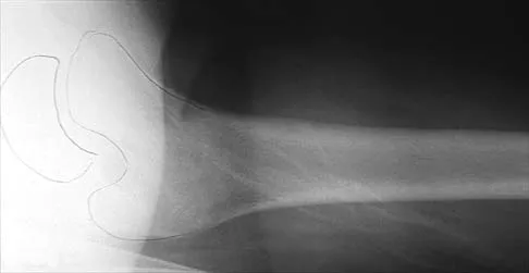
An 8-year-old boy has had pain and swelling around the right knee for the past 4 weeks. He recalls bumping it about 4 weeks ago. He has no pain in other joints, and denies any fevers, chills, or other symptoms. A radiograph is shown in Figure 13. Laboratory studies show a WBC count of 9,700/mm3, an erythrocyte sedimentation rate of 18 mm/h, and a C-reactive protein level of 3.7 mg/L. What is the next most appropriate step in management?


Explanation

The history and laboratory findings are consistent with osteomyelitis of the patella. The radiograph reveals bone destruction in the patella; therefore, the next most appropriate step is open biopsy and debridement of the site. Aspiration of the knee joint may be needed to rule out septic arthritis prior to patellar debridement. With this amount of bone destruction, surgical debridement is helpful to obtain cultures and to remove necrotic material. Administering antibiotics without any prior culture increases the risk of negative cultures later and a potentially incorrect choice of antibiotic. A neoplasm should be included in the differential. It would be inappropriate to initiate chemotherapy and radiation therapy without a biopsy-confirmed diagnosis. A bone scan is likely to demonstrate uptake, but radiographs have already localized the abnormality to the patella. Morrisy RT: Bone and joint sepsis, in Morrissy RT, Weinstein SL (eds): Lovell and Winter's Pediatric Orthopaedics, ed 5. Philadelphia, PA, Lippincott Williams & Wilkins, 2001, pp 466-470.

Question 4

A 2-year-old child is being evaluated for limb-length and girth discrepancy. As a newborn, the patient was large for gestational age and had hypoglycemia. Current examination shows enlargement of the entire right side of the body, including the right lower extremity and foot. The skin shows no abnormal markings, and the neurologic examination is normal. The spine appears normal. Radiographs confirm a 2-cm discrepancy in the lengths of the lower extremities. Additional imaging studies should include


Explanation

The patient may have Beckwith-Wiedemann syndrome (BWS), which consists of exophthalmos, macroglossia, gigantism, visceromegaly, abdominal wall defects, and neonatal hypoglycemia. Hemihypertrophy develops in approximately 15% of patients with BWS. Patients with hemihypertrophy that is the result of BWS have a 40% chance of developing malignancies such as Wilms' tumor or hepatoblastoma; therefore, frequent ultrasound screening is recommended until about age 7 years. The absence of nevi and vascular markings helps to rule out other causes of hemihypertrophy, such as neurofibromatosis, Proteus syndrome, and Klippel-Trenaunay syndrome. Bone age estimations are not accurate at this young age but may become more useful later to help predict the timing of epiphysiodesis procedures. DeBaun MR, Tucker MA: Risk of cancer during the first four years of life in children from The Beckwith-Wiedemann Syndrome Registry. J Pediatr 1998;132:398-400. Ballock RT, Wiesner GL, Myers MT, et al: Hemihypertrophy concepts and controversies. J Bone Joint Surg Am 1997;79:1731-1738.

Question 5

A 12 1/2-year-old boy reports intermittent knee pain and limping that interferes with his ability to participate in sports. He actively participates in football, basketball, and baseball. He denies any history of injury. Examination shows full range of motion without effusion. Radiographs reveal an osteochondritis dissecans (OCD) lesion on the lateral aspect of the medial femoral condyle. MRI scans are shown in Figures 14a and 14b. Initial treatment should consist of


Explanation

14b This skeletally immature patient has a small OCD lesion that appears stable, and he has not undergone any treatment. Therefore, a trial of immobilization until pain resolves is the best initial choice. Thereafter, cessation of sport activities for 4 to 6 months may allow healing of the lesion. Surgical treatment of juvenile OCD lesions is reserved for unstable lesions, patients who have not shown radiographic evidence of healing and are still symptomatic after 6 months of nonsurgical management, or patients who are approaching skeletal maturity. Good results with stable in situ lesions that have failed to respond to nonsurgical management have been reported with both transarticular and retroarticular drilling. Results after excision alone are poor at 5-year follow-up, and it is unclear if microfracture will improve the long-term outcome. Mosaicplasty may be the next best option for patients who remain or become symptomatic after excision of the fragment and microfracture. Wall E, Von Stein D: Juvenile osteochondritis dissecans. Orthop Clin North Am 2003;34:341-353.

Question 6

A 14-year-old boy undergoes application of a circular frame with tibial and fibular osteotomy for gradual limb lengthening. He initiates lengthening 7 days after surgery. During the first week of lengthening, he reports that turning of the distraction devices is becoming increasingly difficult. On the 9th day of lengthening, he is seen in the emergency department after feeling a pop in his leg and noting the acute onset of severe pain. What complication has most likely occurred?


Explanation

Incomplete corticotomy may result from osteotomy with limited soft-tissue stripping and exposure. When the patient begins distraction, tension develops at all wire/half-pin and bone interfaces, leading to increasing difficulty in distraction and limb pain. Sudden spontaneous completion of the osteotomy with continued tension applied by the fixator results in acute distraction of the osteotomy with severe pain. Premature consolidation is unlikely this early following the initial surgery. Birch JG, Samchukov ML: Use of the Ilizarov method to correct lower limb deformities in children and adolescents. J Am Acad Orthop Surg 2004;12:144-154.

Question 7

What is the most common primary malignant bone or cartilage tumor in children?


Explanation

Osteosarcoma is the most common primary malignant bone tumor (5.6 per 1 million children younger than age 15 years), and Ewing's sarcoma is second (2.1 per 1 million children). Giant cell tumor and chondrosarcoma are rare in children. Osteochondroma is more common than any of the above tumors in children, but it is not malignant. Himelstein BP, Dormans JP: Malignant bone tumors of childhood. Pediatr Clin North Am 1996;43:967-984. Pierz KA, Womer RB, Dormans JP: Pediatric bone tumors: Osteosarcoma, Ewing's sarcoma, and chondrosarcoma associated with multiple hereditary osteochondromatosis. J Pediatr Orthop 2001;21:412-418.

Question 8

What is the peak period of onset in children with pauciarticular juvenile rheumatoid arthritis?


Explanation

Approximately one half of patients with juvenile rheumatoid arthritis (JRA) have the pauciarticular form, which by definition includes only patients with fewer than five joints involved. The peak period of onset is between the ages of 2 and 4 years, with half of the affected children coming to medical attention before age 4 years. The knee is most often affected, with the ankle-subtalar and elbow joints next in frequency. The average duration of the disease is 2 years and 9 months, with half the cases lasting less than 2 years. Arthritis, in Herring JA (ed): Tachdjian's Pediatric Orthopaedics, ed 3. St Louis, MO, WB Saunders, 2002, pp 1811-1839.

Question 9

A 10-year-old girl who is Risser stage 0 has back deformity associated with neurofibromatosis type 1 (NF1). She has no back pain. Examination shows multiple cafe-au-lait nevi with normal lower extremity neurologic function and reflexes. Standing radiographs of the spine show a short 50-degree right thoracic scoliosis with a kyphotic deformity of 55 degrees (apex T8). A 10-degree progression in scoliosis has occurred during the past 1 year. There is no cervical deformity. MRI shows mild dural ectasia, primarily in the upper lumbar region. Management should consist of


Explanation

Scoliotic deformities in patients with NF1 are often dysplastic with short, angular curves. Posterior arthrodesis is made more difficult by the presence of kyphosis and of weak posterior elements caused by dural ectasia. Combined anterior and posterior spinal arthrodesis is generally preferred for progressive dysplastic curves to maximize deformity correction and to decrease the risk of pseudarthrosis. Anterior fusion may also prevent crankshaft phenomenon in young children. Brace treatment is not effective for large, rigid, or dysplastic curves. Kim HW, Weinstein SL: Spine update: The management of scoliosis in neurofibromatosis. Spine 1997;22:2770-2776.

Question 10

In obstetrical brachial plexus palsy, which of the following signs is associated with the poorest prognosis for recovery in a 2-month-old infant?


Explanation

Persistent Horner's sign (ptosis, myosis, and anhydrosis) is a sign of proximal injury, usually avulsion of the roots from the cord which disrupts the sympathetic chain. Root rupture or avulsion proximal to the myelin sheath has less chance of healing. Two-month-old infants with persistent weakness in the other areas described may still have a good prognosis for recovery. Concurrent clavicle fracture has been shown to have no prognostic value. Clarke HM, Curtis CG: An approach to obstetrical brachial plexus injuries. Hand Clin 1995;11:563-581.

Question 11

A 6-year-old boy with acute hematogenous osteomyelitis of the distal femur is being treated with intravenous antibiotics. The most expeditious method to determine the early success or failure of treatment is by serial evaluations of which of the following studies?


Explanation

Successful antibiotic treatment of osteomyelitis should lead to a rapid decline in the CRP. The CRP should decline after 48 to 72 hours of appropriate treatment. Imaging studies will take much longer to show resolution of bone infection. Unkila-Kallio L, Kallio MJ, Eskola J, et al: Serum C-reactive protein, erythrocyte sedimentation rate, and white blood cell count in acute hematogenous osteomyelitis of children. Pediatrics 1994;93:59-62.

Question 12

A 6-year-old girl has a painless spinal deformity. Examination reveals 2+ and equal knee jerks and ankle jerks, negative clonus, and a negative Babinski. The straight leg raising test is negative. Abdominal reflexes are asymmetrical. PA and lateral radiographs are shown in Figures 15a and 15b. What is the next most appropriate step in management?


Explanation

15b The patient has an abnormal neurologic exam as shown by the abnormal abdominal reflexes. Furthermore, she has a significant curve and is younger than age 10 years. These findings are not consistent with idiopathic scoliosis. MRI will best rule out syringomyelia or an intraspinal tumor. Bracing and surgery are not indicated for this small curvature prior to obtaining an MRI scan. Ginsburg GM, Bassett GS: Back pain in children and adolescents: Evaluation and differential diagnosis. J Am Acad Orthop Surg 1997;5:67-78.

Question 13

Figure 16 shows the radiograph of a 7-year-old boy who sustained a pathologic fracture of the left humerus 1 day ago. Initial management should consist of


Explanation

The radiograph shows a pathologic fracture through a unicameral (simple) bone cyst (UBC). This is the most common location and presentation of a UBC. Less than 10% of UBCs heal spontaneously following a fracture. Urgent biopsy is not indicated because the lesion appears benign and the histology of fracture callus may be misinterpreted as osteosarcoma. After the fracture heals with the use of a sling and swathe, the UBC may be treated with a minimally invasive procedure such as injection of bone marrow and/or demineralized bone matrix. The chance for success is relatively low in an active cyst located adjacent to the physis. More invasive procedures, such as curettage, Rush rod fixation, or cannulated screw decompression, have been described but are rarely necessary for treatment of upper extremity cysts. Rougraff BT, Kling TJ: Treatment of active unicameral bone cysts with percutaneous injection of demineralized bone matrix and autogenous bone marrow. J Bone Joint Surg Am 2002;84:921-929. Robosch A, Saraph V, Linhart WE: Flexible intramedullary nailing for the treatment of unicameral bone cysts in long bones. J Bone Joint Surg Am 2000;82:1447-1453.

Question 14

Figure 17 shows the AP radiograph of a 5-year old child who has mild short stature and a painless bilateral gluteus medius lurch. Initial work-up should include


Explanation

Bilateral flattening of the femoral heads suggests multiple epiphyseal dysplasia; therefore, a skeletal survey is indicated to look for involvement of other epiphyses. Unilateral flattening of the femoral head would suggest Legg-Perthes disease. Sponseller PD: Skeletal dysplasias, in Morrissy RT, Weinstein SL (eds): Lovell and Winter's Pediatric Orthopaedics, ed 5. Philadelphia, PA, Lippincott Williams & Wilkins, 2001, pp 269-270.

Question 15

A 7-year-old girl with spinal muscular atrophy (SMA) type II has popping of the left hip. Examination reveals painless subluxation of the joint in adduction with palpable reduction in abduction. Radiographs show coxa valga, subluxation of the left hip, and pelvic obliquity with elevation of the left hemipelvis. Treatment should consist of


Explanation

Observation is the treatment of choice. Hip subluxation and dislocation are not uncommon in patients with SMA type II who are unlikely to be ambulatory. Scoliosis occurs in these patients 100% of the time and frequently creates pelvic obliquity. However, in long-term follow-up, patients with SMA type II and hip dislocations had little associated pain or functional limitations because of hip instability. In addition, recurrent hip subluxation after surgical treatment has been documented. Given the rarity of symptoms from hip instability in long-term follow-up, and the possibility of recurrent dislocation, surgical intervention for hip instability may expose SMA type II patients to undue surgical risk for minimal if any functional gain. Sporer SM, Smith BG: Hip dislocation in patients with spinal muscular atrophy. J Pediatr Orthop 2003;23:10-14.

Question 16

A newborn with myelomeningocele has no movement below the waist and has bilateral hips that dislocate with provocative flexion and adduction. What is the best treatment option for the hip instability?


Explanation

The status of the hips (located or dislocated) in children with thoracic-level myelomeningocele has no effect on the functional outcome of these patients. Management of unstable hips in this population should be limited to treatment of the contractures that may lead to poor limb positioning in either braces or a wheelchair. The use of the Pavlik harness and/or spica cast is contraindicated because they would promote flexion and abduction contractures. In the past, open reduction either through an anterior or medial approach had been performed with a high incidence of redislocation and other complications, with little functional gain for the child. Gabriel KG: Natural history of hip deformity in spina bifida, in Sarwark JR, Lubicky JP (eds): Caring for the Child With Spina Bifida. Rosemont, IL, American Academy of Orthopaedic Surgeons, 2001, pp 89-103.

Question 17

A 14-year-old boy reports a 4-month history of increasing backache with difficulty walking long distances. His parents state that he walks with his knees slightly flexed and is unable to bend forward and get his hands to his knees. He denies numbness, tingling, and weakness in his legs and denies loss of bladder and bowel control. A lateral radiograph of the lumbosacral spine is shown in Figure 18. What is the best surgical management for this condition?


Explanation

The patient has a grade 4 spondylolisthesis. Optimal surgical management is posterior spinal fusion from L4 to the sacrum. The use of instrumentation is controversial. Vertebrectomy is typically reserved for spondylo-optosis (grade 5) cases. Spinal fusion from L5 to S1 usually is not successful for a slip that is greater than 50%. Isolated anterior spinal fusion has not been successful, and direct repair of the pars defect is only useful for spondylolysis without spondylolisthesis. Lenke LG, Bridwell KH: Evaluation and surgical treatment of high-grade isthmic dysplastic spondylolisthesis. Instr Course Lect 2003;52:525-532.

Question 18

Duchenne's muscular dystrophy is a genetic disorder that is transmitted by which of the following modes of inheritance?


Explanation

Patients with Duchenne's muscular dystrophy show progressive muscular weakness because of the absence of dystrophin and have the clinical picture of progressive muscle weakness. The condition is an X-linked genetic disease. Fitzgerald RH, Kaufer H, Malkani AL: Orthopaedics. St Louis, MO, Mosby Year Book, 2002, pp 1573-1583.

Question 19

A 4-month-old infant is referred for evaluation of congenital scoliosis. The child has no congenital heart anomalies, and a renal ultrasound shows that he has one kidney. Examination reveals mild scoliosis and a large hairy patch on the child's back. Neurologic evaluation is normal for his age. A clinical photograph and radiograph are shown in Figures 19a and 19b. Initial management should consist of


Explanation

19b Congenital anomalies of the spine, including failure of formation and failure of segmentation, are associated with other anomalies in other organ systems that develop at the same time. These include anomalies in the genitourinary system, cardiac anomalies, Sprengel's deformity, radial hypoplasia, and gastrointestinal anomalies including imperforate anus and trachealesophageal fistula. Spinal dysraphism is the most common associated abnormality. McMaster found an 18% incidence before the common use of MRI. Bradford and associates reported on 16 of 42 patients with congenital spinal anomalies and spinal dysraphism using MRI. Neural axis lesions may be associated with visible midline abnormalities such as a hairy patch or nevus. The child has already had a cardiac and renal work-up, and based on the findings of the hairy patch and congenital vertebral anomalies, MRI of the entire spine is prudent at this time. Spinal fusion is indicated for progressive congenital scoliosis or kyphosis. Physical therapy does not affect the natural history of congenital scoliosis. McMaster MJ: Occult intraspinal anomalies and congenital scoliosis. J Bone Joint Surg Am 1984;66:588-601. Bradford DS, Heithoff KB, Cohen M: Intraspinal abnormalities and congenital spine deformities: A radiographic and MRI study. J Pediatr Orthop 1991;11:36-41.

Question 20

A 12-year-old boy reports limping and chronic knee pain that is now inhibiting his ability to participate in sports. Clinical examination and radiographs of the knee are normal. Additional evaluation should include


Explanation

While all of the answers may be appropriate, radiating pain from hip pathology must be excluded. At this age, a slipped capital femoral epiphysis is likely. Therefore, the hip must be examined. Kocher MS, Bishop JA, Weed B, et al: Delay in diagnosis of slipped capital femoral epiphysis. Pediatrics 2004;113:322-325.

Question 21

In children with moderate to severe osteogenesis imperfecta (OI), intravenous pamidronate therapy has been shown to increase the thickness of cortical bone. This occurs primarily as a consequence of


Explanation

Histologic studies have shown that increased bone turnover is the rule in OI. Pamidronate (and all bisphosphonates) reduce osteoclast-mediated bone resorption. Osteoblastic new bone formation on the periosteal surface of long bones is minimally impaired. With inhibition of osteoclastic bone resorption on the endosteal surface, the cortex of the bone can begin to thicken as it does with normal growth in individuals unaffected by OI. Mineralization and collagen matrix organization are not directly affected by pamidronate. Zeitlin L, Fassier F, Glorieux FH: Modern approach to children with osteogenesis imperfecta. J Pediatr Orthop B 2003;12:77-87. Falk MJ, Heeger S, Lynch KA, et al: Intravenous bisphosphonate therapy in children with osteogenesis imperfecta. Pediatrics 2003;111:573-578.

Question 22

Split posterior tibial tendon transfer is used in the treatment of children with cerebral palsy. Which of the following patients is considered the most appropriate candidate for this procedure?


Explanation

Split posterior tibial tendon transfers are best performed in patients with spastic cerebral palsy who are between the ages of 4 and 7 years and have flexible equinovarus deformities. Rigid deformities typically require bony reconstruction procedures. Tendon transfers in patients with athetosis are unpredictable. Green NE, Griffin PP, Shiavi R: Split posterior tibial-tendon transfer in spastic cerebral palsy. J Bone Joint Surg Am 1983;65:748-754.

Question 23

Late surgical treatment of posttraumatic cubitus varus (gunstock deformity) is usually necessitated by the patient reporting problems related to


Explanation

Cubitus varus, elbow hyperextension, and internal rotation are all typical components of the gunstock deformity. This deformity results from malunion of a supracondylar fracture of the humerus. All of the problems listed above have been reported as sequelae of a gunstock deformity, although the malunion usually causes no functional limitations. Unacceptable appearance is the most common reason why patients or parents request corrective osteotomy. O'Driscoll SW, Spinner RJ, McKee MD, et al: Tardy posterolateral rotatory instability of the elbow due to cubitus varus. J Bone Joint Surg Am 2001;83:1358-1369. Gurkan I, Bayrakci K, Tasbas B, et al: Posterior instability of the shoulder after supracondylar fractures recovered with cubitus varus deformity. J Pediatr Orthop 2002;22:198-202.

Question 24

An 11-year-old boy sustained an ankle injury while playing football. Figure 20 shows an AP radiograph obtained the day of injury. Treatment should consist of


Explanation

The child has an injury involving both the growth plate and the articular surface of the ankle. Because of the significant displacement, open reduction and internal fixation is indicated to realign the physis and joint surface. The best method of fixation to avoid growth arrest is one that does not cross the physis. This is usually achieved by a transverse epiphyseal screw parallel to the physis. If the metaphyseal fragment was large enough, a transverse metaphyseal screw could be used instead. The incidence of growth arrest following physeal ankle injuries is as high as 50%, and long-term follow-up is indicated. Cass JR, Peterson HA: Salter-Harris Type-IV injuries of the distal tibial epiphyseal growth plate, with emphasis on those involving the medial malleolus. J Bone Joint Surg Am 1983;65:1059-1070.

Question 25

A 3-year-old child has bilateral genu varum and short stature. Radiographs show physeal widening and generalized osteopenia. The femora and tibiae show anterolateral bowing. Laboratory studies show low normal serum calcium values, significantly decreased serum phosphate levels, and normal parathyroid hormone (PTH), alkaline phosphatase, and vitamin-D levels. These findings are consistent with


Explanation

Children with vitamin D-resistant rickets are short in stature and have genu varum, physeal widening, and generalized osteopenia. The abnormality in inherited vitamin D-resistant rickets is the renal tubule's inability to resorb phosphate leading to hypophosphatemia. Laboratory findings in the condition are normal or near normal serum calcium values, significantly decreased serum phosphate levels, elevated alkaline phosphatase levels, and normal PTH and vitamin-D levels. The most common form is inherited as an X-linked dominant trait. Nutritional rickets has a normal or low serum phosphate levels, normal or low serum calcium values, and decreased levels of 25(OH) vitamin D and 1,25-dihydroxyvitamin D. Hypophosphatasia is a rare condition characterized by a deficiency of alkaline phosphatase in the serum and tissues, leading to generalized abnormal mineralization of bone. Primary hyperparathyroidism usually is caused by a parathyroid adenoma, and the child generally has abdominal problems and hypercalcemic crisis. Laboratory findings include elevated serum calcium values, alkaline phosphatase levels, and PTH levels, and decreased serum phosphate levels. Children with renal osteodystrophy tend to have genu valgum, and laboratory findings include elevated serum phosphate, alkaline phosphatase, and PTH levels, and low serum calcium values. Findings of renal disease include elevated BUN and creatinine. Herring JA: Metabolic and endocrine bone diseases, in Herring JA (ed): Tachdjian's Pediatric Orthopaedics, ed 3. Philadelphia, PA, WB Saunders, 2002, pp 1686-1710.

Question 26

A 6-year-old boy falls from the monkey bars and presents with a severely displaced extension-type supracondylar humerus fracture. On examination in the emergency department, his hand is pink, but the radial pulse is not palpable. He is taken to the operating room, where closed reduction and percutaneous pinning are performed. Postoperatively, the hand remains pink and well-perfused, but the radial pulse remains absent. What is the most appropriate next step in management?


Explanation

A 'pink, pulseless' hand following closed reduction and percutaneous pinning of a pediatric supracondylar humerus fracture indicates that collateral circulation is adequate to perfuse the hand. The standard of care is observation and admission, as the radial pulse typically returns within 24 to 48 hours once vasospasm subsides. Vascular exploration is indicated for a 'white, pulseless' hand or if adequate perfusion is lost after reduction.

Question 27

A 6-week-old female infant is being treated with a Pavlik harness for an easily reducible dislocated left hip. At her 2-week follow-up appointment, the parents report that she is no longer kicking her left leg as much as her right. On physical examination, she demonstrates decreased active knee extension on the left, though she does not appear to be in pain. What is the most likely cause of this clinical finding?


Explanation

Femoral nerve palsy is a known complication of Pavlik harness treatment and is typically caused by excessive hip flexion. It presents as an inability to actively extend the knee. The appropriate management is to temporarily discontinue the harness or adjust the anterior straps to decrease hip flexion until the palsy completely resolves.

Question 28

A 13-year-old obese male presents to the emergency department after a minor slip. He is unable to bear weight on his right leg, even with crutches. Radiographs reveal a severe slipped capital femoral epiphysis (SCFE). Which of the following interventions during surgical treatment has been shown to potentially decrease the risk of osteonecrosis in this specific clinical scenario?


Explanation

The patient has an unstable SCFE, defined by the inability to bear weight. Unstable SCFE has a significantly higher rate of osteonecrosis (up to 50%) compared to stable SCFE. Urgent capsular decompression (capsulotomy) to relieve intracapsular hematoma tamponade, followed by gentle or no reduction prior to pinning, has been shown to lower the risk of avascular necrosis. Forceful closed reduction is strictly contraindicated as it further disrupts the delicate blood supply.

Question 29

A 4-year-old boy who was treated successfully for idiopathic clubfoot during infancy with the Ponseti method presents with a dynamic supination deformity of the foot during the swing phase of gait. On examination, his ankle passive dorsiflexion is 15 degrees past neutral with the knee extended, and the foot is completely flexible. What is the most appropriate management for this patient?


Explanation

The child presents with a relapsing clubfoot characterized by a dynamic supination deformity. This is common after Ponseti casting and is driven by a strong tibialis anterior muscle overpowering the weak evertors. For a flexible deformity with adequate passive dorsiflexion, a complete tibialis anterior tendon transfer to the lateral cuneiform is the gold standard treatment to balance the foot. A split transfer is not recommended in idiopathic clubfoot relapse.

Question 30

An 8-year-old boy is diagnosed with Legg-Calve-Perthes disease. Anteroposterior pelvic radiographs demonstrate sclerosis and fragmentation of the left capital femoral epiphysis. According to the Herring lateral pillar classification, which of the following radiographic findings indicates a Group C classification, which is associated with the poorest prognosis?


Explanation

The Herring lateral pillar classification assesses the height of the lateral pillar of the capital femoral epiphysis on the AP radiograph during the fragmentation phase. Group A has no involvement; Group B maintains >50% of the lateral pillar height; Group C maintains <50% of the lateral pillar height. Group C is associated with the poorest clinical and radiographic outcomes, and patients in this group who are >8 years of age typically benefit from surgical containment.

Question 31

A 14-year-old boy twists his ankle while playing basketball. Radiographs demonstrate a Salter-Harris type III fracture of the anterolateral distal tibial epiphysis. What is the primary mechanism and deforming force responsible for this specific fracture pattern?


Explanation

A juvenile Tillaux fracture is a Salter-Harris type III fracture of the anterolateral distal tibial epiphysis. It occurs during adolescence (typically 12-14 years of age) when the medial aspect of the distal tibial physis has closed but the lateral aspect remains open. External rotation of the foot causes the anterior inferior tibiofibular ligament (AITFL) to tension and avulse the anterolateral epiphysis.

Question 32

A 5-year-old boy with a history of multiple low-energy fractures, blue sclerae, and dentinogenesis imperfecta is being evaluated for surgical intervention of a severely bowed femur. He receives intravenous bisphosphonate therapy every 3 months. What is the primary mechanism of action of bisphosphonates in this condition?


Explanation

Osteogenesis imperfecta is most commonly caused by a genetic defect in type I collagen synthesis. Bisphosphonates are a mainstay of medical management to increase bone mineral density and reduce fracture rates. They function primarily by binding to hydroxyapatite crystals in bone and inhibiting osteoclast-mediated bone resorption, which leads to a relative increase in bone mass, despite the collagen matrix remaining abnormal.

Question 33

A 6-year-old girl with spastic quadriplegic cerebral palsy, GMFCS level V, undergoes routine hip surveillance. Anteroposterior pelvic radiographs reveal a migration percentage (Reimers' index) of 45% in the right hip. She experiences minimal pain, but hip abduction is limited to 15 degrees bilaterally. What is the most appropriate next step in surgical management?


Explanation

In children with cerebral palsy, a migration percentage > 40% indicates structural hip subluxation with a high risk of progression to dislocation, particularly in non-ambulatory patients (GMFCS IV/V). While soft tissue releases are appropriate for at-risk hips with < 30% migration, once significant structural dysplasia and subluxation (>40%) occur, bony reconstruction is required. This typically consists of a proximal femoral varus derotational osteotomy (VDRO) combined with a pelvic osteotomy (e.g., Dega or San Diego) to restore joint congruity and prevent painful dislocation.

Question 34

A 12-year-old boy presents with a history of recurrent ankle sprains and a painful, rigid flatfoot. On examination, subtalar motion is severely restricted, and there is palpable spasm of the peroneal muscles. Oblique radiographs of the foot demonstrate an 'anteater nose' sign. Which of the following is the most likely diagnosis?


Explanation

The clinical presentation of a rigid flatfoot, restricted subtalar motion, and peroneal spasticity in an adolescent is classic for a tarsal coalition. The 'anteater nose' sign seen on a 45-degree internal oblique radiograph of the foot is pathognomonic for a calcaneonavicular coalition. It represents an elongated anterior process of the calcaneus approaching the navicular. A talocalcaneal coalition typically presents with a 'C-sign' on a lateral radiograph.

Question 35

A 3-year-old girl is diagnosed with infantile idiopathic scoliosis. Radiographs reveal a primary left thoracic curve of 35 degrees. The rib-vertebra angle difference (RVAD) of Mehta is measured at 25 degrees. What is the most appropriate initial management for this patient to control deformity progression?


Explanation

Infantile idiopathic scoliosis with a curve > 30 degrees and an RVAD > 20 degrees has a high likelihood of rapid progression. Serial elongation-derotation-flexion (EDF) casting, also known as Mehta casting, is the gold standard initial management for progressive early-onset scoliosis in this age group. It has been shown to effectively control curve progression, allow for pulmonary development, and in some cases, completely resolve the deformity. Bracing is less effective in controlling severe rotation in infantile progressive curves.

Question 36

A 3-year-old girl is diagnosed with a neglected developmental dysplasia of the left hip. Radiographs show a high dislocation of the femoral head. What is the standard surgical management for this patient?


Explanation

In children older than 2 to 3 years with a neglected developmental dysplasia of the hip, the risk of redislocation and avascular necrosis is high due to soft tissue contractures and secondary acetabular dysplasia. The standard of care typically involves an open reduction, a femoral shortening osteotomy (to relieve soft tissue tension and reduce the risk of AVN), and a pelvic osteotomy (e.g., Dega or Salter) to address acetabular dysplasia and provide stable coverage.

Question 37

A 2-year-old boy who was treated for idiopathic clubfoot with the Ponseti method presents with recurrent equinovarus deformity. What is the most common cause of relapse in this clinical scenario?


Explanation

Non-compliance with the foot abduction orthosis (FAO) is widely recognized as the most common cause of relapse in clubfoot treated with the Ponseti method. Bracing protocols typically require 23-hour wear for 3 months, followed by nighttime wear until 4-5 years of age. Without bracing, relapse rates can exceed 80%.

Question 38

A 14-year-old boy is evaluated for hip pain and severe stiffness 6 months after undergoing in situ pinning for a stable slipped capital femoral epiphysis (SCFE) of the right hip. On examination, he has globally restricted range of motion of the right hip. Radiographs reveal narrowing of the joint space to less than 3 mm, osteopenia, and no evidence of hardware penetration into the joint. What is the most likely diagnosis?


Explanation

Chondrolysis is characterized by acute cartilage necrosis, presenting with severe stiffness, pain, and globally restricted range of motion. Radiographically, it is defined by a joint space of less than 3 mm. Although historically associated with unrecognized hardware penetration, it can occur in unpinned SCFEs or following in situ pinning without joint penetration. AVN typically presents with sclerosis and collapse of the femoral head rather than isolated symmetric joint space narrowing.

Question 39

A 6-year-old boy sustains a completely displaced extension-type supracondylar fracture of the humerus. On physical examination in the emergency department, the patient is unable to flex the interphalangeal joint of the thumb and the distal interphalangeal joint of the index finger. Which of the following nerves is most likely injured?


Explanation

The anterior interosseous nerve (AIN) is a branch of the median nerve and is the most commonly injured nerve in extension-type supracondylar humerus fractures. It provides motor innervation to the flexor pollicis longus, the radial half of the flexor digitorum profundus, and the pronator quadratus. Injury results in the inability to form an 'A-OK' sign (loss of IP flexion of the thumb and DIP flexion of the index finger).

Question 40

An 8-year-old boy presents with a painless limp of 3 months' duration. Radiographs show fragmentation of the femoral head consistent with Legg-Calvé-Perthes disease. According to the Herring Lateral Pillar classification, which of the following radiographic findings portends the worst prognosis?


Explanation

The Herring Lateral Pillar classification is strongly correlated with prognosis in Legg-Calvé-Perthes disease. Group C (less than 50% lateral pillar height maintained) indicates severe involvement and carries the worst prognosis for femoral head sphericity and future joint congruity. Age at onset (>8 years) and Group C classification are the strongest predictors of a poor outcome.

Question 41

A 2.5-year-old girl is brought in by her parents for evaluation of bowing of her legs. On standing full-length radiographs, the metaphyseal-diaphyseal angle (Drennan's angle) is measured at 18 degrees bilaterally. What is the most appropriate next step in management?


Explanation

A metaphyseal-diaphyseal angle (Drennan's angle) greater than 16 degrees strongly suggests early infantile Blount's disease (tibia vara) rather than physiologic bowing. In children under 3 years of age with early-stage infantile Blount's disease, a trial of bracing with Knee-Ankle-Foot Orthoses (KAFOs) is indicated. If bracing fails or if the child is older (e.g., >3-4 years) with advanced Langenskiöld stages, proximal tibial osteotomy is warranted.

Question 42

A 4-year-old boy with a history of multiple low-energy long bone fractures, blue sclerae, and dentinogenesis imperfecta is evaluated. A genetic disorder is suspected. The pathophysiology of this condition is primarily related to a defect in the synthesis of which of the following?


Explanation

Osteogenesis imperfecta is primarily caused by autosomal dominant mutations in the COL1A1 or COL1A2 genes, which encode the alpha chains of type I collagen. This leads to defective synthesis of type I collagen, manifesting as bone fragility, blue sclerae, hearing loss, and dentinogenesis imperfecta. Type II collagen defects are associated with spondyloepiphyseal dysplasia. Defective osteoclasts are seen in osteopetrosis. FGFR3 mutations cause achondroplasia. CBFA1 mutations cause cleidocranial dysplasia.

Question 43

A 4-year-old boy who weighs 18 kg (40 lbs) sustains an isolated, closed, length-stable, spiral fracture of the mid-diaphyseal femur after a fall from a playground structure. What is the most appropriate definitive management?


Explanation

For children aged 6 months to 5 years with isolated, closed femoral shaft fractures and acceptable shortening (<2 cm), early spica casting is the standard of care. Flexible intramedullary nailing is typically reserved for children aged 5-11 years or weighing more than 50 lbs (23 kg). Rigid nailing is contraindicated in this age group due to the risk of avascular necrosis of the femoral head and growth arrest from injury to the trochanteric apophysis.

Question 44

A 14-year-old girl sustains an ankle injury while playing soccer. Radiographs reveal a Salter-Harris III fracture of the anterolateral distal tibial epiphysis. The mechanism of injury involves avulsion by which of the following structures?


Explanation

A juvenile Tillaux fracture is a Salter-Harris III fracture of the anterolateral distal tibial epiphysis. It occurs in adolescents due to the asymmetrical closure pattern of the distal tibial physis, which closes first centrally, then medially, and finally laterally. An external rotation force causes the anterior inferior tibiofibular ligament (AITFL) to avulse the anterolateral epiphysis, which is the last portion of the physis to close.

Question 45

A 13-year-old boy with a BMI of 35 presents to the emergency department unable to bear weight on his left leg after a minor slip. Radiographs confirm an acute, severe left slipped capital femoral epiphysis (SCFE). He cannot bear weight even with crutches. What is the current consensus regarding surgical treatment for this patient to minimize complications?


Explanation

The patient has an unstable SCFE (defined by the inability to bear weight with or without crutches). Unstable SCFE is associated with a high risk of avascular necrosis (AVN). Current evidence suggests that urgent or emergent surgical stabilization (often within 24 hours), combined with capsular decompression (to reduce intracapsular tamponade), minimizes the risk of AVN. Forceful closed reduction is contraindicated as it increases the risk of AVN.

Question 46

A 4-year-old boy who was successfully treated for bilateral idiopathic clubfeet as an infant using the Ponseti method presents with returning deformity in the right foot. During the swing phase of gait, the right foot supinates dynamically. Passive range of motion demonstrates fully correctable deformities without residual equinus. What is the most appropriate next step in surgical management?


Explanation

Relapse in clubfoot treated with the Ponseti method often presents as dynamic supination during the swing phase of gait due to relative overactivity of the tibialis anterior muscle compared to the evertors. When the foot is passively correctable, the treatment of choice is the transfer of the entire tibialis anterior tendon to the lateral cuneiform (often preceded by a brief period of corrective casting). Unlike in cerebral palsy, where a SPLATT is common, the entire tendon is transferred in clubfoot relapse.

Question 47

A 9-year-old boy weighing 55 kg (121 lbs) sustains a closed, length-unstable, midshaft femur fracture. Among the following options, which surgical intervention carries the highest risk of complication, specifically loss of reduction and malunion, in this specific patient?


Explanation

Titanium elastic nails (TENs) are a standard of care for pediatric femur fractures in children aged 5 to 11 years. However, a patient weight exceeding 49 kg (approx 100-110 lbs) is a recognized relative contraindication due to a significantly higher risk of biomechanical failure, leading to loss of reduction, malunion, and prominent hardware. For this patient, submuscular plating or a rigid lateral-entry nail would be more appropriate.

Question 48

A 6-week-old female is being treated with a Pavlik harness for a developmental dysplasia of the hip (DDH) that was dislocated but reducible on exam. At her 1-week follow-up, the parents report she has stopped kicking her left leg. On clinical examination, she holds the left knee in extension and does not actively contract her quadriceps with tickling. Ultrasound confirms the hip is currently reduced. What is the most appropriate next step in management?


Explanation

The clinical scenario describes a femoral nerve palsy, a known complication of treating DDH with a Pavlik harness caused by hyperflexion of the hip. Presenting signs include absent active knee extension and loss of quadriceps function. The appropriate initial management is to discontinue the harness to relieve pressure on the femoral nerve and allow for neurologic recovery, which typically occurs over a few days to weeks. Tightening straps or ignoring the palsy risks permanent nerve injury.

Question 49

A 7-year-old boy with spastic quadriplegic cerebral palsy (GMFCS Level IV) presents with a slowly progressing scissoring gait, pain with diaper changes, and difficulty sitting in his wheelchair. A recent AP pelvis radiograph demonstrates a right hip Reimers migration percentage of 55%. The acetabulum is dysplastic, but there is no evidence of degenerative arthritis. What is the most appropriate definitive management?


Explanation

In non-ambulatory children with cerebral palsy, a hip migration percentage greater than 40-50% indicates significant subluxation with a high risk of progression to painful dislocation. Soft tissue releases alone (tenotomies) are insufficient at this stage and age. In the absence of severe degenerative changes, comprehensive bony reconstruction consisting of a varus derotational osteotomy (VDRO), pelvic osteotomy (e.g., Dega or San Diego), and often open reduction is the gold standard to provide a painless, stable, and locatable hip.

Question 50

A 13-year-old boy with a BMI in the 99th percentile presents to the emergency department with acute left groin pain. He states he twisted his leg getting out of bed. On examination, he is completely unable to bear weight on the left leg, even with the use of crutches. Radiographs demonstrate a severe posterior and inferior displacement of the left capital femoral epiphysis. According to the Loder classification, his inability to bear weight puts him at highest risk for which of the following complications?


Explanation

The Loder classification divides Slipped Capital Femoral Epiphysis (SCFE) into stable and unstable based strictly on the ability to bear weight (with or without crutches). An unstable SCFE (inability to bear weight) has a significantly higher rate of avascular necrosis (up to 50%), whereas AVN is exceedingly rare in stable SCFE. Chondrolysis is more commonly associated with unrecognized hardware penetration into the joint space.

Question 51

A 6-year-old girl falls from monkey bars and sustains a widely displaced, extension-type supracondylar humerus fracture. On initial presentation, she has no palpable radial pulse, but the hand is warm, pink, and has a brisk capillary refill of less than 2 seconds. She is taken urgently to the operating room, where an anatomic closed reduction is achieved and stabilized with three divergent lateral pins. After pinning, the hand remains pink and warm, but the radial pulse remains absent. What is the most appropriate next step in management?


Explanation

The management of a 'pulseless, pink' hand following adequate reduction and pinning of a supracondylar humerus fracture is observation and close monitoring. The pink color and brisk capillary refill indicate that collateral circulation is providing adequate perfusion to the hand. Routine vascular exploration is not indicated unless the hand becomes cool, pale, and ischemic (a 'pulseless, white' hand) after reduction.

Question 52

An 8-year-old boy presents with a 4-month history of a painless limp. Radiographs demonstrate fragmentation and sclerosis of the left proximal femoral epiphysis consistent with Legg-Calvé-Perthes disease. The lateral one-third of the femoral head exhibits a 60% loss of height compared to the contralateral normal hip. According to the Herring lateral pillar classification, what is his expected prognosis without surgical intervention?


Explanation

The Herring lateral pillar classification predicts outcome in Perthes disease based on the height of the lateral column of the epiphysis during the fragmentation stage. Group C implies >50% collapse of the lateral pillar. Patients in Group C generally have a poor prognosis with a high likelihood of aspherical congruency and early osteoarthritis, especially in children older than 8 years, regardless of surgical containment.

Question 53

A 14-year-old boy presents with chronic, vague lateral foot pain and a history of multiple ankle sprains. On physical examination, he has a rigid pes planovalgus deformity with absent subtalar motion. A lateral radiograph of the foot demonstrates an elongated anterior process of the calcaneus projecting toward the navicular. To properly characterize this anatomy prior to surgical intervention, what is the best diagnostic imaging modality?


Explanation

The clinical presentation of rigid flatfoot, absent subtalar motion, and an elongated anterior process of the calcaneus ('anteater sign') strongly suggests a calcaneonavicular tarsal coalition. A CT scan is the gold standard imaging modality to fully evaluate the size and extent of the coalition, assess for degenerative changes in adjacent joints, and rule out concurrent talocalcaneal coalitions prior to planning a surgical resection.

Question 54

A 12-year-old female presents to the clinic with an adolescent idiopathic scoliosis (AIS) right thoracic curve of 28 degrees. When counseling her parents about the risk of curve progression, you explain the concept of peak height velocity (PHV). Which of the following maturity indicators most closely corresponds to the peak velocity of growth in a patient with AIS?


Explanation

The peak height velocity (PHV) represents the phase of maximum linear growth and is the period of highest risk for curve progression in AIS. It typically occurs just before menarche. The Sanders maturity scale, which assesses the ossification of hand epiphyses, identifies Stage 3 (adolescent rapid-early) and Stage 4 (adolescent rapid-late) as the exact periods correlating with PHV. Triradiate cartilage closure is closely associated but Sanders 3 is the most precise indicator of the peak.

Question 55

A 4-year-old girl whose weight is above the 95th percentile presents with a waddling gait and progressive bowing of both legs. Standing long-leg radiographs show medial metaphyseal beaking of the proximal tibiae and a metaphyseal-diaphyseal angle of 18 degrees. What physiological principle best explains the progression of her proximal medial tibial physeal deformity?


Explanation

The patient has infantile Blount's disease (tibia vara), which is characterized by a high metaphyseal-diaphyseal angle (>16 degrees) and progressive varus deformity. The underlying mechanism is explained by the Heuter-Volkmann principle, which states that excessive compressive forces across a physis inhibit longitudinal growth, whereas tensile forces stimulate it. In obese or early-walking children, excessive mechanical compression on the posteromedial proximal tibial physis inhibits its growth, exacerbating the varus deformity.

Question 56

A 3-month-old infant with developmental dysplasia of the hip (DDH) is being treated with a Pavlik harness. During a follow-up visit, the parents report that the infant is not moving the right leg as much as the left. On physical examination, the right knee is held in extension, and the patellar reflex is absent on the right side. What is the most likely cause of this clinical finding?


Explanation

Femoral nerve palsy is a known complication of the Pavlik harness, typically caused by excessive flexion of the hip which compresses the femoral nerve against the inguinal ligament. Clinical signs include an inability to actively extend the knee and an absent patellar reflex. The treatment is adjusting the harness to decrease the amount of flexion or temporarily removing it until nerve function recovers.

Question 57

A 12-year-old boy presents with right hip pain and an antalgic gait. He is diagnosed with a severe right slipped capital femoral epiphysis (SCFE). His weight is greater than the 95th percentile for his age. You plan to perform in situ pinning of the right hip. Which of the following is considered the strongest indication for prophylactic pinning of the contralateral asymptomatic hip?


Explanation

Prophylactic pinning of the contralateral hip is heavily debated, but universally accepted indications include the presence of an underlying endocrine disorder (e.g., hypothyroidism, renal osteodystrophy, panhypopituitarism) or a history of radiation therapy, as these patients have an exceptionally high risk of developing a contralateral slip. Other factors that lower the threshold for prophylactic pinning include an inability to reliably follow up, open triradiate cartilage, and young chronological age at presentation.

Question 58

A 3-year-old girl is evaluated for progressive bowing of both lower extremities. She is at the 95th percentile for weight. Standing long-leg radiographs show significant genu varum with a metaphyseal-diaphyseal angle (Drennan angle) of 18 degrees bilaterally, as well as prominent medial metaphyseal beaking. What is the most appropriate initial management for this patient?


Explanation

The patient's presentation and radiographic findings (metaphyseal-diaphyseal angle >16 degrees, medial metaphyseal beaking) are diagnostic of infantile Blount's disease (tibia vara). For a 3-year-old child (typically Langenskiöld stage I or II), the initial treatment consists of a trial of bracing with Knee-ankle-foot orthoses (KAFOs) worn during ambulation or waking hours. Surgical intervention (tibial osteotomy) is generally reserved for children who fail bracing or present after 4 years of age.

Question 59

A 6-year-old boy falls from the monkey bars and sustains a widely displaced, extension-type supracondylar humerus fracture. On presentation to the emergency department, his hand is pink and warm with brisk capillary refill, but the radial pulse is absent. Neurological examination reveals weakness in flexing the interphalangeal joint of the thumb and the distal interphalangeal joint of the index finger. What is the most appropriate next step in management?


Explanation

This patient has a "pink, pulseless" hand associated with a displaced supracondylar humerus fracture, along with an anterior interosseous nerve (AIN) palsy. The standard of care is urgent closed reduction and percutaneous pinning (CRPP). If the hand remains pink and well-perfused (warm with good capillary refill) after reduction, observation without vascular exploration is the accepted management, even if the radial pulse does not immediately return. Vascular exploration is indicated only if the hand becomes or remains white/ischemic after reduction.

Question 60

An 8-year-old boy is diagnosed with Legg-Calvé-Perthes disease (LCPD). While evaluating his plain radiographs, you are assessing the "head-at-risk" signs described by Catterall, which are indicative of a poorer prognosis. Which of the following radiographic findings represents a Catterall "head-at-risk" sign?


Explanation

Catterall described five "head-at-risk" signs for Legg-Calvé-Perthes disease, which portend a poorer prognosis due to the increased risk of femoral head deformation. These include the Gage sign (a V-shaped radiolucency in the lateral epiphysis and adjacent metaphysis), lateral subluxation of the femoral head, calcification lateral to the epiphysis, horizontal growth plate, and metaphyseal cysts. Medial subluxation and central physeal arrest are not part of the Catterall risk criteria.

Question 61

A 2-week-old infant is undergoing serial casting for a severe right idiopathic clubfoot using the Ponseti method. After 5 weeks of weekly cast changes, the cavus, adductus, and varus deformities have been fully corrected, but there is residual equinus of 15 degrees. What is the most appropriate next step in management?


Explanation

In the Ponseti method, the components of the clubfoot deformity are corrected sequentially: Cavus, Adductus, Varus, and finally Equinus. Once the forefoot and midfoot are fully abducted (typically ~70 degrees) and the heel is in valgus, residual equinus is usually present and cannot be fully corrected with casting alone without causing a iatrogenic rocker-bottom foot deformity. The standard treatment for this residual equinus is a percutaneous Achilles tenotomy, followed by the application of a final long-leg cast with the foot in maximum dorsiflexion and abduction for 3 weeks.

Question 62

A 2-year-old child with a known diagnosis of neurofibromatosis type 1 (NF1) presents with anterolateral bowing of the tibia that has progressed to a complete fracture. Radiographs confirm congenital pseudarthrosis of the tibia (CPT). Which of the following biological adjuncts is most strongly supported in the surgical management of this condition to improve bone union rates?


Explanation

Congenital pseudarthrosis of the tibia (CPT) is extremely challenging to treat, with high rates of nonunion and refracture. Surgical management typically involves resection of the pseudarthrosis, rigid stabilization (often combining intramedullary fixation with an Ilizarov external fixator), and extensive bone grafting. The use of recombinant human bone morphogenetic protein-2 (rhBMP-2) as an adjunct has been shown in multiple studies to significantly increase union rates and decrease the time to union in children with CPT.

Question 63

A 14-year-old female gymnast complains of lower back pain that worsens with extension and twisting movements. Physical examination reveals tight hamstrings and pain elicited on the single-leg hyperextension test (Stork test). Standing AP and lateral radiographs of the lumbar spine show no distinct abnormalities. If advanced imaging is to be ordered to confirm an acute pars interarticularis stress reaction, which imaging modality is highly sensitive and best avoids ionizing radiation?


Explanation

Magnetic Resonance Imaging (MRI) of the lumbar spine, specifically utilizing T2-weighted and STIR sequences, is highly sensitive for detecting bone marrow edema indicative of an acute pars stress reaction (early spondylolysis). Unlike SPECT scans or CT scans, MRI has the significant advantage of not exposing the pediatric patient to ionizing radiation, making it the preferred advanced imaging modality for evaluating suspected acute pars pathology in young athletes when plain radiographs are negative.

Question 64

A 6-year-old boy with spastic quadriplegic cerebral palsy (GMFCS Level V) is undergoing routine orthopedic surveillance. According to established hip surveillance guidelines for children with cerebral palsy, which of the following radiographic measurements is the most critical parameter to track for evaluating the risk and progression of hip displacement?


Explanation

Reimer's migration percentage is the universally accepted standard measurement for monitoring hip displacement in children with cerebral palsy. It measures the percentage of the ossified femoral head that is laterally uncovered by the ossified lateral margin of the acetabulum. A migration percentage of >30% represents subluxation and is generally considered an indication for closer surveillance or possible surgical intervention (e.g., adductor/psoas lengthening or varus derotational osteotomy), especially in non-ambulatory children (GMFCS levels IV and V) who are at the highest risk.

Question 65

A 13-year-old boy presents with a 6-month history of frequent ankle sprains, difficulty running, and lateral foot pain. On physical examination, he has a rigid flatfoot with marked restriction of subtalar motion, and the peroneal tendons appear spastic. A lateral radiograph of the foot reveals a distinct "C-sign". Which of the following is the most likely primary diagnosis, and what is the best initial advanced imaging modality to clearly delineate the extent of the pathology for preoperative planning?


Explanation

The clinical presentation of a rigid flatfoot, limited subtalar motion, and peroneal spasticity in an adolescent is highly suggestive of a tarsal coalition. The "C-sign" on a lateral radiograph is a classic finding for a talocalcaneal (subtalar) coalition, representing the continuous bony outline of the medial talar dome and the sustentaculum tali. Calcaneonavicular coalitions are typically identified by the "anteater nose" sign on an oblique view. A CT scan is the gold standard imaging modality to definitively define the location, extent, and osseous bridging of the coalition for accurate surgical planning.

Question 66

A 12-year-old boy who is at the 99th percentile for BMI presents with right thigh pain and an antalgic gait. Radiographs confirm a mild right slipped capital femoral epiphysis (SCFE). Which of the following is the most reliable radiographic predictor that this patient will subsequently develop a contralateral SCFE?


Explanation

The most significant predictor of a future contralateral SCFE is skeletal immaturity at the time of the initial presentation. An open triradiate cartilage, which corresponds to a modified Oxford bone age score of less than 16, indicates substantial remaining growth and a high risk of developing a contralateral slip. Therefore, prophylactic in situ pinning of the contralateral hip is strongly considered in these patients. Slip severity and BMI are factors, but the status of the triradiate cartilage is the most specific predictor of a subsequent contralateral slip.

Question 67

A 4-year-old boy, successfully treated for an idiopathic left clubfoot as an infant using the Ponseti method, presents with recurrence. On physical examination, the foot is plantigrade at rest, but he exhibits dynamic supination during the swing phase of gait. Passive range of motion is full, and the heel easily rests in a neutral position. What is the most appropriate management for this patient?


Explanation

Dynamic supination during gait in a relapsed Ponseti-treated clubfoot is a classic presentation caused by an overpowering anterior tibial tendon without a strong evertor counterbalance. The standard treatment described by Ponseti for this specific dynamic deformity is a full transfer of the anterior tibial tendon to the third (lateral) cuneiform. A split anterior tibial tendon transfer (SPLATT) is typically reserved for spastic conditions, such as cerebral palsy or adult stroke, not idiopathic clubfoot relapse.

Question 68

A 6-year-old girl sustains a severely displaced, extension-type supracondylar humerus fracture. On initial presentation in the emergency department, her hand is pale and pulseless. Following urgent closed reduction and percutaneous pinning in the operating room, the fracture is anatomically aligned, but the hand remains pale, cold, and pulseless after 15 minutes of observation. What is the next most appropriate step in management?


Explanation

A pulseless, pale (white) hand following anatomical reduction and stabilization of a supracondylar humerus fracture is an absolute indication for immediate anterior vascular exploration. The brachial artery may be incarcerated in the fracture site, kinked, or transected. A pulseless but well-perfused (pink, warm) hand can often be closely observed, but a pale and pulseless hand mandates surgical exploration to restore perfusion and prevent ischemic contracture.

Question 69

A 7-year-old boy with spastic quadriplegic cerebral palsy (GMFCS Level V) is evaluated in the clinic. He is non-ambulatory. Pelvic radiographs demonstrate a Reimers Migration Percentage of 55% on the right and 45% on the left, with an intact Shenton's line and minimal degenerative changes. On physical exam, his hips can be abducted to 30 degrees bilaterally. What is the most appropriate surgical intervention?


Explanation

In older children (typically >4-5 years) with spastic cerebral palsy and a Reimers Migration Percentage greater than 40-50%, soft tissue releases alone (like adductor tenotomies) have an unacceptably high failure rate. Bony reconstruction is indicated, which consists of proximal femoral varus derotational osteotomies (VDRO) frequently combined with pelvic osteotomies (such as Dega or San Diego) to comprehensively address the dysplasia and subluxation before irreversible joint degeneration occurs.

Question 70

A 9-year-old boy presents with an 8-month history of a painless limp. Radiographs reveal fragmentation of the left femoral head. According to the Herring lateral pillar classification, the hip is categorized as Type B. Range of motion is well preserved. Based on current evidence, which of the following treatments provides the best long-term radiographic outcome for this patient?


Explanation

The Herring lateral pillar classification and the patient's age at onset dictate treatment in Legg-Calvé-Perthes disease. Multiple multicenter studies have demonstrated that children who are 8 years or older at the onset of symptoms and have a Lateral Pillar Type B or B/C border hip achieve significantly better radiographic and clinical outcomes with surgical containment (such as a proximal femoral osteotomy or pelvic osteotomy) compared to nonoperative management. Those under 8 years generally do well without surgery for Type B, and Type C does poorly regardless of treatment.

Question 71

A 15-year-old severely obese male presents with worsening bilateral bowing of his legs. Standing radiographs reveal severe varus, procurvatum, and internal rotation deformities. The medial proximal tibial physes appear to be prematurely closed, and the mechanical axis passes entirely medial to the knee joints. Which of the following is the most appropriate definitive management?


Explanation

The patient has adolescent Blount disease with severe multiplanar deformity (varus, procurvatum, internal rotation) and closed (or nearing closure) medial physes. Because growth is essentially complete, guided growth (hemiepiphysiodesis) will not be effective in correcting the deformity. The definitive treatment requires multiplanar proximal tibial and fibular osteotomies (often stabilized with an external fixator to allow gradual correction) to restore the mechanical axis.

Question 72

A 24-month-old girl presents with a waddling gait. An anteroposterior radiograph of the pelvis demonstrates a completely dislocated left hip. The acetabular index on the left is 42 degrees, compared to 20 degrees on the right. She has not had any prior treatment. What is the most appropriate management strategy?


Explanation

In a child of this age (24 months) presenting late with Developmental Dysplasia of the Hip (DDH), closed reduction has an unacceptably high rate of failure and avascular necrosis. The standard of care requires open reduction to clear intra-articular obstacles. Because of the significant secondary acetabular dysplasia (acetabular index >40 degrees), a pelvic osteotomy (e.g., Salter or Pemberton) is necessary. Furthermore, due to severe soft tissue contractures that occur in late presentations, a femoral shortening osteotomy is indicated to relieve joint pressure during reduction, drastically decreasing the risk of avascular necrosis.

Question 73

A 13-year-old boy presents with a history of recurrent ankle sprains and an increasingly rigid, painful right flatfoot. Peroneal spasm is noted on physical examination. A lateral radiograph of the foot demonstrates a continuous osseous bridge connecting the talar dome to the sustentaculum tali (the 'C-sign'). What is the initial treatment of choice for this condition?


Explanation

The presence of a 'C-sign' on the lateral radiograph strongly suggests a talocalcaneal (subtalar) coalition. The patient exhibits the classic presentation of a rigid, peroneal spastic flatfoot. Despite the anatomical abnormality, the initial treatment for symptomatic tarsal coalition is always nonoperative, aimed at breaking the pain cycle and reducing inflammation. A period of immobilization in a short leg walking cast or a rigid CAM boot for 4 to 6 weeks is the standard first-line therapy before considering surgical resection.

Question 74

A 14-year-old girl sustains an inversion injury to her right ankle while playing soccer. Radiographs demonstrate an isolated Salter-Harris type III fracture of the anterolateral aspect of the distal tibial epiphysis. The mechanism of this specific fracture pattern is primarily due to avulsion by which of the following ligamentous structures?


Explanation

This patient has a juvenile Tillaux fracture. This injury occurs in older adolescents whose distal tibial physis is partially closed. The typical closure pattern begins centrally, progresses medially, and finally closes laterally. An external rotation force causes the intact anterior inferior tibiofibular ligament (AITFL) to avulse the still-open anterolateral portion of the distal tibial epiphysis, resulting in a Salter-Harris III fracture.

Question 75

A 13-year-old obese male presents with a 1-day history of extreme left groin pain following a minor fall. He is completely unable to bear weight on the left leg, even with the assistance of crutches. Radiographs reveal a severe left slipped capital femoral epiphysis (SCFE). He is scheduled for urgent surgical stabilization. Which of the following surgical steps is associated with the highest increased risk of postoperative avascular necrosis (AVN) in this patient?


Explanation

This patient has an unstable SCFE, defined clinically by the inability to bear weight even with assistive devices. Unstable SCFE has a significantly high baseline risk for avascular necrosis (up to 47%). Performing forceful or repeated closed reduction maneuvers drastically increases this risk by further disrupting the already tenuous retinacular blood supply to the epiphysis. Current recommendations advise against forceful reduction; incidental repositioning may occur by simply placing the patient on the fracture table. A capsulotomy (option 0) is actually advocated by many surgeons to decrease intracapsular pressure and potentially reduce AVN risk.

Question 76

A 6-year-old child with spastic quadriplegic cerebral palsy (GMFCS level V) presents for routine follow-up. An AP pelvis radiograph demonstrates a migration percentage of 48% bilaterally. What is the most appropriate management?


Explanation

In children with cerebral palsy, a migration percentage (Reimer's migration index) greater than 40-50% typically warrants bony reconstruction to prevent painful dislocation and ensure seating of the femoral head. Soft tissue releases (adductor tenotomy) are generally insufficient once the migration percentage exceeds 40% in older children. Bony reconstruction typically involves a proximal femoral varus derotational osteotomy (VDRO) combined with a pelvic osteotomy (e.g., Dega or San Diego).

Question 77

A 5-year-old girl sustains a severely displaced extension-type supracondylar humerus fracture. On arrival in the emergency department, her hand is pink and well-perfused, but the radial pulse is not palpable. After prompt closed reduction and percutaneous pinning in the operating room, the hand remains pink and warm with a capillary refill of less than 2 seconds, but the pulse remains non-palpable. What is the next most appropriate step?


Explanation

The 'pink, pulseless' hand after reduction and pinning of a pediatric supracondylar humerus fracture should be managed with close observation and admission for 24-48 hours. The collateral circulation in these cases is sufficient to perfuse the hand. Most pulses return within a few days to weeks. Immediate vascular exploration is indicated for a 'white, pulseless' (ischemic) hand that does not improve after fracture reduction.

Question 78

A 6-week-old female infant is diagnosed with a completely dislocated but reducible left hip (Developmental Dysplasia of the Hip). She is placed in a Pavlik harness. At the 3-week follow-up ultrasound, the left hip remains dislocated within the harness. What is the next best step in management?


Explanation

If a Pavlik harness fails to reduce a dislocated hip within 3-4 weeks, continuing its use is contraindicated due to the risk of 'Pavlik harness disease' (damage to the posterior acetabular wall) and avascular necrosis. The generally accepted next step is a trial of a rigid abduction orthosis. If the rigid orthosis also fails, the next step would be closed reduction and spica casting under anesthesia, potentially with an arthrogram.

Question 79

A 12-year-old obese boy presents with left groin and knee pain for 2 months. Radiographs confirm a mild, stable slipped capital femoral epiphysis (SCFE) on the left. In which of the following scenarios is prophylactic percutaneous pinning of the contralateral (asymptomatic) right hip most strongly indicated?


Explanation

Prophylactic pinning of the contralateral hip is strongly considered in patients at a high risk of developing bilateral SCFE. Classic indications for prophylactic pinning include underlying endocrine disorders (such as hypothyroidism, renal osteodystrophy, or panhypopituitarism), a history of previous radiation therapy, and a young age at presentation (typically < 10 years for boys, < 11 years for girls) with widely open triradiate cartilage.

Question 80

A 7-year-old boy presents with a painless limp of 3 months' duration. Radiographs show sclerosis, fragmentation, and lateral displacement of the right capital femoral epiphysis, consistent with Legg-Calvé-Perthes disease. Which of the following radiographic findings represents one of Catterall's 'head-at-risk' signs, indicating a poorer prognosis?


Explanation

Catterall's 'head-at-risk' signs for Legg-Calvé-Perthes disease identify patients with a poorer prognosis and a higher likelihood of significant femoral head deformity. These signs include: calcification lateral to the epiphysis, Gage sign (a V-shaped radiolucency in the lateral portion of the epiphysis/metaphysis), lateral subluxation of the femoral head, horizontal orientation of the growth plate, and metaphyseal cysts.

Question 81

A 2-week-old infant with idiopathic bilateral clubfoot is undergoing serial casting using the Ponseti method. After correcting the cavus, adductus, and varus deformities, the foot demonstrates 15 degrees of residual equinus. What is the next most appropriate step?


Explanation

In the Ponseti method for treating clubfoot, deformities are corrected in the sequence of Cavus, Adductus, Varus, and finally Equinus (CAVE). Once the midfoot and forefoot are appropriately abducted and the heel is in valgus, the residual equinus is addressed. The vast majority of infants (over 80-90%) require a percutaneous Achilles tenotomy to achieve dorsiflexion, followed by a final cast applied for 3 weeks.

Question 82

A 14-year-old male basketball player presents after feeling a 'pop' in his right knee while jumping. Radiographs reveal a displaced Ogden Type III tibial tubercle avulsion fracture with intra-articular extension. He has significant tense swelling over the anterior aspect of the leg. Which of the following complications is most critically associated with this specific injury pattern?


Explanation

Tibial tubercle avulsion fractures, particularly when displaced (Ogden Type II or III), have a well-documented risk of tearing the anterior tibial recurrent artery. This leads to bleeding directly into the anterior compartment of the leg, placing the patient at high risk for acute compartment syndrome. Clinicians must have a high index of suspicion and perform serial compartment examinations.

Question 83

A 12-year-old premenarchal female (Risser stage 0) presents with adolescent idiopathic scoliosis. Standing posteroanterior radiographs reveal a primary right thoracic curve measuring 32 degrees and a compensatory left lumbar curve of 20 degrees. What is the most appropriate management recommendation?


Explanation

The indications for bracing in Adolescent Idiopathic Scoliosis (AIS) are a growing child (Risser 0-2, premenarchal or < 1 year postmenarchal) with a curve between 25 and 40 degrees, or curve progression of > 5 degrees over 6 months in a curve initially measuring 20-29 degrees. Because this patient is Risser 0 with significant growth remaining and a 32-degree curve, full-time bracing with a TLSO (typically prescribed for 16-23 hours/day) is indicated. The BrAIST trial confirmed full-time bracing decreases the risk of curve progression to surgical magnitude.

Question 84

A 4-year-old boy presents with right hip pain, an acute limp, and refusal to bear weight for the past 24 hours. His oral temperature is 38.6°C (101.5°F). Laboratory tests show a WBC count of 13,500/mm3, an ESR of 45 mm/hr, and a CRP of 2.5 mg/dL. He has no history of recent trauma. Based on the Kocher criteria, what is the approximate predicted probability that this child has a septic arthritis of the hip?


Explanation

The Kocher criteria differentiate septic arthritis from transient synovitis of the hip in children. The four classic criteria are: inability to bear weight, temperature > 38.5°C, ESR > 40 mm/hr, and WBC > 12,000/mm3. This patient meets all four criteria. The predicted probability of septic arthritis based on the number of positive criteria is approximately: 1 criterion = 3%, 2 criteria = 40%, 3 criteria = 93%, and 4 criteria = 99%.

Question 85

A 3-year-old obese female presents with progressive bilateral bowing of her legs. Radiographs reveal an abrupt varus deformity at the proximal tibial metaphyses with a metaphyseal-diaphyseal angle (Drennan's angle) of 20 degrees bilaterally. Beaking of the medial metaphysis is also noted. What is the most appropriate initial management for this patient?


Explanation

This presentation is classic for infantile Blount disease (tibia vara), supported by the patient's age, obesity, metaphyseal beaking, and a metaphyseal-diaphyseal angle greater than 16 degrees. For children younger than 3-4 years old with Langenskiöld stage I or II disease, the initial treatment of choice is nonoperative management with bilateral KAFOs (worn either full-time or during weight-bearing activities). Surgery (valgus producing osteotomy) is reserved for older children, those who fail brace treatment, or higher Langenskiöld stages.

Question 86

A 3-year-old boy, previously treated with serial casting and Achilles tenotomy for idiopathic clubfoot, presents with a relapsed deformity. On examination, he exhibits dynamic supination of the foot during the swing phase of gait. Passive dorsiflexion is 15 degrees past neutral. What is the most appropriate next step in management?


Explanation

Relapse of a clubfoot presenting as dynamic supination during the swing phase in a toddler with preserved passive dorsiflexion is best treated with a tibialis anterior tendon transfer (ATTT) to the lateral cuneiform. This addresses the muscular imbalance. Repeated casting is indicated if there is fixed stiffness or loss of dorsiflexion before tendon transfer.

Question 87

A 5-year-old girl with spastic quadriplegic cerebral palsy (GMFCS level V) undergoes routine hip surveillance. Radiographs demonstrate a migration percentage of 45% in the right hip. Clinical examination reveals 20 degrees of hip abduction bilaterally with a positive Thomas test. Which of the following is the most appropriate management?


Explanation

In children with severe cerebral palsy (GMFCS IV or V) and a hip migration percentage greater than 40-50%, soft tissue releases alone (e.g., adductor tenotomies) have an unacceptably high failure rate. Bony reconstruction with a proximal femoral varus derotational osteotomy (VDRO), often combined with a pelvic osteotomy, is the standard of care to achieve and maintain hip reduction.

Question 88

A 6-week-old female infant is placed in a Pavlik harness for treatment of developmental dysplasia of the hip. At the 1-week follow-up, the parents note that she is no longer kicking her left leg. On examination, the infant lacks active knee extension on the left and the patellar reflex is absent. Which of the following harness configurations is the most likely cause of this complication?


Explanation

Femoral nerve palsy is a well-known complication of Pavlik harness treatment and typically presents with decreased active knee extension and an absent patellar reflex. It is caused by excessive flexion of the hip. Treatment involves temporarily discontinuing or adjusting the harness to decrease hip flexion, which usually leads to spontaneous resolution. In contrast, excessive abduction places the infant at high risk for avascular necrosis (AVN).

Question 89

A 12-year-old boy with a BMI in the 99th percentile undergoes in situ pinning of a severe left slipped capital femoral epiphysis (SCFE). Which of the following factors most strongly indicates the need for prophylactic pinning of the contralateral, asymptomatic right hip?


Explanation

Prophylactic pinning of the contralateral hip in SCFE is controversial for idiopathic cases but is universally recommended for patients with an underlying endocrinopathy (such as hypothyroidism, renal osteodystrophy, or growth hormone deficiency) due to the exceedingly high risk of bilateral involvement. Age <10 years or the inability to follow up reliably are also relative indications, whereas severity of the primary slip and BMI are not independent absolute indications.

Question 90

A 3-year-old girl is evaluated for bilateral bowlegs. Clinical examination reveals a lateral thrust during gait and a prominent medial metaphyseal beak of the proximal tibia. Radiographs demonstrate a metaphyseal-diaphyseal angle of 20 degrees bilaterally. Based on her age and radiographic findings, what is the most appropriate initial management?


Explanation

The patient exhibits classic signs of infantile Blount's disease (age < 4 years, pronounced medial metaphyseal beak, metaphyseal-diaphyseal angle > 16 degrees). For children under 3-4 years of age with early Langenskiöld stages (I or II), non-operative treatment with daytime bracing using KAFOs is the standard initial management. Surgery is indicated if bracing fails or if the child presents at an older age with advanced disease.

Question 91

A 14-year-old boy presents with a 6-month history of frequent right ankle sprains and lateral foot pain. Examination reveals a rigid flatfoot with restricted subtalar motion. Radiographs show a prominent 'C sign' on the lateral view. A CT scan confirms a talocalcaneal coalition involving 65% of the posterior facet with early degenerative changes. He has failed a 6-month trial of casting and orthotics. What is the most appropriate surgical treatment?


Explanation

The 'C sign' is a radiographic marker of a talocalcaneal coalition. While simple resection (with or without interposition) is effective for small coalitions without arthritis, a subtalar arthrodesis is the treatment of choice for a talocalcaneal coalition that involves >50% of the posterior facet, is associated with degenerative changes, or occurs in an older adolescent. Triple arthrodesis is reserved for more extensive hindfoot arthritis or multiple coalitions.

Question 92

A 6-year-old boy sustains a widely displaced, extension-type supracondylar humerus fracture. In the emergency department, his hand is pale, pulseless, and cool. Following urgent closed reduction and percutaneous pinning, the hand becomes pink and warm with brisk capillary refill (<2 seconds), but the radial pulse remains absent. What is the most appropriate next step in management?


Explanation

The management of a 'pink, pulseless' hand following the reduction of a pediatric supracondylar humerus fracture is observation. Because the hand is well-perfused (warm, pink, brisk capillary refill), collateral circulation is adequate. The radial pulse typically returns over hours to weeks as vasospasm resolves. Vascular exploration is only indicated for a persistently 'white, pulseless' hand after adequate reduction.

Question 93

An 8-year-old boy presents with a limp and right hip pain for 3 months. Radiographs reveal fragmentation of the right capital femoral epiphysis consistent with Legg-Calvé-Perthes disease. The lateral pillar height is evaluated and found to be 40% of the normal contralateral side. Based on the Herring Lateral Pillar Classification, what is the prognosis and recommended treatment?


Explanation

According to the Herring Lateral Pillar Classification, a lateral pillar height <50% places the hip in Group C. The prospective multicenter study by Herring et al. demonstrated that for Group C hips, surgical containment (osteotomy) does not significantly improve the final Stulberg radiographic outcome compared to non-operative treatment, as these hips tend to have poor results regardless of intervention. In contrast, children >8 years old with Group B or B/C border hips benefit significantly from surgical containment.

Question 94

A 10-year-old girl is evaluated for worsening mid-thigh pain that awakens her at night. Radiographs show a permeative, destructive diaphyseal lesion of the femur with a prominent 'onion skin' periosteal reaction. Core needle biopsy confirms a dense proliferation of uniform, small, round blue cells. Which of the following chromosomal translocations is most characteristic of this malignancy?


Explanation

The clinical, radiographic, and histologic descriptions are classic for Ewing sarcoma. The hallmark genetic abnormality of Ewing sarcoma is the t(11;22)(q24;q12) translocation, which creates the EWS-FLI1 fusion protein, found in approximately 85% to 90% of cases. t(9;22) is associated with chronic myelogenous leukemia or myxoid chondrosarcoma; t(12;16) with myxoid liposarcoma; t(2;13) with alveolar rhabdomyosarcoma; and t(X;18) with synovial sarcoma.

Question 95

A 9-month-old male infant with a known mutation in the FGFR3 gene presents for routine evaluation. The parents report increased irritability, poor head control, and recent episodes of apnea during sleep. On physical examination, the child has generalized hypotonia but demonstrates hyperreflexia and sustained ankle clonus. Which of the following is the most critical diagnostic study to perform next?


Explanation

The child has achondroplasia (due to an FGFR3 mutation). Infants with achondroplasia are at high risk for foramen magnum stenosis, which can lead to life-threatening cervicomedullary compression. Symptoms of upper motor neuron compression include hypotonia, hyperreflexia, sleep apnea, and potentially sudden death. MRI of the brain and cervicomedullary junction is the modality of choice to evaluate the severity of stenosis and the presence of cord signal changes, dictating the need for urgent suboccipital decompression.

Question 96

A 12-year-old boy with a body mass index (BMI) in the 99th percentile undergoes uneventful in situ single-screw fixation for a stable right slipped capital femoral epiphysis (SCFE). The parents inquire about the necessity of prophylactic fixation for the asymptomatic left hip. Which of the following patient factors most strongly supports proceeding with prophylactic pinning of the contralateral hip?


Explanation

The decision to perform prophylactic pinning of the contralateral hip in SCFE is based on the risk of a subsequent slip. An open triradiate cartilage (or a modified Oxford bone age score of 16 or lower) is one of the strongest radiographic predictors for a future contralateral slip. Other significant risk factors include endocrine disorders (e.g., hypothyroidism), renal osteodystrophy, history of radiation therapy, and younger age (typically boys < 10-12 years or girls < 10 years). While obesity is a risk factor for SCFE generally, the biologic immaturity indicated by an open triradiate cartilage is a more specific and potent indicator for prophylactic fixation.

Question 97

A 5-year-old boy sustains a completely displaced, extension-type supracondylar fracture of the humerus. On initial evaluation, his hand is pink and warm, but the radial pulse is not palpable. Neurologic examination reveals an inability to actively flex the interphalangeal joint of the thumb. The patient undergoes urgent closed reduction and percutaneous pinning in the operating room. Postoperatively, the hand remains well-perfused, pink, and warm, with a capillary refill time of less than 2 seconds, but the radial pulse remains non-palpable by Doppler. What is the most appropriate next step in management?


Explanation

The clinical scenario describes a 'pulseless, pink' hand following closed reduction and percutaneous pinning (CRPP) of a supracondylar humerus fracture, accompanied by an anterior interosseous nerve (AIN) palsy (indicated by the inability to flex the IP joint of the thumb). The AIN is the most commonly injured nerve in extension-type supracondylar fractures. Current AAOS guidelines and pediatric orthopaedic consensus dictate that if the hand remains pink and well-perfused (capillary refill < 2 seconds, warm) after anatomic reduction and stabilization, the most appropriate management is observation and close clinical monitoring. Arterial exploration is indicated for a 'pulseless, white' (ischemic) hand that does not improve after fracture reduction.

Question 98

An infant is undergoing treatment with a Pavlik harness for developmental dysplasia of the hip (DDH). During a routine follow-up evaluation, the parents report that the child has stopped kicking the affected leg. On physical examination, the affected knee is held in extension and there is an absence of active quadriceps contraction. This specific complication is most directly caused by which of the following positioning errors within the harness?


Explanation

Femoral nerve palsy is a well-documented complication of Pavlik harness treatment for DDH, typically presenting as an inability to actively extend the knee or a decrease in spontaneous kicking on the affected side. It is caused by hyperflexion of the hips, which compresses the femoral nerve against the inguinal ligament. Management involves adjusting the anterior straps to decrease hip flexion, which almost universally results in spontaneous resolution of the nerve palsy within a few days to weeks. Conversely, excessive abduction in the harness increases the risk of avascular necrosis (AVN) of the femoral head.

Question 99

A 9-year-old boy presents to the emergency department with acute right arm pain after throwing a baseball. Radiographs demonstrate a centrally located, expansile, purely lytic lesion in the proximal humeral metaphysis that involves the entire medullary canal. A cortical fragment is seen resting in the dependent portion of the cystic cavity. There is a minimally displaced pathologic fracture through the lesion. What is the most appropriate initial management?


Explanation

The radiographic description of a centrally located, lytic metaphyseal lesion involving the entire width of the bone with a dependent cortical fragment ('fallen leaf' or 'fallen fragment' sign) is pathognomonic for a unicameral bone cyst (UBC). The most appropriate initial management of a pathologic fracture through a UBC in the upper extremity is immobilization (e.g., sling or splint) to allow the fracture to heal. Surgery or adjuvant treatments (such as steroid or bone marrow aspirate injections) are not indicated acutely. Up to 15-20% of UBCs may heal spontaneously following a fracture, though many will require subsequent treatment if the cyst persists and poses a continued fracture risk.

Question 100

An 8.5-year-old boy presents with a 4-month history of a painless limp. Radiographs demonstrate Legg-Calvé-Perthes disease in the fragmentation stage. Analysis of the anteroposterior pelvis radiograph shows that the lateral pillar of the affected capital femoral epiphysis maintains 60% of its normal height. Based on the Herring lateral pillar classification and prospective multicenter data, which of the following statements most accurately reflects the current consensus on his management and prognosis?


Explanation

According to the Herring lateral pillar classification for Legg-Calvé-Perthes disease, a femoral head that maintains >50% of its lateral pillar height is classified as Group B. The prospective multicenter study by Herring et al. demonstrated that for children older than 8 years of age at the time of disease onset with Group B or Group B/C border involvement, surgical containment (e.g., femoral varus osteotomy or pelvic osteotomy) results in significantly better long-term radiographic outcomes (measured by the Stulberg classification) compared to nonoperative management. Patients under age 8 with Group B generally have favorable outcomes regardless of treatment, whereas Group C patients over age 8 often have poor outcomes despite surgical intervention.

None

Clinic OS
Medically Verified Content by
Prof. Clinic OS
Consultant Orthopedic & Spine Surgeon
Chapter Index