العربية
Part of the Master Guide

AAOS Pediatric Orthopedic MCQs (Set 2): DDH, SCFE & Spinal Deformities | Board Review

Orthopedic Pediatrics 2026 MCQs: Board Review Questions & Answers (Part 3)

23 Apr 2026 88 min read 80 Views
Figure for Pediatrics 2007 MCQs - Part 3 - Question 51

Key Takeaway

This article provides essential research regarding Orthopedic Pediatrics 2026 MCQs: Board Review Questions & Answers (Part 3). Top-rated Orthopedic Pediatrics 2026 MCQs bank. Practice with clinical case questions, orthopedic surgery board review, and evidence-based answers updated for 2026.

Orthopedic Pediatrics 2026 MCQs: Board Review Questions & Answers (Part 3)

Comprehensive 100-Question Exam


00:00

Start Quiz

Question 1

A 14-year-old girl has a painful hallux valgus deformity that has not responded to shoe modifications. Figure 21 shows a standing AP radiograph. What is the most appropriate surgical procedure?


Explanation

The radiograph reveals an increased first-second intermetatarsal angle and a congruent metatarsophalangeal joint with an abnormal distal metatarsal articular angle. Correction of both of these abnormalities requires a proximal and distal first metatarsal osteotomy. Coughlin M: Juvenile bunions, in Coughlin MJ, Mann RA (eds): Surgery of the Foot and Ankle, ed 6. St Louis, MO, Mosby, 1993, pp 297-339.

Question 2

Figure 22 shows the radiograph of a 7-year-old boy who underwent retrograde elastic nailing of a femoral shaft fracture. What is the most common problem following this procedure?


Explanation

Several large clinical studies have shown that the most common problem after elastic nailing of a femoral shaft fracture is persistent pain and irritation at the nail insertion site. Unacceptable shortening and malunion are very rare in a 7-year-old patient. Rotational malalignment also is unusual. Osteonecrosis has been reported in solid antegrade nailing but not with elastic nailing of femoral shaft fractures in skeletally immature patients. Flynn JM, Luedtke LM, Ganley TJ, et al: Comparison of titanium elastic nails with traction and a spica cast to treat femoral fractures in children. J Bone Joint Surg Am 2004;86:770-777. Flynn JM, Hresko T, Reynolds RA, et al: Titanium elastic nails for pediatric femur fractures: A multicenter study of early results with analysis of complications. J Pediatr Orthop 2001;21:4-8.

Question 3

A newborn girl with an isolated unilateral dislocatable hip is placed in a Pavlik harness with the hips flexed 100 degrees and at resting abduction. Figure 23 shows an ultrasound obtained 2 weeks later. What is the next step in management?


Explanation

The infant has a well-positioned hip in the Pavlik harness and treatment should be continued in the current position. The success rate is over 90% with the use of this device for a dislocatable hip. Ultrasound is a useful tool to confirm appropriate positioning of the cartilaginous femoral head during treatment. If the femoral head is not reduced after 2 to 3 weeks in the harness, this mode of treatment should be abandoned. Forceful extreme abduction can cause osteonecrosis of the femoral epiphysis and should be avoided. Closed reduction, arthrography, and spica casting are indicated if the hip cannot be maintained in a reduced position with the harness. Lehmann HP, Hinton R, Morello P, et al: Developmental dysplasia of the hip practice guideline: Technical report. Committee on Quality Improvement, and Subcommittee on Developmental Dysplasia of the Hip. Pediatrics 2000;105:E57.

Question 4

During the early swing phase of the normal gait cycle, what lower extremity muscle is primarily contracting?


Explanation

Electromyography during walking reveals the tibialis anterior muscle is active during early swing, allowing the foot to clear the ground. All of the other muscles are quiet, as the limb moves forward through space with minimal muscular effort. The other muscles are primarily active during weight acceptance or push-off. Gage JR: An overview of normal walking. Instr Course Lect 1990;39:291-303.

Question 5

A 6-month-old child is seen in the emergency department with a spiral fracture of the tibia. The parents are vague about the etiology of the injury. There is no family history of a bone disease. In addition to casting of the fracture, initial management should include


Explanation

Unwitnessed spiral fractures should raise the possibility of child abuse, especially prior to walking age. With nonaccidental trauma being considered in the differential diagnosis, a skeletal survey is indicated to determine if there are other fractures in various stages of healing. Kempe CH, Silverman FN, Steele BF, et al: The battered-child syndrome. JAMA 1962;181:17-24.

Question 6

What is the primary indication for performing an arthroscopic synovectomy on a patient with hemophilia that is the result of factor VIII deficiency?


Explanation

Improved medical management has changed musculoskeletal outcomes for individuals with hemophilia. Patients with severe hemophilia receiving prophylactic administration of factor VIII may never develop a target joint that requires further orthopaedic intervention. Patients with moderate hemophilia and those patients with severe hemophilia not receiving prophylactic treatment will still develop joints that have recurrent hemarthroses. When recurrent hemarthrosis continues despite optimal medical management, synovectomy is indicated. While synovectomy is predictable in its ability to decrease joint bleeding, it does not necessarily improve joint range of motion or prevent the development of hemophilic arthropathy over time. It will not reverse articular damage to the joint once it has developed. Dunn AL, Busch MT, Wyly JB; et al: Arthroscopic synovectomy for hemophilic joint disease in a pediatric population. J Pediatr Orthop 2004;24:414-426.

Question 7

The rate of complications after in situ pinning of a chronic slipped capital femoral epiphysis is highest with placement of the screw in what quadrant of the femoral head?


Explanation

The rate of complications increases as the pin moves farther from the ideal position, which is the center of the head. This is the strongest argument for the use of a single pin. The highest rate of complications, primarily osteonecrosis and pin penetration, is associated with pin placement in the anterior superior quadrant. Raney EM, Ogden JA: Slipped capital femoral epiphysis. Current Ortho 1995;9:111-116.

Question 8

What is the incidence and significance of anterior cruciate ligament laxity following tibial eminence fractures in skeletally immature individuals?


Explanation

Measurable anterior cruciate ligament laxity, while frequently seen after tibial eminence fractures, usually does not cause symptoms. It is found even in patients whose fractures have been anatomically reduced and fixed, leading to speculation that it is due to stretching of the ligament at the time of injury. Willis R, Blokker C, Stall TM, et al: Long-term follow-up of anterior eminence fractures. J Pediatr Orthop 1993;13:361-364.

Question 9

A full-term newborn has webbing at the knees, rigid clubfeet, a Buddha-like posture of the lower extremities, and no voluntary or involuntary muscle action at and below the knees. Radiographs of the spine and pelvis reveal an absence of the lumbar spine and sacrum. What maternal condition is associated with this diagnosis?


Explanation

The history, physical examination, and radiographic findings are consistent with type IV sacral agenesis or caudal regression syndrome. These children are born with no lumbar spine or sacrum. The T12 vertebra is often prominent posteriorly. Popliteal webbing and knee flexion contractures are common with this diagnosis. There is a higher incidence of this diagnosis when the mother has diabetes mellitus. Maternal drug abuse and alcoholism can produce phenotypically unique children but without the findings described here. Maternal idiopathic scoliosis is not associated with caudal regression syndrome. Chan BW, Chan KS, Koide T, et al: Maternal diabetes increases the risk of caudal regression caused by retinoic acid. Diabetes 2002;51:2811-2816.

Question 10

Figure 24 shows the sitting AP and lateral spinal radiographs of a nonambulatory 12½-year-old boy with Duchenne muscular dystrophy who is being evaluated for scoliosis. The lumbar curve from T12 to L5 measures 36 degrees, and the thoracic curve from T3 to T12 measures 24 degrees on the AP radiograph. He has 5 degrees of pelvic obliquity. His forced vital capacity is 45% of predicted for height and weight. What is the most appropriate treatment for the spinal deformity?


Explanation

Posterior spinal fusion is the treatment of choice for scoliosis in patients with Duchenne muscular dystrophy once they are no longer able to walk. This treatment improves quality of life and upright wheelchair positioning. Its effect on pulmonary function is less clear, as pulmonary function will continue to decline because of the underlying muscle disease. While bracing and wheelchair modifications may slow the progression of the curve, progression will continue. Surgical intervention at this stage does not have to include the pelvis, which, in general, is indicated in curves of greater than 40 degrees, and when pelvic obliquity is greater than 10 degrees. Fixation to the pelvis should also be considered in lumbar curves where the apex is lower than L1. Surgical treatment usually can be safely performed if the vital capacity is greater than 35%. Hahn GV, Mubarak SJ: Muscular dystrophy, in Weinstein SL (ed): The Pediatric Spine, ed 2. Philadelphia, PA, Lippincott Williams & Wilkins, 2001, pp 819-832.

Question 11

A 3-year-old child has refused to walk for the past 2 days. Examination in the emergency department reveals a temperature of 102.2 degrees F (39 degrees C) and limited range of motion of the left hip. An AP pelvic radiograph is normal. Laboratory studies show a WBC count of 9,000/mm3, an erythrocyte sedimentation rate (ESR) of 65 mm/h, and a C-reactive protein level of 10.5 mg/L (normal < 0.4). What is the next most appropriate step in management?


Explanation

Examination reveals an irritable hip, creating a differential diagnosis of transient synovitis versus pyogenic hip arthritis. Kocher and associates described four criteria to help predict the presence of infection: inability to bear weight, fever, ESR of more than 40 mm/h, and a peripheral WBC count of more than 12,000/mm3. This patient meets three of the four criteria, with a positive predictive value of 73% to 93% for joint infection. Therefore, aspiration of the hip is warranted, with a high likelihood that emergent hip arthrotomy will be indicated. Ideally, intravenous antibiotics should be administered after culture material has been obtained from needle aspiration of the hip. An urgent bone scan is better indicated as a screening test for sacroiliitis or diskitis. If the arthrocentesis proves negative, CT or MRI of the pelvis may be indicated to rule out a pelvic or psoas abscess. Del Beccaro MA, Champoux AN, Bockers T, et al: Septic arthritis versus transient synovitis of the hip: The value of screening laboratory tests. Ann Emerg Med 1992;21:1418-1422. Kocher MS, Mandiga R, Zurakowski D, et al: Validation of a clinical prediction rule for the differentiation between septic arthritis and transient synovitis of the hip in children. J Bone Joint Surg Am 2004;86:1629-1635.

Question 12

A 2-year-old girl has had a swollen right knee for the past 7 weeks. There is no history of significant trauma, and she has not had a fever or been ill. Her parents report that she is stiff in the morning but otherwise does not report pain. A CBC count and erythrocyte sedimentation rate are normal. Treatment with naproxen at appropriate doses for the past 2 weeks has resulted in some improvement. Radiographs show only soft-tissue swelling. Examination reveals a healthy-appearing child with a warm and swollen right knee that is only slightly tender but lacks full extension by 20 degrees. What is the next most appropriate step in management?


Explanation

Up to 30% of children with juvenile rheumatoid arthritis (increasingly known now as juvenile idiopathic arthritis or JIA) already have potentially damaging uveitis at the time of diagnosis. This patient has typical oligoarticular JRA (JIA) and therefore is at significant risk for uveitis. MRI, radioisotope scanning, or an ACE level most likely would not provide additional useful diagnostic information because intra-articular derangement, osteomyelitis, or sarcoidosis are all unlikely. Arthrocentesis and triamcinolone hexacetonide joint injection might be indicated if continued use of nonsteroidal medication does not result in improvement, but should be held off for at least an additional 4 to 6 weeks to see if continued use of naproxen results in control of the arthritis. Wolf MD, Lichter PR, Ragsdale CG: Prognostic factors in the uveitis of juvenile rheumatoid arthritis. Ophthalmology 1987;94:1242. Cassidy JT, Petty RE: Textbook of Pediatric Rheumatology. Philadelphia, PA, WB Saunders, 2001, p 220.

Question 13

A 12-year-old girl has back pain after falling 20 feet and landing in the sitting position. She has no fractures or other injuries, and her neurologic examination is normal. A lateral radiograph, transverse CT scan, and reformatted sagittal CT scan are shown in Figures 25a through 25c. Which of the following methods is associated with the best long-term outcome?


Explanation

25b 25c The patient has a displaced burst fracture. Fusion with instrumentation has shown better results than casting alone. Posterior fusion with instrumentation, with sagittal plane correction, yields the best results. Decompression occurs indirectly with correction of the kyphosis. Anterior decompression is unnecessary. Lalonde F, Letts M, Yang JP, et al: An analysis of burst fractures of the spine in adolescents. Am J Orthop 2001;30:115-120. Clark P, Letts M: Trauma to the thoracic and lumbar spine in the adolescent. Can J Surg 2001;44:337-345.

Question 14

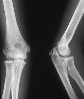
Figure 26 shows the radiograph of an otherwise healthy Caucasian 5-year-old boy who has a painless limp. What is the best treatment option?


Explanation

The prognosis of Legg-Perthes disease in children younger than age 6 years is good. There is no indication that surgical treatment will improve the outcome. Range-of-motion exercises to prevent contracture may be helpful. Herring JA, Kim HT, Browne R: Legg-Calve-Perthes disease: Prospective multicenter study of the effect of treatment on outcome. J Bone Joint Surg Am 2004;86:2121-2134.

Question 15

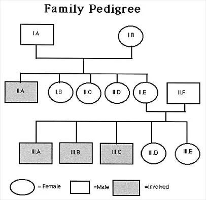
A 12-year-old girl who has a history of frequent tripping and falling also has bilateral symmetric hand weakness, high arched feet, absent patellar and Achilles tendon reflexes, and excessive wear on the lateral border of her shoes. She reports that she has multiple paternal family members with similar deformities. She most likely has a defect of what protein?


Explanation

The girl shows clinical features of hereditary motor sensory neuropathy type 1, Charcot-Marie-Tooth disease. The most common type of this autosomal-dominant disease is due to an underlying defect in the gene coding for peripheral myelin protein-22 on chromosome 17. Many other less common mutations have been identified in this family of neuropathies. Dystrophin is a protein that is abnormal in Duchenne's muscular dystrophy, which affects males and is diagnosed earlier. Type I collagen is defective in osteogenesis imperfecta. Alpha-L-iduronidase is defective in mucopolysaccharidosis type I, Hurler's syndrome. Defective cartilage oligomeric matrix protein is associated with some forms of multiple epiphyseal dysplasia. Patel PI, Roa BB, Welcher AA, et al: The gene for the peripheral myelin protein PMP-22 is a candidate for Charcot-Marie-Tooth disease type 1A. Nat Genet 1992;1:159-165.

Question 16

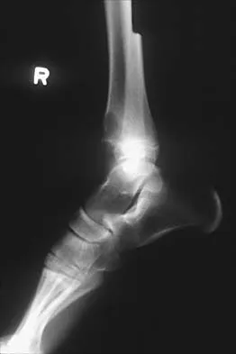
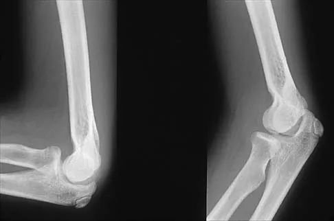
A 12-year-old boy with an ankle fracture undergoes closed reduction under sedation in the emergency department. Figure 27 shows a lateral radiograph of the ankle after two attempts at closed reduction. Based on these findings, treatment should now consist of


Explanation

The widening of the physis associated with incomplete reduction of this fracture suggests that periosteum is interposed at the fracture site. Clinical and animal study findings suggest that the interposed periosteum may lead to premature physeal closure. Repeated forceful attempts at reduction may subject the physis to further injury and should be avoided. Growth problems are common in children with Salter-Harris type I fractures of the lower extremities. Nonunions are rare in children with Salter-Harris type I fractures. Barmada A, Gaynor T, Mubarak SJ: Premature physeal closure following distal tibial physeal fractures: A new radiographic predictor. J Pediatr Orthop 2003;23:733-739.

Question 17

A 9-year-old girl has pain over the fifth toe that is aggravated by shoe wear. Clinical photographs are shown in Figures 28a and 28b. Treatment of this deformity should consist of


Explanation

28b The major obstacle to overcome in the surgical treatment of this cock-up deformity is recurrence. Dorsal releases can be performed; however, chronic dislocation of the fifth MTP joint usually needs to be addressed with plantar release as well. Chronic dorsal soft-tissue contractures may be overcome with translation of the toe into a plantar-based incision, as described originally by Cockin and accredited to Butler. This is the treatment of choice. Resection of the proximal phalanx improves symptoms but induces a secondary deformity; this procedure is usually reserved for skeletally mature individuals. Black GB, Grogan DP, Bobechko WP: Butler arthroplasty for correction of adducted fifth toe: A retrospective study of 36 operations between 1968 and 1982. J Pediatr Orthop 1985;5:439-441. Paton RW: V-Y plasty for correction of varus fifth toe. J Pediatr Orthop 1990;10:248-249.

Question 18

What acetabular procedure for developmental dysplasia of the hip does not require a concentric reduction of the femoral head in the acetabulum?


Explanation

All of the reorientation innominate osteotomies require a concentric reduction of the hip. The Staheli shelf procedure may be performed even with the hip subluxated, but it is a salvage procedure that covers a portion of the femoral head with capsular fibrocartilage rather than hyaline cartilage. Staheli LT, Chew DE: Slotted acetabular augmentation in childhood adolescence. J Pediatr Orthop 1992;12:569-580.

Question 19

Figure 29 shows the AP radiograph of a 14-year-old boy. The radiographic findings are most consistent with what pathologic process?


Explanation

The severe depression of the proximal medial tibial epiphysis is most consistent with the diagnosis of neglected infantile Blount's disease. Blount's disease in adolescents produces a deformity in the metaphyseal region. Septic arthritis and JRA affect both sides of the joint. Hemophilia produces a characteristic widening of the intercondylar notch. Thompson GH, Carter JR: Late-onset tibia vara (Blount's Disease). Clin Orthop 1990;255:24-35.

Question 20

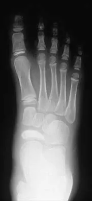
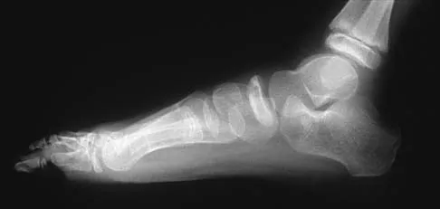
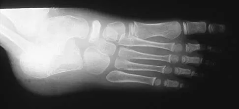
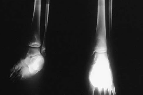
A 5-year-old boy has had pain in the right foot for the past month. Examination reveals tenderness and mild swelling in the region of the tarsal navicular. Radiographs are shown in Figure 30. Management should consist of


Explanation

The child has the classic findings of Kohler's disease or osteochondrosis of the tarsal navicular. The cause of this condition is not known, but osteonecrosis and mechanical compression have been proposed. Children generally report midfoot pain over the tarsal navicular and limping. Physical findings include tenderness, swelling, and occasionally redness in the region of the tarsal navicular. Radiographs show sclerosis and narrowing of the tarsal navicular. The natural history of the condition is spontaneous resolution and reconstitution of the navicular. Symptomatic treatment with restriction of weight bearing or casting is recommended. Karp M: Kohler's disease of the tarsal scaphoid. J Bone Joint Surg 1937;19:84-96.

Question 21

A 9-year-old child sustained a fracture-dislocation of C-5 and C-6 with a complete spinal cord injury. What is the likelihood that scoliosis will develop during the remaining years of his growth?


Explanation

The incidence of late spinal deformity after complete spinal cord injury in children depends on the level of the spinal cord injury and the age of the patient at the time of injury. If a cervical level injury occurs before age 10 years, paralytic scoliosis will develop in virtually 100% of patients. Brown JC, Swank SM, Matta J, et al: Late spinal deformity in quadriplegic children and adolescents. J Pediatr Orthop 1984;4:456-461. Lancourt JE, Dickson JH, Carter RE: Paralytic spinal deformity following traumatic spinal-cord injury in children and adolescents. J Bone Joint Surg Am 1981;63:47-53.

Question 22

Figures 31a and 31b show the radiograph and MRI scan of an otherwise normal 3-month-old infant who has a spinal deformity. MRI reveals no intraspinal anomalies. What is the next step in management?


Explanation

31b Congenital scoliosis in an infant warrants evaluation of the renal, cardiac, and neurologic systems because frequently there is concurrent pathology. Progression in this instance is possible but not certain; therefore, progression must be documented prior to any surgical intervention. Close observation with serial radiographs every 4 to 6 months is appropriate. All of the surgical options listed may be reasonable choices in the future, but cardiac evaluation is the most important issue at this time. Basu PS, Elsebaie H, Noordeen MH: Congenital spinal deformity: A comprehensive assessment at presentation. Spine 2002;27:2255-2259.

Question 23

A 22-month-old girl has cerebral palsy. Which of the following findings is a good prognostic indicator of the child's ability to walk in the future?


Explanation

For the parachute test, the examiner holds the child prone and then lowers the child rapidly toward the floor. The parachute reaction is normal or positive if the child reaches toward the floor. The Moro or startle reflex should not be present beyond age 6 months. Asymmetric tonic neck reflex, extensor thrust, and absent foot placement are abnormal findings at any age. Bleck EE: Orthopaedic Management in Cerebral Palsy. Lavenham, Suffolk, The Lavenham Press, 1987, pp 121-139.

Question 24

The husband of a 22-year-old woman has hypophosphatemic rickets. The woman has no orthopaedic abnormalities, but she is concerned about her chances of having a child with the same disease. What should they be told regarding this disorder?


Explanation

Hypophosphatemia is a rare genetic disease usually inherited as an X-linked dominant trait. The fact that the woman has no skeletal manifestations would indicate that the husband has the X-linked mutation. The disease is more severe in boys than it is in girls. The husband will not transmit the disease to his sons. However, all of their daughters will be affected either with the disease or as carriers. If the woman has the disease or the trait, there is a 50% chance that her sons will inherit the disease and a 50% chance that her daughters will be carriers or have a milder form of the disease. Parents should be advised to have genetic counseling so they can be informed when deciding whether to have children. Herring JA: Metabolic and endocrine bone diseases, in Tachdjian's Pediatric Orthopaedics, ed 3. New York, NY, WB Saunders, 2002, pp 1685-1743. Sillence DO: Disorders of bone density, volume, and mineralization, in Rimoin DL, Conner JM, Pyerite RE, et al (eds): Principles and Practice of Medical Genetics, ed 4. New York, NY, Churchill Livingstone, 2002.

Question 25

A 9-year-old boy sustained a traumatic brain injury and right lower extremity trauma in an accident involving a motor vehicle and a pedestrian. Initial evaluation in the emergency department reveals an obtunded patient who is breathing spontaneously and withdraws appropriately to painful stimuli. After initial resuscitation and stabilization, a CT scan reveals a right parietal intracranial hemorrhage. Radiographs of the swollen right thigh are shown in Figures 32a and 32b. Management of the fractured femur should ultimately consist of


Explanation

32b A child with a traumatic brain injury generally achieves significant neurologic recovery and has a more favorable prognosis than an adult. Early stabilization of fractures facilitates transportation of the child for diagnostic tests and decreases the incidence of shortening and malunion. Surgical treatment of the fracture is indicated when cerebral perfusion pressure has stabilized. Casting or traction is not the most appropriate treatment of a femoral fracture in a child of this age with a brain injury. Fracture reduction is difficult to maintain if the brain injury leads to spasticity, and transportation within the hospital for tests is more difficult. Insertion of a reamed antegrade intramedullary nail inserted at the piriformis fossa is associated with a small risk of osteonecrosis of the femoral head. The transverse femoral fracture in this patient is ideally suited for stabilization with flexible intramedullary nails. Ligier and associates treated 123 femoral shaft fractures in children with flexible intramedullary nails, including 35 patients with head injury. In one patient with hemiplegia and a urinary tract infection, a deep wound infection developed, necessitating nail removal. The remaining patients all healed without major complications. Heinrich and associates treated 78 diaphyseal femoral fractures with flexible intramedullary nails, including 14 with head injury. No major complications were reported and all fractures healed. Tolo VT: Management of the multiply injured child, in Rockwood CA, Wilkins KE, Beaty JH (eds): Fractures in Children, ed 4. Philadelphia, PA, Lippincott-Raven, 1996, pp 83-95. Ligier JN, Metaizeau JP, Prevot J, et al: Elastic stable intramedullary nailing of femoral shaft fractures in children. J Bone Joint Surg Br 1988;70:74-77. Heinrich MS, Drvaric DM, Darr K, et al: The operative stabilization of pediatric diaphyseal femur fractures with flexible intramedullary nails: A prospective analysis. J Pediatric Orthop 1994;14:501-507.

Question 26

A 5-year-old boy falls from monkey bars and presents to the emergency department. Radiographs reveal a completely displaced, extension-type supracondylar humerus fracture. On examination, his hand is pink and warm, but the radial pulse is not palpable. Capillary refill is brisk (< 2 seconds). He is unable to make an 'OK' sign, but finger extension is intact. What is the most appropriate initial management?


Explanation

The clinical presentation is a 'pulseless, pink hand' associated with a completely displaced supracondylar humerus fracture, along with an anterior interosseous nerve (AIN) palsy (unable to make an OK sign). The initial management for a well-perfused, pulseless hand in this setting is urgent closed reduction and percutaneous pinning (CRPP). Often, the pulse returns following reduction as the kinked or compressed brachial artery is relieved. If the hand remains pink and well-perfused with brisk capillary refill after stabilization, continued observation is appropriate without immediate vascular exploration. Vascular exploration is indicated if the hand is poorly perfused (pale, cold, pulseless) before reduction and remains so after reduction, or if it becomes poorly perfused after reduction.

Question 27

A 12-year-old boy weighing 95 kg presents to the emergency department with severe left hip pain and inability to bear weight after tripping two days ago. He reports mild, intermittent left knee pain over the preceding month. Radiographs demonstrate a severe left slipped capital femoral epiphysis (SCFE). Based on the Loder classification, what is the most significant risk factor for developing avascular necrosis (AVN) in this patient?


Explanation

The Loder classification categorizes Slipped Capital Femoral Epiphysis (SCFE) into stable and unstable based on the patient's clinical ability to bear weight (with or without crutches). An unstable SCFE (inability to bear weight) is associated with a much higher rate of avascular necrosis (AVN), historically reported as up to 47%, compared to nearly 0% in stable SCFE. Thus, instability is the most profound prognostic factor for AVN in these patients.

Question 28

A 4-year-old boy with a history of bilateral idiopathic clubfeet treated successfully with the Ponseti method during infancy presents with recurrent intoeing on the right. During gait evaluation, he demonstrates dynamic supination of the right foot in the swing phase. Passive range of motion shows a supple foot that is easily correctable to neutral. Radiographs are unremarkable. What is the most appropriate next step in management?


Explanation

This child has a classic presentation of clubfoot relapse following Ponseti casting, characterized by dynamic supination during the swing phase of gait. In a child older than 2.5 to 3 years with a supple foot, the treatment of choice is the transfer of the anterior tibial tendon (ATT) to the third (lateral) cuneiform. This converts the ATT from a supinator to a straight dorsiflexor, rebalancing the foot and preventing further recurrence. If fixed equinus or cavus is present, preliminary re-casting or a concurrent Achilles tendon lengthening may be required.

Question 29

A 13-year-old boy presents with chronic, vague midfoot aching and a history of frequent ankle sprains. Examination reveals a rigid, flat right foot with severely limited subtalar motion. Forced inversion elicits significant pain. Lateral foot radiographs demonstrate a 'C-sign'. A subsequent CT scan confirms a coalition involving less than 50% of the posterior facet with no degenerative changes. After failing 6 months of nonoperative management, including immobilization in a short leg cast, what is the recommended surgical procedure?


Explanation

The 'C-sign' on a lateral radiograph is formed by the continuous outline of the medial border of the talar dome and the sustentaculum tali, strongly indicating a talocalcaneal coalition. Since the coalition involves less than 50% of the posterior facet and the patient has no secondary degenerative changes, the appropriate surgical management after failing conservative treatment is resection of the talocalcaneal coalition. Interposition of a fat graft (or bone wax) is recommended to reduce the risk of recurrence. If the coalition was >50% or if advanced arthritis was present, a subtalar or triple arthrodesis would be indicated.

Question 30

A 14-year-old girl sustains an ankle injury while playing soccer. Radiographs show a displaced Salter-Harris III fracture of the anterolateral distal tibia. Which of the following best describes the biomechanical mechanism and developmental etiology of this specific fracture?


Explanation

This is a juvenile Tillaux fracture, which is a Salter-Harris III fracture of the anterolateral distal tibia. It typically occurs in adolescents around 12 to 14 years of age. The distal tibial physis closes in an asymmetric, predictable pattern: central, then anteromedial, then posteromedial, and finally lateral (anterolateral). Because the anterolateral physis is the last to close, it is susceptible to avulsion forces. The mechanism is external rotation of the foot within the mortise, causing the anterior inferior tibiofibular ligament (AITFL) to avulse the anterolateral epiphyseal fragment.

Question 31

An 8-year-old boy is diagnosed with Legg-Calvé-Perthes disease. AP and frog-leg lateral pelvis radiographs show the hip is currently in the fragmentation stage. According to the Herring lateral pillar classification, which of the following radiographic features is the most critical for determining the long-term prognosis?


Explanation

The Herring Lateral Pillar Classification is evaluated on the AP radiograph during the fragmentation stage of Legg-Calvé-Perthes disease and is widely considered the most reliable prognostic indicator for future sphericity of the femoral head. It specifically evaluates the degree of height loss in the lateral pillar (the lateral 15% to 30% of the femoral head). Group A has no height loss, Group B has less than 50% height loss, and Group C has greater than 50% height loss (which carries the worst prognosis).

Question 32

An 18-month-old girl presents with a painless limp. Examination demonstrates a positive Trendelenburg sign on the left and a leg length discrepancy. Pelvic radiographs confirm a completely dislocated left hip with an acetabular index of 42 degrees and a broken Shenton's line.

What is the most appropriate definitive management?


Explanation

In a child of ambulatory age (typically > 18 months) presenting with a missed or late-diagnosed Developmental Dysplasia of the Hip (DDH) that is completely dislocated, secondary adaptive changes such as severe soft tissue contractures, acetabular dysplasia, and excessive femoral anteversion/coxa valga have occurred. An open reduction is typically necessary to clear obstacles (e.g., inverted limbus, hypertrophied pulvinar, intact transverse acetabular ligament). A femoral shortening osteotomy is frequently performed to decompress the joint, allowing reduction without excessive pressure on the cartilage, thereby minimizing the risk of avascular necrosis (AVN). A concurrent pelvic osteotomy (e.g., Salter or Pemberton) is necessary to correct the severe acetabular dysplasia (acetabular index of 42 degrees).

Question 33

A 3-year-old boy presents with a femur fracture following minimal trauma. This is his fourth long bone fracture. Clinical examination reveals blue sclerae and dentinogenesis imperfecta. Genetic testing confirms a mutation in the COL1A1 gene. He is started on intravenous pamidronate. What is the primary mechanism of action of this pharmacological therapy?


Explanation

The patient has Osteogenesis Imperfecta (OI), typically caused by an autosomal dominant mutation in type I collagen genes (COL1A1 or COL1A2). Intravenous bisphosphonates, such as pamidronate, are the standard of care for moderate to severe OI to reduce fracture frequency and improve bone density. Bisphosphonates function primarily by attaching to hydroxyapatite binding sites on bony surfaces, leading to the inhibition of osteoclast-mediated bone resorption. They do not repair the underlying genetic defect in collagen synthesis.

Question 34

A 7-year-old girl with spastic quadriplegic cerebral palsy (GMFCS Level V) is evaluated during routine hip surveillance. Her parents report increasing difficulty with perineal hygiene and positioning her in her wheelchair. An AP pelvis radiograph demonstrates a right hip Reimers migration percentage of 65% with significant coxa valga and an intact acetabular teardrop.

What is the recommended surgical management to provide a stable, concentric hip?


Explanation

Children with severe cerebral palsy (GMFCS IV and V) are at high risk for progressive hip displacement due to spasticity and muscle imbalance, leading to coxa valga and secondary acetabular dysplasia. A Reimers migration percentage (MP) greater than 50% generally indicates the need for comprehensive bony reconstruction. The standard of care to achieve a stable, concentric, and pain-free hip in this scenario is a one-stage reconstruction involving a varus derotational osteotomy (VDRO) of the proximal femur to correct the coxa valga and anteversion, combined with a pelvic osteotomy (e.g., Dega, San Diego, or Pemberton) to address acetabular dysplasia, often alongside soft-tissue releases. Salvage procedures (like resection arthroplasty) are reserved for painful, chronically dislocated hips with severe degenerative changes that cannot be reconstructed.

Question 35

A 10-year-old boy with spinal muscular atrophy (SMA) type II presents with a progressive, collapsing thoracolumbar neuromuscular scoliosis measuring 85 degrees. He is non-ambulatory, has pelvic obliquity, and his forced vital capacity (FVC) is 40% of predicted. He underwent placement of magnetically controlled growing rods at age 5, which have now reached their maximum excursion. What is the most definitive surgical option at this stage?


Explanation

This patient has reached the stage where his growth-friendly instrumentation (magnetic rods) has 'graduated' or 'maxed out.' In a 10-year-old with a severe, progressive collapsing neuromuscular scoliosis (like in SMA) and pelvic obliquity, sufficient spinal length has usually been achieved. The definitive and standard management upon graduation from a growing construct is a definitive posterior spinal fusion (PSF). For non-ambulatory patients with neuromuscular scoliosis and pelvic obliquity, the fusion typically extends from the upper thoracic spine down to the pelvis to restore sitting balance, correct the pelvic obliquity, and provide a stable spine for pulmonary function and wheelchair seating.

Question 36

A 6-year-old boy sustains a completely displaced Gartland type III supracondylar humerus fracture. He undergoes prompt closed reduction and percutaneous pinning. Postoperatively, the radial pulse remains unpalpable, but the hand is warm with a brisk capillary refill of less than 2 seconds. Pulse oximetry on the index finger shows a strong waveform and 99% oxygen saturation.

What is the most appropriate next step in management?


Explanation

In the setting of a supracondylar humerus fracture with a "pulseless but pink" hand following satisfactory closed reduction and pinning, the standard of care is observation and admission for serial clinical examinations. The collateral circulation in pediatric patients is typically robust enough to maintain adequate perfusion even if the brachial artery is in spasm, contused, or tethered. Arterial exploration is strictly indicated if the hand is persistently ischemic (white, cool, lack of capillary refill) after reduction.

Question 37

A 2.5-year-old girl is brought in for a persistent, painless limp. Physical examination reveals asymmetric thigh folds, limited abduction of the left hip, and a positive Galeazzi sign on the left. Radiographs confirm a dislocated left hip with an acetabular index of 42 degrees and a delayed ossification center of the femoral head.

What is the most appropriate surgical management for this patient?


Explanation

By the age of 2.5 years, conservative measures such as a Pavlik harness or closed reduction are inappropriate due to the severity of adaptive changes and capsular constriction. This patient has Developmental Dysplasia of the Hip (DDH) with significant acetabular dysplasia (acetabular index > 30 degrees). The standard of care for a walking child older than 18-24 months is an open reduction combined with a pelvic osteotomy (e.g., Salter or Pemberton) to correct the dysplasia and provide anterior/lateral coverage, often accompanied by a femoral shortening osteotomy depending on the degree of proximal migration.

Question 38

A 12-year-old boy is diagnosed with a stable slipped capital femoral epiphysis (SCFE) of the left hip. He denies any right hip pain. Which of the following is considered the most widely accepted absolute indication for prophylactic in-situ pinning of his contralateral asymptomatic right hip?


Explanation

While prophylactic pinning of the contralateral hip in SCFE is controversial for idiopathic cases, there is broad consensus that patients with atypical SCFE—specifically those with underlying endocrine disorders (e.g., hypothyroidism, panhypopituitarism, renal osteodystrophy) or prior radiation therapy—should undergo prophylactic contralateral pinning. These patients have a significantly higher risk of bilateral involvement (up to 100% in some endocrine cohorts) compared to those with idiopathic SCFE.

Question 39

A 4-year-old boy who was successfully treated for an idiopathic clubfoot with the Ponseti method presents with a relapse. His parents report that he walks on the outside of his foot. On examination, he demonstrates dynamic supination of the foot during the swing phase of gait. However, his passive ankle dorsiflexion is 15 degrees with the knee extended, and his heel is in neutral alignment. What is the most appropriate next step in management?


Explanation

Dynamic supination during the swing phase in a toddler with a previously corrected clubfoot is a classic presentation of a localized relapse caused by a strong over-pull of the tibialis anterior muscle. Because passive dorsiflexion is well maintained (no fixed equinus), an Achilles tenotomy is not required. The treatment of choice in children (typically aged 2.5 to 5 years) is a full transfer of the anterior tibial tendon (TATT) to the lateral (third) cuneiform. A split transfer (SPLATT) is generally reserved for adult patients with upper motor neuron lesions (e.g., stroke, traumatic brain injury) and is not the standard of care for pediatric clubfoot relapses.

Question 40

An 8-year-old boy is diagnosed with Legg-Calvé-Perthes disease. According to the modified lateral pillar (Herring) classification, which of the following radiographic findings signifies the poorest prognosis for long-term hip congruency?


Explanation

The Herring (Lateral Pillar) classification evaluates the height of the lateral pillar of the femoral head during the fragmentation stage of Legg-Calvé-Perthes disease. Group A has 100% height maintenance, Group B has >50% maintenance, and Group C has <50% maintenance of the lateral pillar height. Group C carries the worst prognosis for maintaining a spherical, congruent hip joint at skeletal maturity, particularly in patients who present over the age of 8. Group B/C (the modified addition) also represents a borderline poor prognosis.

Question 41

A 10-year-old premenarcheal girl is incidentally found to have a right thoracic adolescent idiopathic scoliosis (AIS). Upright standing radiographs demonstrate a Cobb angle of 26 degrees. Her Risser stage is 0. Based on standard prognostic criteria (Lonstein and Carlson), what is the approximate risk that this curve will progress to a surgical or bracing threshold (>50 degrees or requiring intervention)?


Explanation

The Lonstein and Carlson progression factor evaluates the risk of curve progression in AIS based on the Cobb angle, Risser stage, and chronological age. A young, premenarcheal patient (Risser 0 or 1) presenting with a curve between 20 and 29 degrees has a high risk of progression, calculated to be approximately 68%. This patient clearly meets the indications for bracing (curve > 25 degrees in an immature patient).

Question 42

An 11-year-old boy weighing 65 kg (143 lbs) sustains an isolated, closed, transverse midshaft femur fracture during a football game. What is the most appropriate definitive surgical management to minimize complications while maximizing functional outcome?


Explanation

For pediatric femoral shaft fractures, patient weight and age dictate the optimal implant. Children weighing over 50 kg (110 lbs) or older than 11 years have unacceptably high rates of loss of fixation, malunion, and hardware prominence when treated with flexible titanium elastic nails (TENs). Rigid intramedullary nailing is indicated; however, the piriformis fossa entry point is strictly avoided in children due to the high risk of iatrogenic avascular necrosis of the femoral head. A lateral trochanteric entry point rigid nail avoids the vascular supply to the femoral head and is the standard of care for this demographic.

Question 43

A 7-year-old boy with spastic quadriplegic cerebral palsy (GMFCS Level V) presents for routine orthopedic surveillance. His parents report increased difficulty with perineal care. Pelvic radiographs demonstrate a Reimers migration percentage of 55% in the right hip. Clinical exam reveals hip abduction is limited to 15 degrees bilaterally with the hips flexed.

What is the most appropriate management?


Explanation

Hip displacement is a common and morbid complication in children with severe cerebral palsy (GMFCS IV and V). A Reimers migration percentage greater than 40-50% indicates significant subluxation that has progressed beyond the capacity of isolated soft-tissue releases (which are typically indicated for migration percentages between 30% and 40%). Reconstructive surgery consisting of a proximal femoral varus derotational osteotomy (VDRO) combined with a pelvic osteotomy (e.g., Dega or San Diego) is the gold standard to achieve a concentric, painless, and stable hip in this setting.

Question 44

A 13-year-old boy presents with an insidious onset of right lateral foot pain and a history of recurrent ankle sprains. Examination shows a rigid flatfoot on the right side. A lateral radiograph demonstrates an elongated anterior process of the calcaneus (the "anteater nose" sign). Which of the following physical examination findings is most specific to this diagnosis?


Explanation

The "anteater nose" sign is pathognomonic for a calcaneonavicular tarsal coalition. Tarsal coalitions mechanically block normal subtalar motion, resulting in a peroneal spastic flatfoot. During a normal single-limb heel rise (tiptoe stance), the windlass mechanism and subtalar joint mechanics cause the hindfoot to invert (correct into varus). In a rigid flatfoot caused by a tarsal coalition, the hindfoot remains fixed in valgus during heel rise.

Question 45

A 2-year-old boy, who is above the 95th percentile for weight, presents with bilateral bowing of his legs. Standing AP radiographs show a metaphyseal-diaphyseal angle (MDA) of 18 degrees bilaterally, with early beaking of the medial proximal tibial metaphysis. Which of the following is the most appropriate initial management?


Explanation

This presentation is highly characteristic of infantile Blount disease (tibia vara), defined by pathological bowing and a metaphyseal-diaphyseal angle (MDA, or Drennan angle) greater than 16 degrees. While physiological bowing resolves spontaneously and typically has an MDA < 11 degrees, an MDA > 16 degrees demands intervention. The accepted first-line treatment for infantile Blount disease in a child under the age of 3 is bracing with knee-ankle-foot orthoses (KAFOs). Surgery is reserved for those who fail bracing or who present at an older age (typically >4 years) with progressive deformity.

Question 46

A 4-year-old boy treated previously for idiopathic clubfoot with the Ponseti method presents with a relapsed dynamic supination deformity during the swing phase of gait. His passive ankle dorsiflexion is 15 degrees, and the hindfoot is flexible. What is the most appropriate next step in management?


Explanation

For a relapsed dynamic supination deformity in a child who has previously undergone successful Ponseti casting and has flexible deformity and adequate dorsiflexion, a tibialis anterior tendon transfer (TATT) to the lateral cuneiform is the treatment of choice.

Question 47

A 6-week-old female infant is placed in a Pavlik harness for a dislocated left hip. After 3 weeks of proper wear, ultrasound demonstrates that the hip remains persistently dislocated. What is the most appropriate next step in management?


Explanation

Continuation of a Pavlik harness in a persistently dislocated hip beyond 3 to 4 weeks increases the risk of 'Pavlik harness disease' (damage to the posterior wall of the acetabulum) and avascular necrosis. The harness should be discontinued, and the infant should be scheduled for a closed reduction and spica casting.

Question 48

A 14-year-old boy presents with a painful, swollen ankle after a skateboarding fall. Radiographs demonstrate an intra-articular fracture of the anterolateral distal tibial epiphysis. What ligament is responsible for the avulsion of this fracture fragment?


Explanation

A juvenile Tillaux fracture is a Salter-Harris III fracture of the anterolateral distal tibial epiphysis. It is caused by an avulsion force transmitted through the anterior inferior tibiofibular ligament (AITFL) during external rotation of the foot. It occurs in adolescents because the distal tibial physis closes from central to anteromedial to posteromedial to lateral, leaving the anterolateral physis open and vulnerable.

Question 49

A 12-year-old boy with a BMI of 32 undergoes in situ pinning of a stable slipped capital femoral epiphysis (SCFE) with a single cannulated screw. Postoperatively, he has persistent severe pain, limited range of motion, and joint stiffness. Radiographs show joint space narrowing and subchondral radiolucencies. What is the most likely diagnosis?


Explanation

Chondrolysis is a devastating complication of SCFE characterized by acute loss of articular cartilage, leading to rapid joint space narrowing, stiffness, and severe pain. It is associated with unrecognized screw penetration into the joint. Avascular necrosis typically presents with sclerosis, cysts, and eventual collapse of the femoral head rather than diffuse joint space narrowing early on.

Question 50

A 6-year-old girl sustains an extension-type completely displaced supracondylar humerus fracture. Examination reveals she is unable to flex the interphalangeal joint of her thumb and the distal interphalangeal joint of her index finger. Which nerve is most likely injured?


Explanation

The anterior interosseous nerve (AIN), a branch of the median nerve, is the most commonly injured nerve in extension-type supracondylar humerus fractures. Injury to the AIN results in the inability to flex the interphalangeal joint of the thumb (flexor pollicis longus) and the distal interphalangeal joint of the index finger (flexor digitorum profundus).

Question 51

A 5-year-old boy with spastic quadriplegic cerebral palsy (GMFCS Level V) is evaluated in the clinic. His bilateral hip migration percentages are calculated to be 45%. He has limited hip abduction to 20 degrees bilaterally. What is the most appropriate management?


Explanation

In non-ambulatory children (GMFCS IV and V) with cerebral palsy, a migration percentage (MP) > 40% usually indicates progressive hip displacement that is unlikely to respond to soft-tissue releases (adductor tenotomies) alone. The standard of care for a hip with an MP > 40% and significant dysplasia is bony reconstructive surgery, which typically involves a varus derotational osteotomy (VDRO) of the proximal femur, often combined with a pelvic osteotomy (e.g., Dega or San Diego).

Question 52

A 3-year-old girl with recurrent long bone fractures, blue sclerae, and dentinogenesis imperfecta is diagnosed with osteogenesis imperfecta. She is started on a medical therapy that aims to increase bone mineral density and reduce the fracture rate. What is the primary mechanism of action of this medication class?


Explanation

The patient has osteogenesis imperfecta and is being treated with bisphosphonates (e.g., pamidronate or zoledronic acid). Bisphosphonates inhibit osteoclast-mediated bone resorption by inducing osteoclast apoptosis, which leads to increased bone mineral density, a reduced rate of fractures, and improvement in vertebral body shape in children with OI.

Question 53

A 5-year-old boy falls off monkey bars and sustains a laterally displaced pediatric elbow fracture.

Radiographs demonstrate a fracture of the lateral condyle with 4 mm of displacement. What is the most appropriate management?


Explanation

Lateral condyle fractures of the humerus with > 2 mm of displacement are generally treated with open reduction and internal fixation (ORIF) to ensure anatomic restoration of the articular surface and physis, minimizing the risk of nonunion, malunion, and late tardy ulnar nerve palsy. Closed reduction and pinning can be attempted for less displaced fractures, but 4 mm requires direct visualization to ensure the articular surface is congruent.

Question 54

A 7-year-old boy presents with a painless limp of 3 months duration. Radiographs confirm the diagnosis of Legg-Calve-Perthes disease. Which of the following is considered a 'head at risk' sign indicating a poorer prognosis and potential need for surgical intervention?


Explanation

Catterall described several 'head at risk' signs in Legg-Calve-Perthes disease that indicate a poor prognosis and a higher likelihood of femoral head deformity. These include Gage sign (a V-shaped radiolucency in the lateral portion of the epiphysis/metaphysis), lateral subluxation of the femoral head, calcification lateral to the epiphysis, horizontal growth plate, and metaphyseal cysts.

Question 55

A 13-year-old boy complains of recurrent ankle sprains and deep, aching midfoot pain. Physical examination reveals rigid, flat feet and decreased subtalar motion. Radiographs reveal a 'C-sign' on the lateral view. What is the most likely anatomic location of the pathology?


Explanation

The 'C-sign' on a lateral foot radiograph is a classic finding for a talocalcaneal coalition. It represents the continuous bony bridge between the talar dome and the sustentaculum tali. Calcaneonavicular coalitions are best seen on an oblique radiograph and classic findings include the 'anteater nose' sign.

Question 56

A 13-year-old boy presents with severe right hip and thigh pain after a minor slip. He is unable to bear weight on the right leg. He reports a 2-month history of intermittent right knee pain prior to this event. On examination, attempted hip flexion results in obligatory external rotation. Radiographs confirm a displaced slipped capital femoral epiphysis (SCFE).

What is the most appropriate management to minimize the risk of avascular necrosis (AVN) in this patient?


Explanation

This patient has an unstable Slipped Capital Femoral Epiphysis (SCFE), defined by the Loder classification as the inability to bear weight even with crutches. Unstable SCFE carries a significantly higher risk of avascular necrosis (AVN) compared to stable SCFE (up to 50% vs. nearly 0%). Current evidence strongly supports urgent surgical intervention (within 24 hours of symptom onset) via in situ percutaneous pinning or an open procedure (like a modified Dunn) depending on the surgeon's expertise. Urgent decompression/pinning decreases the intracapsular pressure and stabilizes the physis, significantly lowering the AVN rate compared to delayed treatment. Traction and spica casting are historically associated with poor outcomes and are no longer standard of care.

Question 57

A 5-year-old boy sustains a completely displaced supracondylar humerus fracture (Gartland Type III). Upon presentation, his hand is pink, but the radial pulse is absent. He undergoes urgent closed reduction and percutaneous pinning.

In the recovery room, the fracture is well-reduced, the hand remains pink and warm with a capillary refill of less than 2 seconds, and oxygen saturation on the index finger is 99%; however, the radial pulse remains nonpalpable. What is the most appropriate next step in management?


Explanation

The 'pulseless, pink, perfused' hand after adequate closed reduction and percutaneous pinning of a supracondylar humerus fracture is a well-recognized clinical entity. Because the hand is well perfused (capillary refill <2 seconds, good warmth, and normal pulse oximetry), collateral circulation is adequate. The standard of care is admission for 24 to 48 hours for close neurovascular monitoring. Operative exploration of the brachial artery is only indicated if the hand becomes dysvascular (pulseless, pale, cold) after reduction, or if there is impending compartment syndrome.

Question 58

An 18-month-old girl presents with a painless limp. Her parents note that her left leg appears shorter than the right. On examination, Galeazzi sign is positive on the left, and hip abduction is restricted. Radiographs demonstrate a completely dislocated left hip with a broken Shenton's line and an acetabular index of 42 degrees.

Which of the following is the most appropriate definitive management?


Explanation

In a child of walking age (typically older than 18 months) presenting with a newly diagnosed, completely dislocated hip due to Developmental Dysplasia of the Hip (DDH), closed reduction is rarely successful or adequate. Due to secondary adaptive changes, including acetabular dysplasia (evidenced by an acetabular index of 42 degrees) and contractures, open reduction is required. Furthermore, a concomitant pelvic osteotomy (e.g., Salter or Pemberton) is almost always necessary to provide adequate anterolateral coverage for the femoral head and ensure stability. A femoral shortening osteotomy may also be needed to reduce pressure on the proximal femoral physis and decrease the risk of avascular necrosis.

Question 59

A 4-year-old boy with a history of idiopathic congenital talipes equinovarus initially successfully treated with the Ponseti method presents with recurrent in-toeing and lateral foot wear. On examination, he demonstrates a dynamic supination of the foot during the swing phase of gait. His passive range of motion is full, the foot is completely correctable passively, and there is no fixed equinus. What is the most appropriate surgical intervention?


Explanation

The patient presents with dynamic supination and recurrent in-toeing, a common manifestation of a relapsed clubfoot treated with the Ponseti method. Because the deformity is dynamic and passively correctable, bony procedures or extensive soft tissue releases are not indicated. The treatment of choice for a dynamic supination deformity in a relapsed Ponseti clubfoot is the transfer of the entire Tibialis Anterior Tendon (TATT) to the third (lateral) cuneiform. A split anterior tibial tendon transfer (SPLATT) is less predictable in this specific condition and is more commonly used in cerebral palsy or adult acquired deformities.

Question 60

An 8-year-old boy is evaluated for an 8-month history of right hip pain and a painless limp.

AP pelvis radiographs demonstrate fragmentation of the right capital femoral epiphysis consistent with Legg-Calvé-Perthes disease. According to the Herring Lateral Pillar Classification, which of the following defines a Lateral Pillar Group C hip, which carries the poorest prognosis?


Explanation

The Herring Lateral Pillar Classification is the most widely used prognostic radiographic classification for Legg-Calvé-Perthes disease, evaluated during the fragmentation stage on the AP radiograph. Group A involves no radiolucency or loss of height in the lateral third of the epiphysis (lateral pillar). Group B demonstrates a lucency but maintains >50% of the lateral pillar height. Group C involves a loss of >50% of the lateral pillar height (i.e., maintenance of <50%). Group C hips have the poorest prognosis, frequently leading to a flat, aspherical head and early-onset osteoarthritis.

Question 61

A 2-and-a-half-year-old girl is evaluated for bilateral bowlegs. Standing AP radiographs of the lower extremities are obtained.

Which of the following radiographic parameters is most reliable in differentiating infantile Blount disease from physiologic genu varum?


Explanation

The metaphyseal-diaphyseal angle (MDA) of Drennan is the most reliable radiographic parameter used to differentiate infantile Blount disease from physiologic genu varum. An MDA greater than 16 degrees is highly predictive of progressive infantile Blount disease, whereas an MDA less than 11 degrees suggests physiologic bowing that will likely resolve spontaneously. Values between 11 and 16 degrees warrant close observation and serial radiographs. Tibiofemoral angle and mechanical axis deviation do not reliably differentiate the two entities in this age group, as both are present in physiologic bowing.

Question 62

A 12-year-old girl presents for evaluation of a spinal deformity. Standing posteroanterior radiographs demonstrate a right thoracic adolescent idiopathic scoliosis (AIS) with a Cobb angle of 22 degrees.

Which of the following parameters indicates that the patient is currently in the period of maximum risk for rapid curve progression?


Explanation

The risk of progression in adolescent idiopathic scoliosis (AIS) is most strongly correlated with the patient's remaining growth potential. The period of maximum growth—and thus maximum curve progression risk—is during peak height velocity (PHV). PHV typically occurs before menarche, before the closure of the triradiate cartilage, and at Risser stage 0 (typically Sanders stage 3). Risser stage 4, post-menarche status, and Sanders stage 7 all indicate advanced skeletal maturity and a significantly lower risk of curve progression.

Question 63

A 14-year-old boy presents to the emergency department after sustaining a twisting injury to his right ankle while skateboarding. Radiographs and a subsequent CT scan demonstrate a Salter-Harris III fracture of the anterolateral distal tibial epiphysis.

Which of the following ligamentous structures is responsible for avulsing this bony fragment?


Explanation

The patient has a Juvenile Tillaux fracture, which is a Salter-Harris III fracture of the anterolateral distal tibial epiphysis. This injury occurs in adolescents (typically ages 12-14) because the distal tibial physis closes in a predictable pattern: central -> anteromedial -> posteromedial -> anterolateral. During an external rotation injury, the anterior inferior tibiofibular ligament (AITFL) becomes taut and avulses the unfused anterolateral epiphysis. The ATFL, CFL, and Deltoid ligaments do not attach to this specific fragment.

Question 64

A 3-year-old child with a known diagnosis of Osteogenesis Imperfecta Type III is admitted to the hospital for elective placement of telescopic intramedullary rods in bilateral femurs. To decrease fracture burden and improve bone mineral density, the patient receives cyclical intravenous pamidronate therapy. What is the primary cellular mechanism of action of this pharmacological treatment?


Explanation

Pamidronate is a nitrogen-containing bisphosphonate. The primary mechanism of action of all bisphosphonates is the inhibition of osteoclast-mediated bone resorption. Specifically, nitrogen-containing bisphosphonates inhibit farnesyl pyrophosphate synthase in the mevalonate pathway, leading to osteoclast apoptosis and decreased bone turnover. They do not primarily stimulate osteoblasts, alter collagen cross-linking (the primary structural defect in OI), or significantly affect PTH secretion or direct intestinal absorption.

Question 65

A 13-year-old girl presents with a 1-year history of recurrent right ankle sprains and deep lateral hindfoot pain. On physical examination, she has a rigid pes planus deformity with peroneal spasticity and significantly limited subtalar motion.

Oblique radiographs of the right foot reveal an elongated anterior process of the calcaneus, often referred to as the 'anteater nose' sign. What is the most likely diagnosis?


Explanation

The patient's clinical presentation of recurrent sprains, rigid flatfoot, and peroneal spasticity is characteristic of a tarsal coalition. The 'anteater nose' sign on an oblique radiograph is pathognomonic for a calcaneonavicular coalition. This sign represents a tubular elongation of the anterior process of the calcaneus that approaches or fuses with the navicular. Talocalcaneal (subtalar) coalitions are typically visualized on Harris axial or lateral radiographs (e.g., the 'C-sign' of Lateur) and are best confirmed with CT, but the 'anteater nose' specifically defines the calcaneonavicular variant.

Question 66

A 12-year-old obese boy presents with 3 weeks of vague knee pain and a limp. Examination reveals obligate external rotation of the hip during flexion. He is diagnosed with a stable slipped capital femoral epiphysis (SCFE).

What is the most appropriate position to place the hip during in situ single-screw fixation to minimize the risk of osteonecrosis?


Explanation

For a stable slipped capital femoral epiphysis (SCFE), the standard of care is in situ fixation with a single cannulated screw. Attempting to forcefully reduce a stable SCFE significantly increases the risk of osteonecrosis (avascular necrosis) of the femoral head due to disruption of the delicate epiphyseal blood supply. Therefore, the hip should be pinned in its resting position without deliberate attempts at reduction.

Question 67

A 5-year-old girl falls from the monkey bars and sustains a Gartland type III extension-type supracondylar humerus fracture.

On presentation, she has a pulseless, pink hand. After prompt closed reduction and percutaneous pinning, her hand remains warm and pink with a capillary refill of 2 seconds, but the radial pulse is still non-palpable. What is the most appropriate next step in management?


Explanation

A pulseless, pink hand is a well-recognized clinical scenario following a displaced supracondylar humerus fracture. If the hand remains well-perfused (pink, warm, brisk capillary refill) after anatomical reduction and stabilization, collateral circulation is adequate. The standard of care is to admit the patient for close observation and serial neurovascular checks. Open vascular exploration is indicated if the hand is pulseless AND poorly perfused (pale/white) after reduction.

Question 68

A 30-month-old girl is evaluated for worsening bilateral genu varum and an evolving thrust during gait. Standing radiographs demonstrate medial metaphyseal beaking.

The metaphyseal-diaphyseal angle (MDA) is measured at 18 degrees on both sides. What is the most appropriate initial management?


Explanation

The clinical picture and a metaphyseal-diaphyseal angle (MDA) > 16 degrees are diagnostic for infantile Blount disease (tibia vara). For children under 3 years of age with early-stage disease (Langenskiöld stage I or II), the initial treatment is non-operative utilizing knee-ankle-foot orthoses (KAFOs) during weight-bearing. Surgery is reserved for patients who fail bracing or present at an older age (typically >3-4 years).

Question 69

A 4-week-old boy is undergoing serial casting using the Ponseti method for isolated, idiopathic congenital talipes equinovarus.

After 4 weeks of casts, the forefoot has been successfully abducted to 60 degrees. However, the heel remains in 15 degrees of equinus. What is the next most appropriate step in management?


Explanation

In the Ponseti method for clubfoot, the sequence of correction is cavus, adductus, varus, and finally equinus. Once the forefoot is abducted to roughly 60 degrees and the calcaneus is visibly everted, residual equinus (which is present in >90% of cases) is treated with a percutaneous Achilles tenotomy. Forcing dorsiflexion in the cast without tenotomy can lead to a midfoot breach (rocker-bottom foot).

Question 70

A 7-year-old boy presents with a painless limp of 3 months' duration. Radiographs demonstrate sclerosis and fragmentation of the capital femoral epiphysis, leading to a diagnosis of Legg-Calvé-Perthes disease.

Which of the following radiographic findings is a "head-at-risk" sign described by Catterall, indicating a poorer prognosis?


Explanation

Catterall identified specific "head-at-risk" clinical and radiographic signs that portend a worse prognosis and a higher likelihood of femoral head deformation in Perthes disease. The radiographic signs include: 1) Gage sign (a V-shaped radiolucency in the lateral portion of the epiphysis and adjacent metaphysis), 2) calcification lateral to the epiphysis, 3) lateral (not medial) subluxation of the femoral head, 4) horizontal physis, and 5) metaphyseal cysts.

Question 71

A 30-month-old girl is brought in by her parents who noticed she walks with a limp. She has not received any prior orthopedic care. Pelvic radiographs reveal a completely dislocated left hip with acetabular dysplasia and a false acetabulum.

What is the most appropriate definitive management?


Explanation

In a child older than 2 years (24 months) presenting with untreated developmental dysplasia of the hip (DDH) and a high dislocation, closed reduction is rarely successful and carries an unacceptably high risk of osteonecrosis. The gold standard is open reduction. Because the soft tissues are contracted and the acetabulum is dysplastic, a femoral shortening osteotomy (to reduce tension and AVN risk) and a pelvic osteotomy (to address acetabular dysplasia) are typically required simultaneously.

Question 72

A 12-year-old boy presents with a painful, swollen knee after falling from a bicycle. Radiographs reveal a completely displaced (Meyers-McKeever Type III) tibial eminence fracture.

Attempts at closed reduction in full extension fail to anatomically reduce the fragment. Which structure is most commonly entrapped beneath the fragment, blocking reduction?


Explanation

Tibial eminence (spine) fractures represent an avulsion of the anterior cruciate ligament insertion in children. When a Type III (completely displaced) fracture cannot be reduced closed, the most common anatomic block to reduction is the entrapment of the anterior horn of the medial meniscus (or the transverse intermeniscal ligament) under the bony fragment. Operative intervention (arthroscopic or open) is required to free the entrapped tissue and fix the fragment.

Question 73

A 14-year-old boy presents with recurrent ankle sprains and rigid, painful flatfeet. Examination reveals severe restriction of subtalar motion and spasm of the peroneal tendons on forceful inversion. Radiographs show a prominent "C sign" and a talar beak.

Which of the following conditions is most likely present?


Explanation

The clinical presentation is classic for a tarsal coalition (rigid flatfoot, peroneal spasticity). The "C sign" on a lateral radiograph—formed by the continuous outline of the medial outline of the talar dome and the inferior outline of the sustentaculum tali—is a highly specific indicator of a talocalcaneal (subtalar) coalition, most commonly involving the middle facet. Calcaneonavicular coalitions are best seen on oblique views and characteristically show the "anteater nose" sign.

Question 74

A 13-year-old premenarchal girl (Risser 0) presents for evaluation of a spinal deformity. Neurological examination is completely normal. Standing PA spine radiograph reveals a right thoracic curve measuring 35 degrees.

What is the most appropriate next step in management?


Explanation

This patient has Adolescent Idiopathic Scoliosis (AIS). She has significant remaining growth potential (premenarchal, Risser 0) and a curve magnitude between 25 and 45 degrees. According to the Bracing in Adolescent Idiopathic Scoliosis Trial (BRAIST) criteria, the standard of care for a skeletally immature patient with a curve of this magnitude is a rigid brace (e.g., TLSO) worn for at least 18 hours a day to prevent curve progression to surgical magnitude (>50 degrees).

Question 75

A 14-year-old boy presents with chronic anteromedial knee pain. An MRI is obtained which demonstrates a 2.5 x 2.5 cm osteochondritis dissecans (OCD) lesion on the lateral aspect of the medial femoral condyle. T2-weighted sequences show high signal intensity fluid completely encircling the bony lesion.

Diagnostic arthroscopy reveals a ballottable but macroscopically intact articular cartilage surface. What is the most appropriate surgical treatment?


Explanation

This patient has an unstable but intact osteochondritis dissecans (OCD) lesion. MRI evidence of fluid tracking behind the lesion indicates instability. Because the articular cartilage is intact and the patient is near skeletal maturity (where healing potential decreases compared to younger children), surgical stabilization is indicated. In situ fixation (using bioabsorbable pins/screws or headless metallic screws) promotes healing by compressing the unstable fragment into its bed while preserving the native articular cartilage.

Question 76

A 12-year-old boy presents to the emergency department unable to bear weight on his left leg for the past 2 days after jumping off a swing. He refuses to walk even with crutches. Figure 4 shows the AP pelvis radiograph.

He is diagnosed with a slipped capital femoral epiphysis (SCFE) and undergoes urgent single-screw in situ fixation. Which of the following is the most likely complication associated with this specific type of presentation compared to a patient who is able to bear weight?


Explanation

The patient has an unstable slipped capital femoral epiphysis (SCFE), defined by the Loder classification as the inability to bear weight with or without crutches. The most significant and common severe complication of an unstable SCFE is avascular necrosis (AVN) of the femoral head, with rates historically reported up to nearly 50%, compared to near 0% in stable SCFE. Urgent, gentle reduction or in situ pinning is required, though the risk of AVN remains high.

Question 77

A 2-week-old newborn with idiopathic clubfoot is being treated with serial casting via the Ponseti method. During the manipulative phase, to correct the deformity, the forefoot must be abducted. To prevent a common technical error and properly correct the deformity, counter-pressure must be applied directly to which of the following structures?


Explanation

The Ponseti method is the gold standard for correcting idiopathic clubfoot. The correction sequence is CAVE (Cavus, Adductus, Varus, Equinus). When correcting the adductus and varus by abducting the forefoot, the fulcrum for correction is the lateral aspect of the talar head. A common error is applying counter-pressure to the calcaneocuboid joint or the base of the fifth metatarsal, which fails to correct the talonavicular subluxation and can cause a spurious correction or midfoot breach.

Question 78

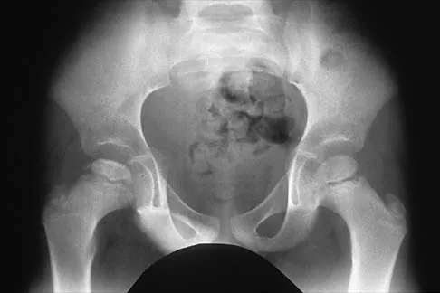
An 18-month-old girl presents with a waddling gait and a painless limp. Figure 10 shows her AP pelvis radiograph demonstrating a dislocated left hip.

She is scheduled to undergo an open reduction of the hip. Which of the following structures represents an EXTRA-articular obstacle to reduction that typically requires division or lengthening?


Explanation

In Developmental Dysplasia of the Hip (DDH), surgical reduction must overcome both extra-articular and intra-articular obstacles. The iliopsoas tendon is a primary extra-articular obstacle, as it tents over the capsule and constricts it (creating an hourglass shape), preventing the femoral head from entering the true acetabulum. Intra-articular obstacles include the pulvinar (fibrofatty tissue), ligamentum teres, inverted limbus, and the transverse acetabular ligament, all of which may need to be excised, incised, or divided to seat the head concentrically.

Question 79

A 6-year-old boy falls from the monkey bars and sustains a painful, swollen elbow. Radiographs demonstrate an extension-type supracondylar humerus fracture with posteromedial displacement of the distal fragment.

Based on the direction of displacement, which of the following nerve injuries is most likely to be present?


Explanation

In extension-type supracondylar humerus fractures, the direction of displacement dictates the structures at risk. With posteromedial displacement of the distal fragment, the proximal fracture spike is driven anterolaterally. This places the radial nerve at the highest risk of injury. Conversely, if the distal fragment is displaced posterolaterally, the proximal spike is driven anteromedially, jeopardizing the median nerve and its anterior interosseous nerve (AIN) branch. The AIN is the most commonly injured nerve overall, but the radial nerve is specifically associated with posteromedial displacement.

Question 80

An 8-year-old boy presents with a 4-month history of a painless right-sided limp. Radiographs demonstrate fragmentation of the capital femoral epiphysis consistent with Legg-Calvé-Perthes disease.

According to the Herring lateral pillar classification, greater than 50% loss of lateral pillar height is noted (Type C). Which of the following factors in this patient is most strongly associated with a poor prognosis and often dictates the need for surgical containment?


Explanation

In Legg-Calvé-Perthes disease, the two most critical prognostic factors are the age at onset and the degree of lateral pillar involvement (Herring classification). Children who develop the disease at 8 years of age or older have less time for the femoral head to remodel before skeletal maturity and generally have worse outcomes, particularly if they have lateral pillar B or C involvement. Surgical containment (e.g., femoral or pelvic osteotomy) is often indicated in this age group to maintain sphericity.

Question 81

A 2.5-year-old girl is evaluated for worsening bilateral genu varum. Standing radiographs reveal a metaphyseal-diaphyseal angle (MDA) of 20 degrees bilaterally with early beaking of the medial proximal tibial metaphysis.

She is diagnosed with infantile Blount disease (Langenskiöld stage II). What is the most appropriate initial management?


Explanation

This child has infantile Blount disease. A metaphyseal-diaphyseal angle (MDA) of >16 degrees is highly predictive of progressive infantile Blount disease rather than physiologic bowing. For children under the age of 3 with Langenskiöld stage I or II, nonoperative treatment with a knee-ankle-foot orthosis (KAFO) is the gold standard initial treatment. Surgery is indicated if bracing fails, if the child is over 4 years old at presentation, or for advanced Langenskiöld stages (III and above).

Question 82

A 3-year-old boy weighing 15 kg sustains an isolated, closed, transverse fracture of the middle third of the right femur after a fall from a trampoline. Radiographs show 1.5 cm of shortening and 10 degrees of varus angulation. What is the most appropriate definitive management for this patient?


Explanation

The treatment of pediatric femoral shaft fractures is age and weight dependent. In children aged 6 months to 5 years with isolated femur fractures and less than 2 cm of shortening, early spica casting is the standard of care and provides excellent outcomes. The Pavlik harness is indicated for infants 0-6 months old. Titanium elastic nails are indicated for children 5-11 years old (or heavier children). Rigid intramedullary nails are used in adolescents near skeletal maturity.

Question 83

A 13-year-old girl presents with ankle pain after a twisting injury while skateboarding. Radiographs and a subsequent CT scan reveal a Salter-Harris III fracture of the anterolateral distal tibial epiphysis with 3 mm of displacement.

Which of the following ligaments is responsible for the avulsion of this fracture fragment?


Explanation

The patient has a juvenile Tillaux fracture, which is a Salter-Harris III fracture of the anterolateral distal tibial epiphysis. This injury occurs during the transitional period of physeal closure (which closes centrally, then medially, then laterally). The mechanism involves an external rotation force applied to the foot, causing the anterior inferior tibiofibular ligament (AITFL) to avulse the anterolateral aspect of the epiphysis, which is the last portion to fuse.

Question 84

A 14-year-old male athlete presents with lower back pain and notably tight hamstrings. A lateral lumbar radiograph reveals a grade II isthmic spondylolisthesis at L5-S1.

He has failed 6 months of nonoperative management, including bracing, physical therapy, and activity modification. What is the most appropriate surgical treatment?


Explanation

For pediatric and adolescent patients with symptomatic low-grade (Grade I or II) isthmic spondylolisthesis who fail comprehensive conservative management, L5-S1 in situ posterolateral arthrodesis is the surgical standard of care. Pars repairs (e.g., Buck or Scott wiring) are generally reserved for young patients with a pars defect but minimal or no slip (typically L1-L4, not L5-S1). Laminectomy alone is contraindicated in children as it promotes instability and further slippage.

Question 85

A 12-year-old boy presents with vague, poorly localized knee pain and occasional catching. Radiographs demonstrate an osteochondritis dissecans (OCD) lesion.

In which of the following anatomic locations is an OCD lesion of the knee most classically found?


Explanation

Osteochondritis dissecans (OCD) of the knee is most commonly found on the lateral aspect of the medial femoral condyle. This classic location accounts for approximately 70-80% of all knee OCD lesions. A helpful mnemonic is 'LAME': Lateral Aspect of the Medial Epicondyle/condyle. The lateral femoral condyle is the second most common site, usually on the inferocentral aspect.

Question 86

A 12-year-old obese boy presents with 3 weeks of right knee pain and a limp. Examination demonstrates obligate external rotation with hip flexion. An AP pelvis radiograph is shown in Figure 1.

He is diagnosed with a stable slipped capital femoral epiphysis (SCFE) and is scheduled for in situ pinning. What is the most reliable technical maneuver to prevent the devastating complication of chondrolysis during this procedure?


Explanation

The most common cause of chondrolysis following SCFE fixation is unrecognized intra-articular hardware penetration. The approach-withdrawal technique utilizes continuous live fluoroscopy while rotating the hip to dynamically verify that the screw tip remains entirely within the bone and has not violated the joint space, thus preventing chondrolysis. Pin placement should ideally be in the center-center position, not anterosuperior.

Question 87

A 5-year-old girl sustains a severely displaced, extension-type supracondylar humerus fracture.

On presentation, her hand is pink and warm, but the radial pulse is absent. After urgent closed reduction and percutaneous pinning, her hand remains pink and warm with brisk capillary refill, but the radial pulse is still not palpable. What is the most appropriate next step in management?


Explanation

In the setting of a 'pink, pulseless' hand following the reduction and stabilization of a pediatric supracondylar humerus fracture, current AAOS guidelines and pediatric orthopaedic consensus recommend close observation rather than immediate vascular exploration. Excellent collateral circulation around the elbow often maintains adequate perfusion. Open exploration is indicated if the hand becomes pale, cold, or loses perfusion.

Question 88

An infant is undergoing serial casting for a right idiopathic clubfoot using the Ponseti method.

After 5 weeks of casting, the cavus, adductus, and varus deformities have been fully corrected. However, on examination, there is only 5 degrees of passive ankle dorsiflexion. What is the most appropriate next step?


Explanation

In the Ponseti method, once the cavus, adductus, and varus have been corrected (typically indicated by 60 degrees of foot abduction), the equinus contracture is addressed. If ankle dorsiflexion is less than 15 degrees, a percutaneous Achilles tendon lengthening (TAL) is indicated. Over 80% of idiopathic clubfeet treated with the Ponseti method require a TAL. Casting alone will not adequately correct persistent equinus, and attempting to force dorsiflexion through casting can cause a rocker-bottom deformity.

Question 89

A 2-year-old girl presents with bilateral genu varum. Her parents are concerned about her bowed legs. To distinguish between physiologic bowing and early infantile Blount disease, a standing AP radiograph of the lower extremities is obtained. Which of the following radiographic findings is most predictive of progression to infantile Blount disease?


Explanation

The metaphyseal-diaphyseal angle (MDA), as described by Levine and Drennan, is the most reliable radiographic parameter to differentiate physiologic bowing from infantile Blount disease in a young child. An MDA > 16 degrees is highly predictive of progression to Blount disease, whereas an angle < 10 degrees typically resolves spontaneously (physiologic bowing).

Question 90

A 14-year-old girl presents with adolescent idiopathic scoliosis (AIS). Upright radiographs demonstrate a right thoracic curve of 55 degrees and a left lumbar curve of 35 degrees. On supine side-bending radiographs, the thoracic curve reduces to 40 degrees, and the lumbar curve reduces to 15 degrees. The T2-T5 kyphosis is +15 degrees. According to the Lenke classification, what type of curve pattern does she have?


Explanation

The Lenke classification system defines structural curves based on their flexibility. A minor curve is considered non-structural if it bends out to less than 25 degrees on side-bending films and has normal sagittal alignment. In this patient, the lumbar curve reduces to 15 degrees (non-structural), and the T2-T5 kyphosis is normal (not structural). Thus, only the main thoracic curve is structural, classifying this as a Lenke Type 1 (Main Thoracic) curve.

Question 91

An 18-month-old boy presents with a painless limp and leg length discrepancy. Examination reveals a positive Galeazzi sign on the right and limited right hip abduction. Pelvic radiographs demonstrate a completely dislocated right hip with a dysplastic acetabulum (acetabular index of 38 degrees).

What is the most recommended treatment plan for this child?


Explanation

In a walking child older than 18 months with developmental dysplasia of the hip (DDH), closed reduction has a high failure rate and an increased risk of avascular necrosis. Additionally, the remaining potential for acetabular remodeling is significantly diminished. Therefore, the standard of care is open reduction combined with a pelvic osteotomy (e.g., Salter or Pemberton) to correct the acetabular dysplasia, often accompanied by a femoral shortening osteotomy to reduce joint reaction forces and AVN risk.

Question 92

A 13-year-old boy presents with recurrent left ankle sprains and a painful rigid flatfoot. On examination, he has significantly decreased subtalar motion and peroneal spasticity.

A lateral radiograph of the foot reveals an elongated anterior process of the calcaneus (the 'anteater' sign). What is the most likely diagnosis?


Explanation

The clinical presentation of a rigid, painful flatfoot with peroneal spasticity and recurrent sprains in an adolescent is classic for a tarsal coalition. The 'anteater nose' sign on a lateral radiograph is highly specific for a calcaneonavicular coalition. This condition is best visualized on a 45-degree internal oblique radiograph of the foot. Talocalcaneal coalitions often present slightly later (ages 12-16) and are associated with the 'C-sign' on lateral radiographs.

Question 93

A 6-year-old boy with a history of multiple low-energy fractures, blue sclerae, and dentinogenesis imperfecta is diagnosed with Osteogenesis Imperfecta (OI) Type I.

This condition is primarily caused by a genetic mutation affecting the synthesis or structure of which of the following?


Explanation

Osteogenesis Imperfecta is a genetic disorder of connective tissue characterized by fragile bones. It is primarily caused by autosomal dominant mutations in the COL1A1 or COL1A2 genes, which encode the alpha chains of Type I collagen. Type I collagen is the major structural protein in bone, sclerae, and dentin. Type II collagen defects cause spondyloepiphyseal dysplasia; FGFR3 mutations cause achondroplasia; COMP mutations cause pseudoachondroplasia; and fibrillin-1 mutations cause Marfan syndrome.

Question 94

A 6-year-old boy falls on an outstretched hand and sustains a lateral condyle fracture of the distal humerus that is displaced 3 mm. The fracture is managed non-operatively, but 6 months later, radiographs show a definitive nonunion. Which of the following is the most common long-term clinical consequence if this nonunion is left untreated?


Explanation

Lateral condyle fractures are Salter-Harris IV equivalent injuries that are intra-articular. Because the fracture fragment is bathed in synovial fluid and has a tenuous blood supply, it is prone to nonunion if displaced > 2 mm and inadequately stabilized. An established nonunion leads to proximal migration of the lateral condyle, resulting in a progressive cubitus valgus deformity. This valgus deformity stretches the ulnar nerve behind the medial epicondyle, ultimately leading to tardy ulnar nerve palsy years later.

Question 95

A 5-year-old girl with spastic quadriplegic cerebral palsy (GMFCS Level V) is undergoing routine orthopedic hip surveillance. She has bilateral hip flexion and severe adductor contractures. Her Reimers migration percentage on a recent surveillance AP pelvis radiograph is calculated at 48% bilaterally. What is the most appropriate orthopedic management?


Explanation

In cerebral palsy hip surveillance, a Reimers migration percentage (MP) greater than 40-50% in a child 4 years or older indicates significant subluxation that is unlikely to respond to soft-tissue release alone. At an MP of 48% in a 5-year-old GMFCS V patient, the standard of care is bony reconstruction consisting of proximal femoral varus derotational osteotomies (VDRO) frequently combined with pelvic osteotomies (e.g., Dega or San Diego) to definitively restore joint containment and prevent painful dislocation.

Question 96

A 12-year-old boy presents to the emergency department with severe left hip pain and an inability to bear weight on the affected limb for 2 days. He reports a preceding 2-month history of mild, intermittent groin pain. AP and frog-leg lateral radiographs demonstrate a slipped capital femoral epiphysis (SCFE) with a 60% displacement. Which of the following is the most significant risk factor for the development of avascular necrosis (AVN) in this patient?


Explanation

Instability is the most significant risk factor for the development of avascular necrosis (AVN) in SCFE. A stable SCFE is defined clinically as the patient being able to bear weight, with or without crutches. An unstable SCFE means the patient is unable to bear weight. The rate of AVN in stable slips is close to 0%, whereas in unstable slips it ranges from 24% to 47%. While the severity of the slip increases the risk of chondrolysis and subsequent cam-type impingement, instability remains the primary predictor of AVN.

Question 97

A 6-year-old boy falls from a playground structure and sustains a widely displaced extension-type supracondylar humerus fracture. On initial presentation, his hand is pink with brisk capillary refill (< 2 seconds), but the radial pulse is absent. He is taken emergently to the operating room. Following a successful closed reduction and percutaneous pinning, the hand remains pink and warm, but the radial pulse remains unpalpable and absent on Doppler ultrasound. What is the most appropriate next step in management?


Explanation

The management of a 'pulseless, pink' hand following adequate reduction and pinning of a pediatric supracondylar humerus fracture is observation. Because the hand is well-perfused (pink, warm, brisk capillary refill), the collateral circulation is sufficient. Vascular exploration is indicated only if the hand becomes poorly perfused (white, cool, pulseless) after reduction. A splint should be applied in mild flexion (45-60 degrees) rather than hyperflexion to avoid vascular compromise and compartment syndrome. The pulse often returns over the next several days as vasospasm resolves.

Question 98

A 4-year-old boy with a history of idiopathic right clubfoot successfully treated with the Ponseti method during infancy presents with a relapsed deformity. His parents report that he frequently trips when running. Gait analysis and clinical examination reveal dynamic supination of the foot during the swing phase of gait. Passive range of motion demonstrates that the deformity is flexible and fully correctable. Which of the following is the most appropriate surgical treatment?


Explanation

Dynamic supination during the swing phase of gait in a relapsed Ponseti-treated clubfoot is a classic indication for a full Tibialis Anterior Tendon Transfer (TATT) to the lateral cuneiform. This procedure transfers the deforming supinatory force of the tibialis anterior and converts it into an eversion force, balancing the foot dynamically. An Achilles tendon lengthening may be performed concurrently if there is residual fixed equinus, but TATT is specifically required to address the dynamic supination. Extensive posteromedial releases are historically associated with severe stiffness and recurrence, and are no longer standard for this presentation.

Question 99

A 14-year-old girl presents with progressive knee pain and swelling. Radiographs reveal an eccentric, expansile, purely lytic lesion in the metaphysis of the distal femur. MRI demonstrates characteristic multiple fluid-fluid levels within the lesion. An incisional biopsy reveals blood-filled spaces lacking an endothelial lining, interspersed with multinucleated giant cells and a spindle cell stroma. Which of the following genetic translocations is most commonly associated with this primary pathology?


Explanation

The clinical, radiographic, and histologic presentation is classic for a primary Aneurysmal Bone Cyst (ABC). The finding of fluid-fluid levels on MRI and blood-filled spaces without an endothelial lining is characteristic. Primary ABCs are true neoplastic processes driven by a highly specific t(16;17) translocation, which leads to the fusion of the USP6 oncogene with various promoter genes (such as CDH11). Identifying this is key for advanced orthopedic board exams. Conversely, t(11;22) is seen in Ewing sarcoma; t(X;18) in synovial sarcoma; t(12;16) in myxoid liposarcoma; and t(9;22) in extraskeletal myxoid chondrosarcoma.

Question 100

A 4-year-old boy presents with progressive left leg bowing. He has a BMI above the 95th percentile. Standing radiographs reveal a metaphyseal-diaphyseal angle of 18 degrees on the left, with prominent medial metaphyseal beaking and focal sclerosis of the proximal tibia consistent with Langenskiöld stage III. He has previously failed conservative management with knee-ankle-foot orthoses (KAFOs). What is the most appropriate surgical intervention to correct the deformity and minimize the risk of recurrence?


Explanation

In infantile Blount's disease presenting at an older age (> 3-4 years) with advanced Langenskiöld staging (stage III or higher), surgical intervention with a proximal tibial and fibular osteotomy is the gold standard. To minimize the high risk of recurrence caused by the persistently abnormal growth forces across the medial physis, the mechanical axis must be overcorrected into 5 to 10 degrees of valgus. Neutral correction has an unacceptably high rate of recurrent varus deformity. Guided medial growth is contraindicated because the medial physis is severely diseased and often incapable of spontaneous recovery in advanced stages.

None

Clinic OS
Medically Verified Content by
Prof. Clinic OS
Consultant Orthopedic & Spine Surgeon
Chapter Index