Dorsal Block Pinning for Proximal Interphalangeal Joint Fracture-Dislocations
Key Takeaway
Welcome to the OR, fellows. Today, we're tackling an unstable proximal interphalangeal (PIP) joint fracture-dislocation using dorsal block pinning. This technique offers stability while promoting early motion. We'll meticulously cover comprehensive anatomy, precise preoperative planning, granular intraoperative execution, and critical pearls to ensure optimal patient outcomes and minimize complications. Prepare for an in-depth, hands-on masterclass.
Comprehensive Introduction and Patho-Epidemiology
The proximal interphalangeal (PIP) joint is an unforgiving articulation, characterized by a highly constrained anatomical architecture that prioritizes stability under immense physiological loads. Often dismissed by the layman or inexperienced practitioner as a mere "jammed finger," the dorsal fracture-dislocation of the PIP joint represents a complex, potentially devastating injury pattern. Sufficient axial and hyperextension forces can lead to a catastrophic sequence of structural failures that, if not treated with meticulous precision, will invariably result in lifelong pain, debilitating stiffness, and profound functional impairment of the entire hand. Our focus in this definitive chapter is the dorsal block pinning technique, a versatile, percutaneous, and highly effective operative method for managing unstable PIP fracture-dislocations that defy conservative management.
The pathogenesis of dorsal PIP fracture-dislocations is rooted in the biomechanical failure of the joint's primary static and dynamic restraints. The most common vector of injury involves simultaneous hyperextension and axial compression forces, a mechanism frequently encountered in athletic endeavors when a projectile impacts the extended fingertip. The sequence of structural failure is predictable and critical to understand for surgical planning. The volar fibrocartilaginous plate, acting as the primary restraint to hyperextension, is typically the first structure to fail. It frequently avulses from its distal insertion on the volar lip of the middle phalanx, often taking a substantial osteochondral fragment of bone with it.
Following the disruption of the volar plate, the radial and ulnar collateral ligaments, which are crucial for mediolateral stability and secondary restraints to dorsal translation, are subjected to overwhelming stress. These ligaments may sustain partial interstitial tearing or complete macroscopic disruption. The resulting articular fragmentation at the base of the middle phalanx compromises the critical "tongue-and-groove" congruity of the joint. With the volar restraints utterly compromised, the unopposed dynamic pull of the central slip of the extensor tendon actively accentuates dorsal displacement and subluxation of the middle phalanx. This dynamic imbalance makes closed reduction exceptionally difficult to maintain without mechanical intervention.
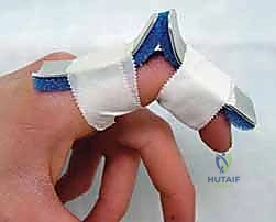
To guide our treatment algorithm, we classify these injuries based on the extent of anatomical disruption and resulting instability. Type I injuries involve partial disruption of the collateral ligaments and the volar plate without significant articular involvement, rendering the joint stable and amenable to non-operative functional taping. Type II injuries represent a more substantial force, leading to longitudinal splitting of the collateral ligaments and complete volar plate rupture; while complete dorsal displacement occurs initially, the joint is typically stable after closed reduction and can be managed with extension block splinting. Type III injuries are severe and involve a significant avulsion fracture of the middle phalanx base. If the fracture involves more than 40% to 50% of the articular surface, the collateral ligament support is critically compromised. The joint exhibits persistent, dynamic dorsal subluxation due to the unopposed pull of the extensor mechanism. It is precisely within this cohort of unstable Type III injuries that operative intervention, specifically dorsal block pinning, becomes an absolute necessity to restore congruity and function.
Detailed Surgical Anatomy and Biomechanics
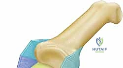
Before initiating any percutaneous intervention or open dissection, a profound and exhaustive understanding of the regional anatomy and biomechanics of the PIP joint is paramount. This anatomical roadmap dictates the safe zones for hardware placement and the biological limits of our reconstructive efforts. The osteology of the PIP joint involves the articulation between the bicondylar head of the proximal phalanx and the reciprocal, biconcave articular surface of the middle phalanx base. The proximal phalanx head features a central trochlear groove flanked by two condyles, creating a highly congruent "tongue-and-groove" configuration with the middle phalanx. The volar lip of the middle phalanx serves as the critical insertion footprint for the volar plate; it is the avulsion of this specific anatomical landmark that precipitates the dorsal fracture-dislocation cascade. Preservation of the delicate articular cartilage during pinning is mandatory, as iatrogenic penetration or thermal necrosis from drilling will inevitably lead to accelerated post-traumatic osteoarthritis.
The ligamentous and capsular structures of the PIP joint form a complex, interdependent network of static restraints. The volar plate is a robust, thick fibrocartilaginous structure situated on the palmar aspect of the joint. It originates proximally from the periosteum of the proximal phalanx via two distinct proximal extensions known as the checkrein ligaments, and it inserts distally into the cartilaginous base of the middle phalanx. The volar plate's primary biomechanical function is to resist hyperextension. The collateral ligament complex consists of the proper collateral ligaments (PCL) and the accessory collateral ligaments (ACL). The PCLs originate from the dorsal-lateral aspect of the proximal phalanx head and insert into the volar-lateral base of the middle phalanx; they are maximally taut in flexion, providing critical mediolateral stability. The ACLs originate more volarly, inserting directly into the lateral margins of the volar plate, and are taut in extension.
The extensor mechanism crossing the PIP joint is an intricate fibrous aponeurosis that profoundly influences the joint's dynamic stability. The central slip, the primary extensor of the PIP joint, inserts directly into the dorsal tubercle at the base of the middle phalanx. In the setting of a volar plate avulsion and collateral ligament disruption, the central slip's unopposed proximal pull becomes the primary deforming force, driving the middle phalanx dorsally and proximally into a subluxated or dislocated position. The lateral bands, which bifurcate from the main extensor tendon, pass laterally to the PIP joint to converge and insert on the distal phalanx, contributing to PIP extension and distal interphalangeal (DIP) flexion. Understanding this dynamic interplay is crucial, as the dorsal block pinning technique relies on neutralizing this specific extensor vector while allowing early controlled flexion.
Finally, the neurovascular anatomy dictates the safe corridors for percutaneous K-wire insertion. The proper digital nerves and arteries course longitudinally along the radial and ulnar aspects of each digit, positioned just volar to the mid-axial line. These critical structures lie deep to the dermis and subcutaneous adipose tissue but superficial to the flexor tendon sheath. When inserting K-wires, particularly from a dorsal-lateral or dorsal-medial trajectory, the surgeon must be acutely aware of their proximity. A misplaced pin, or a pin inserted with excessive thermal energy, can easily cause permanent sensory deficits, painful neuromas, or devastating vascular compromise to the digit. Always visualize the trajectory of the K-wire in three dimensions relative to these neurovascular bundles, utilizing the palpable lateral aspects of the digit to estimate their precise anatomical course.
Exhaustive Indications and Contraindications
The decision to proceed with dorsal block pinning must be based on a rigorous assessment of the injury pattern, the degree of articular involvement, and the inherent stability of the joint following an attempted closed reduction. The primary indication for this technique is an unstable Type III dorsal fracture-dislocation of the PIP joint. Specifically, this applies to injuries where the volar lip fracture encompasses greater than 40% to 50% of the articular surface of the middle phalanx. At this critical threshold, the insertion footprints of the proper collateral ligaments are compromised, rendering the joint profoundly unstable and incapable of maintaining a concentric reduction in functional degrees of extension. Furthermore, dorsal block pinning is indicated when an initial closed reduction cannot achieve a congruent joint space, or when the joint redisplaces into dorsal subluxation at less than 30 degrees of flexion during dynamic fluoroscopic stress testing.
Conversely, dorsal block pinning is contraindicated in several specific clinical scenarios where alternative surgical strategies are mandated. Absolute contraindications include chronic fracture-dislocations presenting more than 6 weeks post-injury. By this stage, profound soft tissue contracture, fibrosis of the collateral ligaments, and early cartilaginous degradation have occurred, rendering percutaneous reduction impossible and necessitating open arthrolysis or salvage procedures. Another critical contraindication is the presence of massive, highly comminuted articular fractures ("pilon-type" fractures of the middle phalanx base) where the dorsal block pin would fail to stabilize the fragmented articular surface. In these instances, dynamic external fixation, open reduction and internal fixation (ORIF) with minifragment screws, or a volar plate arthroplasty are more appropriate interventions. Active localized soft tissue infection or osteomyelitis at the surgical site also strictly precludes the placement of percutaneous hardware.
The choice of dorsal block pinning over non-operative extension block splinting is often driven by patient-specific factors as well as anatomical parameters. While a highly compliant patient with a borderline stable injury (e.g., 30-40% articular involvement) might be managed with a custom orthoplast splint, dorsal block pinning eliminates the variable of patient compliance. It guarantees the mechanical block to extension while permitting immediate, aggressive active flexion protocols. This is particularly vital in laborers, athletes, or individuals with significant digital swelling where external splinting may fail to maintain the precise reduction angle required, leading to insidious subluxation and catastrophic long-term outcomes.
To synthesize the clinical decision-making process, the following table outlines the rigorous indications and contraindications for the dorsal block pinning technique in the management of PIP joint fracture-dislocations.
| Parameter | Indications for Dorsal Block Pinning | Contraindications for Dorsal Block Pinning |
|---|---|---|
| Injury Classification | Unstable Type III dorsal fracture-dislocations. | Type I or stable Type II injuries (manage non-operatively). |
| Articular Involvement | Volar lip fracture involving >40% to 50% of the articular surface. | Massive comminution ("pilon" fractures) requiring ORIF/External Fixation. |
| Joint Stability | Failure to maintain concentric reduction at <30° of flexion. | Joint maintains concentric reduction in full extension or minimal flexion. |
| Chronicity | Acute injuries (ideally <2-3 weeks from the time of injury). | Chronic injuries (>6 weeks) with established fibrosis and contracture. |
| Soft Tissue Envelope | Intact skin, severe swelling precluding reliable external splinting. | Active local infection, compromised dorsal skin bridge. |
| Patient Factors | Anticipated non-compliance with strict extension block splinting protocols. | Medical comorbidities precluding regional or general anesthesia. |
Pre-Operative Planning, Templating, and Patient Positioning
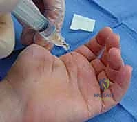
Meticulous preoperative planning is the absolute cornerstone of any successful surgical outcome in hand traumatology, where tolerances are measured in millimeters and functional margins are razor-thin. The process begins with a comprehensive patient history and physical examination to elucidate the exact mechanism of injury, which often provides invaluable clues regarding the magnitude of force and the likely pattern of soft tissue disruption. The time elapsed from injury to presentation is a critical prognostic factor; the likelihood of achieving a near-normal functional result diminishes precipitously after 3 to 4 weeks due to aggressive scar formation, collateral ligament contracture, and potential early articular remodeling. The surgeon must meticulously document the neurovascular status—specifically two-point discrimination and capillary refill—before the administration of any local anesthetic block, as percutaneous pinning carries an inherent risk of iatrogenic nerve injury.
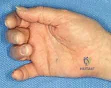
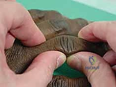
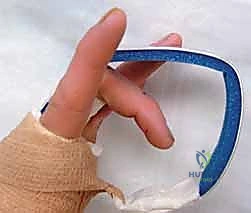
Visual assessment of the injured hand provides immediate diagnostic information. The surgeon should observe the resting cascade of the digits; a disruption of this normal flexion cascade, where the digits progressively flex from the radial to the ulnar side, is pathognomonic for a significant structural injury. Following a comprehensive visual inspection, pinpoint palpation is utilized to localize specific ligamentous or bony pathology. Exquisite tenderness over the volar aspect of the middle phalanx base strongly suggests a volar plate avulsion fracture, while tenderness isolated to the collateral recesses may indicate an isolated ligamentous tear. Once a digital or regional block has been administered to ensure patient comfort, the surgeon must perform a dynamic stability assessment. Gentle manipulation of the PIP joint under live fluoroscopy allows the surgeon to observe the joint under anteroposterior, lateral, and oblique stresses, pinpointing the exact angle of flexion at which dorsal subluxation occurs.
Imaging studies are non-negotiable and must be of the highest radiological quality. Standard radiographs must include a true anteroposterior, a true lateral, and oblique views of the injured digit specifically. A common and dangerous pitfall is relying on a "fanned four-finger lateral" view of the entire hand; this inevitably results in bony overlap and oblique projections that obscure subtle articular step-offs and minimal dorsal subluxations. If the fracture pattern is highly comminuted, or if the degree of articular depression is difficult to quantify on plain radiographs, a dedicated fine-cut Computed Tomography (CT) scan is indicated. The CT scan provides a definitive three-dimensional understanding of the fracture morphology, allowing the surgeon to determine if a simple dorsal block pin will suffice, or if open reduction and internal fixation of a large, depressed articular fragment is required to restore joint congruity.
Patient positioning and operating room setup must be optimized to facilitate seamless fluoroscopic imaging and unhindered access to the surgical field. The patient is typically positioned supine on the operating table, with the affected upper extremity abducted and supported by a radiolucent hand table. A well-padded pneumatic tourniquet is applied to the proximal brachium. Following strict exsanguination of the limb using an Esmarch bandage, the tourniquet is inflated to typically 250 mmHg (or 100 mmHg above the patient's systolic blood pressure) to provide an absolutely bloodless field. The C-arm fluoroscopy unit is positioned parallel to the hand table, allowing the surgeon to obtain perfect AP, lateral, and oblique views of the PIP joint by simply rotating the patient's forearm, without needing to reposition the sterile drapes or the machine itself. The entire hand and forearm are prepped with a standard chlorhexidine or povidone-iodine solution and draped in a standard sterile fashion.
Step-by-Step Surgical Approach and Fixation Technique
With the patient properly anesthetized, the tourniquet inflated, and the sterile field established, the intraoperative execution of the dorsal block pinning technique begins. This is fundamentally a percutaneous, fluoroscopically guided procedure, meaning that formal surgical incisions and deep anatomical dissections are generally avoided. However, this minimally invasive nature demands an exceptional spatial awareness and an uncompromising reliance on high-quality orthogonal imaging. The first step is to perform a final, definitive assessment of joint stability under live fluoroscopy. The surgeon applies longitudinal traction, flexes the PIP joint, and applies volar directed pressure to the middle phalanx to achieve a closed reduction. The joint is then slowly extended while observing the lateral fluoroscopic image. The precise angle at which the middle phalanx begins to subluxate dorsally—the "redisplacement angle"—is meticulously noted. This angle dictates the final position of the dorsal block pin.
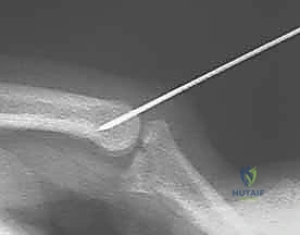
The core principle of the dorsal block pinning technique is to place a rigid mechanical barrier (the K-wire) into the head of the proximal phalanx, which physically prevents the middle phalanx from extending to the redisplacement angle, thereby neutralizing the deforming force of the central slip. A 0.045-inch or 0.035-inch Kirschner wire is typically selected. The entry point is located on the dorsal aspect of the proximal phalanx, approximately 5 to 10 millimeters proximal to the articular surface. The K-wire is driven percutaneously, directed in a dorsal-to-volar and distal-to-proximal trajectory, typically at a 45-degree angle to the longitudinal axis of the proximal phalanx. The pin is advanced through both the dorsal and volar cortices of the proximal phalanx to ensure maximum biomechanical rigidity. Crucially, the distal tip of the pin must protrude sufficiently into the dorsal aspect of the joint space to mechanically block the dorsal lip of the middle phalanx base from extending past the pre-determined safe angle.
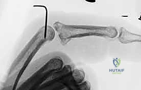
During the insertion of the dorsal block pin, extreme care must be taken to avoid traversing the central slip of the extensor mechanism, as this can lead to severe postoperative stiffness and extension lag. The surgeon should manually retract the extensor mechanism laterally or ensure the pin passes smoothly through the central tendon without causing significant fraying or entrapment. Once the primary block pin is seated, the joint is taken through a range of motion under fluoroscopy. The surgeon must confirm that the joint remains concentrically reduced at the blocked angle of extension, and that full, unhindered flexion is possible. The "V-sign" on the lateral radiograph—a widened dorsal joint space indicating persistent volar plate interposition or incomplete reduction—must be completely absent. The articular surfaces of the proximal and middle phalanges must remain perfectly parallel throughout the permitted arc of motion.
In certain highly unstable cases, particularly those with massive comminution or when the joint tends to subluxate even in deep flexion, a supplementary transarticular K-wire may be required. If deemed necessary, after the dorsal block pin is placed and the joint is concentrically reduced, a second 0.035-inch K-wire is driven obliquely across the PIP joint, starting from the lateral base of the middle phalanx and engaging the contralateral cortex of the proximal phalanx head. This transarticular pin completely immobilizes the joint and is typically removed much earlier than the block pin (usually at 2 to 3 weeks) to initiate early motion. Once all hardware is confirmed to be in optimal position via orthogonal fluoroscopy, the pins are bent to 90 degrees outside the skin to prevent migration, cut short, and capped. A sterile, non-adherent dressing is applied, and the digit is protected in a custom thermoplastic splint.
Complications, Incidence Rates, and Salvage Management
Despite meticulous surgical technique and rigorous adherence to biomechanical principles, dorsal block pinning of the PIP joint is fraught with potential complications. The PIP joint is notoriously prone to profound fibroplasia and scar formation following any traumatic insult, making postoperative stiffness the most ubiquitous complication. The incidence of clinically significant stiffness—defined as a permanent loss of more than 20 degrees of extension or the inability to flex beyond 80 degrees—can approach 30% to 40% even in expertly managed cohorts. This stiffness arises from a combination of capsular contracture, collateral ligament fibrosis, and adhesions within the flexor tendon sheath or extensor mechanism. Prevention relies entirely on flawless concentric reduction and the immediate initiation of a highly supervised, aggressive early active motion protocol.
Pin tract infections are another frequent complication inherent to any percutaneous hardware technique, occurring in approximately 10% to 15% of cases. These are typically superficial infections caused by normal skin flora such as Staphylococcus aureus. Patients present with localized erythema, induration, and purulent drainage at the pin-skin interface. Management involves immediate initiation of oral antibiotics (e.g., a first-generation cephalosporin) and aggressive local wound care. Deep infections or septic arthritis are rare but catastrophic; if suspected, immediate hardware removal, surgical irrigation and debridement of the joint, and intravenous antibiotics are mandatory to prevent rapid cartilaginous destruction and osteomyelitis.
Loss of reduction or hardware failure is a critical complication that typically results from technical errors during surgery or patient non-compliance postoperatively. If the dorsal block pin is placed at an incorrect angle, lacks bicortical purchase, or if the initial assessment of the redisplacement angle was inaccurate, the unopposed pull of the central slip will drive the middle phalanx into dorsal subluxation against the pin. This insidious subluxation rapidly destroys the remaining articular cartilage. If loss of reduction is identified early on postoperative radiographs, immediate revision surgery is required. Late presentations of subluxation or profound post-traumatic osteoarthritis necessitate complex salvage procedures. Options include volar plate arthroplasty for moderately damaged joints, hemi-hamate osteochondral autograft reconstruction for massive volar lip defects, or, in the most severe cases of painful, stiff, and destroyed joints, formal PIP joint arthrodesis positioned in a functional degree of flexion.
The following table summarizes the most common complications associated with dorsal block pinning, their estimated incidence rates, and the appropriate management or salvage strategies.
| Complication | Estimated Incidence | Etiology/Risk Factors | Management and Salvage Strategies |
|---|---|---|---|
| Postoperative Stiffness | 30% - 40% | Capsular contracture, prolonged immobilization, tendon adhesions. | Aggressive hand therapy, dynamic/static progressive splinting. Late surgical arthrolysis/tenolysis. |
| Pin Tract Infection | 10% - 15% | Percutaneous hardware, poor local hygiene, prolonged pin retention. | Oral antibiotics, local wound care. Immediate pin removal if deep infection/septic arthritis is suspected. |
| Loss of Reduction | 5% - 10% | Incorrect pin placement, inadequate bicortical purchase, patient non-compliance. | Immediate revision pinning or ORIF if acute. Salvage (hemi-hamate, arthroplasty, arthrodesis) if chronic. |
| Post-Traumatic Arthritis | 15% - 25% | Initial articular cartilage damage, chronic subluxation, iatrogenic pin damage. | NSAIDs, intra-articular injections. Salvage procedures (arthroplasty or arthrodesis) for severe, debilitating pain. |
| Neurovascular Injury | < 2% | Direct trauma from K-wire insertion, thermal necrosis during drilling. | Observation for neuropraxia. Surgical exploration and nerve repair if complete transection is suspected. |
Phased Post-Operative Rehabilitation Protocols
The surgical placement of the dorsal block pin is merely the first phase in the comprehensive management of a PIP fracture-dislocation; the ultimate functional outcome is dictated entirely by the rigorous execution of a phased postoperative rehabilitation protocol. The primary objective of rehabilitation is to navigate the delicate balance between protecting the fragile articular reduction and preventing the devastating, ubiquitous complication of joint stiffness. This requires a highly symbiotic relationship between the orthopedic surgeon, a certified hand therapist (CHT), and a motivated, compliant patient. The protocol is generally divided into three distinct phases, meticulously timed to align with the biological stages of soft tissue healing and bony consolidation.
Phase 1: Early Active Motion (Weeks 0 to 3 or 4)
If a solitary dorsal block pin has been utilized (without a transarticular pin), the hallmark of this initial phase is early active motion. Within 3 to 5 days postoperatively, once the acute surgical edema has begun to subside, the patient is fitted with a custom thermoplastic splint that protects the K-wire and supports the digit. Under the direct supervision of the CHT, the patient is instructed to perform active flexion of the PIP joint. The dorsal block pin physically prevents the joint from extending into the unstable zone, allowing the patient to safely pull the middle phalanx into flexion using their flexor digitorum superficialis (FDS) and flexor digitorum profundus (FDP) tendons. This early gliding of the flexor tendons prevents dense adhesions and stimulates cartilaginous nutrition. Extension is allowed actively, but only up to the mechanical limit imposed by the pin. If a transarticular pin was required for profound instability, the joint must remain strictly immobilized during this phase to allow for initial soft tissue bridging.
Phase 2: Hardware Removal and Progressive Mobilization (Weeks 3 to 6)
At approximately 3 to 4 weeks postoperatively, clinical and radiographic assessments are performed. If the fracture demonstrates early callus formation and the joint remains concentrically reduced, the dorsal block pin (and transarticular pin, if present) is removed in the clinic. Following hardware removal, the joint is often stiff, and the focus shifts to restoring full active and passive range of motion. Buddy taping to an adjacent, uninjured digit is immediately instituted to provide dynamic support and encourage functional use during activities of daily living. The CHT will initiate passive range of motion exercises, gentle joint mobilizations, and edema control techniques. Extension block splinting may still be utilized at night or during high-risk activities, gradually decreasing the angle of the extension block by 10 degrees per week until full extension is achieved safely.
Phase 3: Strengthening and Contracture Management (Weeks 6 and Beyond)
By the sixth postoperative week, the fracture is typically clinically united, and the soft tissues possess sufficient tensile strength to withstand progressive loading. The rehabilitation focus transitions to strengthening the intrinsic and extrinsic musculature of the hand. Grip and pinch strengthening exercises are incorporated using therapeutic putty and hand dynamometers. Despite aggressive early motion, many patients will develop a mild to moderate flexion contracture of the PIP joint due to volar plate scarring. To combat this, the CHT will implement static progressive extension splinting or dynamic extension orthoses. These devices apply a low-load, prolonged stretch to the volar structures, effectively remodeling the scar tissue over time. Patients must be counseled that maximal medical improvement may not be achieved for 6 to 12 months following the injury, and a permanent, mild enlargement of the joint contour is an expected, unavoidable sequela.
Summary of Landmark Literature and Clinical Guidelines
The evolution of the dorsal block pinning technique is deeply rooted in decades of rigorous orthopedic research and clinical observation. Historically, the management of unstable PIP fracture-dislocations was fraught with high failure rates, as prolonged immobilization inevitably led to catastrophic stiffness, while early unprotected motion resulted in recurrent subluxation. The paradigm shifted significantly with the landmark work of McElfresh, Dobyns, and O'Brien in the 1970s. Their seminal publications elucidated the biomechanical necessity of blocking extension to neutralize the central slip, initially popularizing the use of custom extension-block splinting. However, they astutely noted that splinting was frequently insufficient for highly unstable injuries or in non-compliant patients, laying the conceptual groundwork for internal mechanical blocks.
The modern percutaneous dorsal block pinning technique, as described in this chapter, was refined and popularized by several key authors in the late 20th and early 21st centuries. Studies by Hastings and others demonstrated that percutaneous K-wire fixation provided superior biomechanical rigidity compared to external splinting, reliably preventing dorsal subluxation while safely permitting the early active flexion that is so critical for cartilage preservation and tendon gliding. Their retrospective cohorts proved that patients treated with precise dorsal block pinning achieved significantly better terminal flexion and lower rates of severe post-traumatic arthritis compared to those managed with prolonged immobilization or attempted open reduction of highly comminuted fragments.
Current clinical guidelines, supported by the American Society
