Arthroscopic Capsular Shift: Comprehensive Surgical Technique and Biomechanical Principles
Key Takeaway
The arthroscopic capsular shift is a cornerstone procedure for managing multidirectional shoulder instability. This technique involves precise capsulolabral plication and rotator interval closure to restore glenohumeral kinematics without overconstraining the joint. By systematically addressing inferior, anterior, and posterior capsular redundancy, surgeons can effectively eliminate symptomatic hyperlaxity. Mastery of portal placement, suture shuttling, and tensioning is critical to avoiding neurovascular complications and optimizing postoperative functional outcomes.
Comprehensive Introduction and Patho-Epidemiology
Multidirectional instability (MDI) of the glenohumeral joint represents one of the most complex and nuanced biomechanical challenges encountered by the orthopedic surgeon. Unlike unidirectional traumatic instability—classically encapsulated by the TUBS acronym (Traumatic, Unidirectional, Bankart, Surgery)—MDI is traditionally conceptualized within the AMBRI paradigm (Atraumatic, Multidirectional, Bilateral, Rehabilitation, Inferior capsular shift). Patients presenting with MDI possess a symptomatic, global capsular laxity that permits excessive translation of the humeral head in anterior, posterior, and inferior vectors. The primary pathological lesion is not an avulsion of the capsulolabral complex from the glenoid rim, but rather an inherently redundant, patulous capsule with an enlarged axillary pouch and an incompetent rotator interval.
The pathophysiology of MDI is multifactorial, often rooted in intrinsic collagenous abnormalities. Histological analyses of the glenohumeral capsule in MDI patients frequently demonstrate an altered ratio of Type I to Type III collagen, alongside increased elastin content, leading to diminished tensile strength and increased viscoelastic creep under physiological loads. This inherent tissue laxity is frequently exacerbated by repetitive microtrauma, particularly in overhead athletes (e.g., swimmers, gymnasts, pitchers), where the repetitive attenuation of the capsular restraints outpaces the biological capacity for tissue remodeling. Consequently, the dynamic stabilizers—namely the rotator cuff and periscapular musculature—become fatigued and dyskinetic, failing to maintain the humeral head within the concavity of the glenoid during active motion.
Epidemiologically, MDI predominantly affects adolescents and young adults, with a slightly higher prevalence in females. It is imperative to distinguish between physiological hyperlaxity—which is asymptomatic and highly prevalent in the general population—and true clinical multidirectional instability, which is defined by symptomatic subluxations, dislocations, and profound apprehension that limits activities of daily living. Furthermore, the surgeon must maintain a high index of suspicion for underlying heritable disorders of connective tissue (HDCT), such as Ehlers-Danlos syndrome or Marfan syndrome. Patients with these systemic conditions present a unique surgical challenge, as their inherent fibroblastic dysfunction significantly compromises the potential for robust biological healing following capsular plication.
Historically, the gold standard for surgical intervention in refractory MDI was the open inferior capsular shift, pioneered by Charles Neer in 1980. While revolutionary for its time, the open approach necessitated detachment of the subscapularis, prolonged rehabilitation, and carried a higher risk of iatrogenic stiffness and subscapularis insufficiency. The advent and refinement of the arthroscopic capsular shift have fundamentally transformed the management of this pathology. By allowing circumferential, 360-degree access to the glenohumeral joint, the arthroscopic approach facilitates precise, titratable plication of the redundant capsule while minimizing surgical morbidity, preserving the subscapularis tendon, and optimizing proprioceptive recovery. The primary objective is to volumetrically reduce the capsular volume and restore the normal tension of the glenohumeral ligaments without causing iatrogenic overconstraint.
Detailed Surgical Anatomy and Biomechanics
A profound mastery of glenohumeral surgical anatomy is the absolute prerequisite for executing a successful arthroscopic capsular shift. The glenohumeral joint relies on a delicate interplay between static and dynamic stabilizers. The static restraints are primarily composed of the capsuloligamentous complex, which includes the superior glenohumeral ligament (SGHL), the middle glenohumeral ligament (MGHL), and the inferior glenohumeral ligament (IGHL) complex. In the context of MDI, the IGHL complex is the most critical anatomical structure. It functions as a hammock-like sling supporting the inferior humeral head, consisting of an anterior band, a posterior band, and the intervening axillary pouch. In a patulous shoulder, this axillary pouch is voluminously enlarged, failing to become taut at the extremes of motion, thereby permitting inferior and bidirectional translation.
The rotator interval is another critical anatomical zone implicated in MDI. This triangular space is bordered superiorly by the anterior margin of the supraspinatus tendon, inferiorly by the superior margin of the subscapularis tendon, and medially by the base of the coracoid process. It contains the coracohumeral ligament (CHL), the SGHL, and the intra-articular portion of the long head of the biceps tendon. Biomechanically, the structures within the rotator interval are the primary restraints to inferior translation when the arm is adducted and in neutral rotation. In patients demonstrating a profound sulcus sign, the rotator interval is invariably incompetent. Surgical closure or imbrication of this interval is therefore a mandatory adjunct to inferior capsular plication to restore stability in the adducted arm.
Neurovascular proximity dictates the safety margins of the arthroscopic capsular shift, with the axillary nerve being the structure at greatest risk. Originating from the posterior cord of the brachial plexus, the axillary nerve traverses the quadrangular space to innervate the deltoid and teres minor. As it courses anterior to posterior, it lies in intimate proximity to the inferior glenohumeral capsule at the 6-o'clock position. Cadaveric studies have demonstrated that the axillary nerve is located, on average, merely 10 to 15 millimeters inferior to the glenoid rim. Furthermore, the position of the arm dynamically alters this distance; placing the arm in internal rotation brings the nerve closer to the capsule, whereas external rotation and mild abduction safely distance the nerve from the operative field during inferior capsular suturing.
The biomechanics of the capsular shift rely on the principles of volumetric reduction and vector-based tensioning. By taking discrete, 1-centimeter bites of the redundant capsule and shifting them superiorly and laterally toward the labrum, the surgeon effectively obliterates the dependent axillary pouch. This "pinch-tuck" method not only reduces the overall capsular volume but also restores the reciprocal tension between the anterior and posterior bands of the IGHL. The superior shift of the capsular tissue re-establishes the hammock effect, ensuring that the IGHL complex becomes appropriately taut during abduction and external rotation, thereby physically blocking pathological translation of the humeral head while preserving physiological kinematics.
Exhaustive Indications and Contraindications
The decision to proceed with an arthroscopic capsular shift must be predicated on a rigorous clinical evaluation and the strict exhaustion of conservative measures. Surgery is indicated only for patients with symptomatic multidirectional instability who have failed a minimum of six to twelve months of a highly structured, supervised physical therapy program. This conservative regimen must specifically target periscapular stabilization, scapular dyskinesia correction, and dynamic rotator cuff strengthening. Patients whose instability manifests primarily as pain, fatigue, and apprehension during activities of daily living, and who demonstrate a positive sulcus sign alongside bidirectional laxity (anterior and posterior) on clinical examination, are the ideal candidates.
Differentiating true MDI from voluntary instability is a critical nuance in patient selection. Voluntary dislocators can be broadly categorized into positional dislocators (who use altered muscle activation to subluxate the shoulder, often as a party trick) and psychiatric dislocators (who subluxate the joint for secondary gain). While positional voluntary dislocators may eventually become candidates for surgery if their instability becomes involuntary and painful over time, psychiatric voluntary instability is an absolute contraindication to surgical intervention. Operating on this latter cohort uniformly results in catastrophic failure, recurrent dislocation, and severe iatrogenic complications, as the underlying muscular dyskinesia and psychiatric drivers remain unaddressed.
Patients with underlying heritable disorders of connective tissue (e.g., Ehlers-Danlos syndrome, Marfan syndrome) require highly specialized consideration. While not an absolute contraindication, the presence of a generalized connective tissue disorder significantly alters the surgical prognosis. The inherent collagenous defect means that the plicated tissue is highly susceptible to stretching out postoperatively, leading to recurrent instability. In these patients, the surgeon must consider whether an arthroscopic approach is sufficient, or if an open capsular shift, potentially augmented with allograft tissue to reinforce the deficient capsule, is warranted. Extensive preoperative counseling regarding the elevated risk of failure is mandatory for this demographic.
Indications and Contraindications Summary
| Category | Clinical Scenarios | Rationale / Surgical Implications |
|---|---|---|
| Absolute Indications | Symptomatic MDI refractory to 6-12 months of targeted physical therapy. | Failure of dynamic stabilizers necessitates surgical restoration of static capsular restraints. |
| Absolute Indications | Disabling apprehension and recurrent subluxations impacting activities of daily living. | Volumetric reduction of the patulous capsule is required to restore joint kinematics and quality of life. |
| Relative Contraindications | Heritable Disorders of Connective Tissue (e.g., Ehlers-Danlos syndrome). | High risk of recurrent laxity due to intrinsic collagen defects; may require allograft augmentation or open approach. |
| Relative Contraindications | Significant glenoid or humeral bone loss (e.g., severe Hill-Sachs or bony Bankart). | MDI is fundamentally a soft-tissue pathology; significant bone loss requires bony augmentation (e.g., Latarjet) rather than isolated capsular shift. |
| Absolute Contraindications | Psychiatric voluntary instability (subluxation for secondary gain). | Surgical failure is virtually guaranteed; treatment must be strictly psychiatric and rehabilitative. |
| Absolute Contraindications | Active joint infection or severe glenohumeral osteoarthritis. | Plication in the setting of OA will exacerbate stiffness and pain; infection precludes elective arthroscopy. |
Pre-Operative Planning, Templating, and Patient Positioning
Thorough preoperative planning relies on advanced advanced imaging modalities to precisely define the capsular anatomy and rule out concomitant pathology. Magnetic Resonance Arthrography (MRA) is the imaging modality of choice for evaluating MDI. The intra-articular contrast distends the joint, allowing for the quantification of capsular volume and the direct visualization of the patulous inferior recess. The surgeon must meticulously evaluate the axial, coronal, and sagittal sequences to confirm the absence of classic traumatic lesions, such as discrete Bankart tears, anterior labral periosteal sleeve avulsions (ALPSA), or clinically significant Hill-Sachs lesions. The presence of a hypoplastic labrum is a common finding in MDI and must be noted, as it provides a diminished footprint for capsular plication and may necessitate the use of smaller, more numerous suture anchors if labral repair is required concurrently.
The Examination Under Anesthesia (EUA) is arguably the most critical phase of the surgical intervention, serving as the definitive template for the capsular shift. Once the patient is completely paralyzed and anesthetized, the surgeon must perform a standardized, bilateral assessment of glenohumeral translation. The Load and Shift test is utilized to grade anterior and posterior translation: Grade 1 indicates translation to the glenoid rim; Grade 2 indicates translation over the rim with spontaneous reduction; and Grade 3 indicates translation over the rim that remains locked out. The Sulcus Sign is evaluated by applying inferior traction to the adducted arm; a sulcus greater than 2 centimeters signifies profound incompetence of the rotator interval and the inferior capsular pouch. The exact degree and vector of hyperlaxity determined during the EUA dictate the precise volume of capsular tissue that must be plicated and the extent of rotator interval closure required.
Patient positioning is a matter of strategic importance to maximize visualization of the inferior and posterior capsular recesses, which are notoriously difficult to navigate. While the beach chair position is utilized by some surgeons for its anatomical orientation, the lateral decubitus position is highly advantageous and generally preferred for a global capsular shift. The lateral decubitus orientation allows for the application of balanced, multi-vector traction, which distracts the joint and opens the inferior axillary pouch, providing unparalleled access to the 6-o'clock position where the foundational plication stitches must be placed.
Meticulous attention to the operating room setup is required to prevent positioning-related complications. The anesthetized patient is placed in the lateral decubitus position and secured with a vacuum beanbag, ensuring strict spinal alignment. All bony prominences, particularly the fibular head and greater trochanter, must be heavily padded to prevent compressive neurapraxia. The operative arm is placed in a sterile traction sleeve and suspended in exactly 45 degrees of abduction and 20 degrees of forward flexion. Balanced traction of approximately 10 to 15 pounds is applied. It is critical to avoid excessive traction, which can precipitate devastating brachial plexus neurapraxia, while insufficient traction will fail to adequately open the joint space, rendering the inferior capsular plication technically impossible.
Step-by-Step Surgical Approach and Fixation Technique
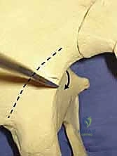
The foundation of an efficacious arthroscopic capsular shift lies in the precise establishment of surgical portals. Poorly placed portals will result in an unfavorable angle of approach, making the complex shuttling of capsular tissue exponentially more difficult. Prior to insufflation, all bony landmarks—the acromion, clavicle, and coracoid process—must be outlined on the skin. The standard posterior viewing portal is established approximately 3 centimeters distal and slightly medial to the posterolateral corner of the acromion. An anterosuperior lateral portal is placed high in the rotator interval, just anterior to the supraspinatus tendon, serving primarily for visualization when working in the posterior compartment. Finally, the critical anterior central (mid-anterior) working portal is established approximately 1 centimeter lateral to the coracoid process, entering the joint just superior to the subscapularis tendon. Large, 8.25-millimeter threaded cannulas are placed in the working portals to facilitate the smooth passage of complex suture shuttles and knot pushers.
Following a comprehensive diagnostic arthroscopy to confirm the voluminous capsular redundancy, meticulous tissue preparation must be undertaken. A capsular shift relies entirely on the biological healing of the plicated capsule to the labrum and glenoid rim; without adequate preparation, the plication will inevitably fail. An arthroscopic rasp or a motorized shaver (operated strictly without suction to avoid inadvertently resecting the intact, albeit hypoplastic, labrum) is introduced. The surgeon must systematically abrade the capsule and the labrum around the entire circumference targeted for plication, typically extending from the 9-o’clock position posteriorly, through the 6-o'clock position inferiorly, and up to the 3-o’clock position anteriorly. This aggressive decortication creates a bleeding, biologically active bed that stimulates a robust fibroblastic healing response, essential for long-term tissue adherence.
The core surgical technique involves the volumetric reduction of the capsule via the "pinch-tuck" method. Starting on the side of the shoulder demonstrating the most profound instability during the EUA, the surgeon utilizes a 45-degree spectrum suture hook or a commercial penetrating suture shuttle. Beginning at the critical 6-o'clock position, the surgeon grasps approximately 1 centimeter of the redundant inferior capsule. The needle is passed up through the capsule, effectively "pinching" the tissue, and then directed superiorly and medially to pass under and through the labrum. This maneuver "tucks" the redundant capsule superiorly, eliminating the dependent axillary pouch. When using nonabsorbable sutures, complex configurations such as mattress or figure-of-eight stitches are highly recommended to create a broad footprint of capsular compression and prevent tissue pull-through in inherently weak tissue.
As the plication advances superiorly, extreme caution must be exercised to protect the axillary nerve. Deep bites in the axillary pouch at the 6-o'clock position carry a high risk of iatrogenic nerve entrapment. The surgeon must ensure that the suture passing device does not penetrate too deeply or too far laterally from the labral footprint. The plication is systematically continued, shifting the capsule superiorly and securing it to the labrum, extending up to the 9-o’clock position posteriorly and the 3-o'clock position anteriorly. Once the inferior and anterior/posterior recesses are obliterated, the procedure concludes with the rotator interval closure. The anterior cannula is withdrawn just outside the joint capsule. Using a suture passing device, a No. 1 PDS suture is passed through the superior portion of the MGHL and retrieved just superior to the SGHL. Tying these sutures imbricates the interval, effectively completely the global volumetric reduction and restoring stability to the adducted arm.
Complications, Incidence Rates, and Salvage Management
Despite meticulous surgical technique, the arthroscopic capsular shift carries a distinct profile of potential complications, driven by the inherent biological deficiencies of the patient cohort and the technical demands of the procedure. Recurrent instability is the most frequently encountered complication, with incidence rates reported between 5% and 15% in long-term follow-up studies. Recurrence is typically multifactorial, stemming from a failure of biological healing at the capsulolabral interface, inadequate initial volumetric reduction, or progressive attenuation of the plicated tissue due to underlying collagenous defects (e.g., undiagnosed Ehlers-Danlos). When recurrent MDI presents, a thorough diagnostic workup including MRA is mandatory to assess capsular volume and tissue integrity. Salvage management often requires an open capsular shift to allow for more aggressive tissue imbrication, and in cases of profound tissue deficiency, augmentation with an Achilles or fascia lata allograft may be necessary to reconstruct the static restraints.
Iatrogenic stiffness and overconstraint represent the opposite end of the complication spectrum, occurring when the capsular volume is reduced excessively or when the rotator interval is closed too tightly. Patients typically present with a severe loss of external rotation and persistent pain due to altered glenohumeral kinematics, which forces the humeral head into non-anatomic contact with the glenoid during motion. The incidence of clinically significant overconstraint is approximately 2% to 5%. Initial management mandates aggressive, supervised physical therapy focusing on capsular stretching and mobilization. If stiffness remains refractory after 6 to 9 months of dedicated rehabilitation, salvage management involves an arthroscopic capsular release, carefully resecting the over-plicated tissue to restore physiological range of motion while taking care not to recreate the initial instability.
Neurologic injury, specifically to the axillary nerve, is a devastating but entirely preventable complication. The axillary nerve's intimate proximity to the inferior capsule at the 6-o'clock position makes it highly vulnerable during the initial stages of the pinch-tuck plication. Deep, blind bites into the axillary pouch can result in direct suture entrapment or transection of the nerve, leading to permanent deltoid paralysis and profound dysfunction. The incidence of axillary nerve injury is exceedingly rare (less than 1%) in the hands of experienced arthroscopists who adhere to strict anatomical landmarks. If a patient presents postoperatively with an isolated deltoid palsy, immediate electromyography (EMG) is indicated. If suture entrapment is confirmed, emergent arthroscopic or open exploration and neurolysis are mandated to salvage nerve function.
Historically, the pursuit of capsular shrinkage led to the widespread use of thermal capsulorrhaphy, utilizing radiofrequency or laser energy to denature collagen and shrink the redundant capsule. This practice has been universally condemned and abandoned due to catastrophic complication rates, specifically thermal chondrolysis and massive capsular necrosis. The thermal energy caused irreversible death of the chondrocytes, leading to rapid, end-stage glenohumeral osteoarthritis in young patients. The incidence of chondrolysis following thermal capsulorrhaphy was unacceptably high, and the salvage management for this devastating complication is exceptionally difficult, often requiring total shoulder arthroplasty in patients under the age of 30. Modern arthroscopic capsular shifts rely strictly on mechanical suture plication and biological healing, entirely avoiding thermal energy.
Complications and Salvage Management Summary
| Complication | Estimated Incidence | Prevention Strategy | Salvage Management |
|---|---|---|---|
| Recurrent Instability | 5% - 15% | Meticulous tissue preparation (rasping); adequate volumetric reduction; strict adherence to post-op rehab. | Open capsular shift; allograft capsular reconstruction for severe tissue deficiency. |
| Iatrogenic Stiffness / Overconstraint | 2% - 5% | Titrated plication based on EUA; avoiding excessive closure of the rotator interval; early passive ROM. | Aggressive physical therapy; arthroscopic capsular release if refractory >6 months. |
| Axillary Nerve Injury | < 1% | Avoiding deep, lateral bites at the 6-o'clock position; keeping the arm in slight ER/abduction during inferior suturing. | Emergent EMG; surgical exploration and neurolysis/repair if suture entrapment is identified. |
| Chondrolysis / Osteoarthritis | Rare (with modern techniques) | Absolute avoidance of thermal capsulorrhaphy; ensuring all suture knots are tied completely off the articular cartilage. | Joint preservation techniques (biologics, osteochondral allograft) or Total Shoulder Arthroplasty for end-stage destruction. |
Phased Post-Operative Rehabilitation Protocols
The ultimate success of an arthroscopic capsular shift is inextricably linked to the patient's strict adherence to a highly structured, phased postoperative rehabilitation protocol. The biological reality of the procedure dictates that the plicated capsular tissue requires significant time to heal to the abraded labrum and glenoid rim. Premature stress on these repairs will inevitably lead to suture pull-through or tissue attenuation, resulting in recurrent instability. Conversely, overly conservative immobilization can lead to debilitating adhesive capsulitis. The rehabilitation protocol must therefore delicately balance the protection of the surgical repair with the progressive restoration of glenohumeral kinematics.
The Immediate Postoperative Phase (Weeks 0 to 6) is characterized by strict immobilization and the protection of the healing biological milieu. In the operating room, the patient's arm is placed in a specialized abduction sling (e.g., Ultrasling) positioned in neutral rotation and slight abduction. This position removes tension from the newly plicated anterior and posterior capsule. The patient is instructed to wear the sling continuously, removing it only for hygiene and specific, approved exercises. During this phase, active range of motion (ROM) of the hand, wrist, and elbow is highly encouraged to prevent distal stiffness and promote venous return. Depending on the surgeon's intraoperative assessment of tissue quality, gentle, passive pendulum exercises may be initiated around week 3 or 4, but active glenohumeral motion is strictly prohibited.
The Intermediate Phase (Weeks 6 to 12) marks the transition from strict immobilization to the controlled restoration of motion. The sling is formally discontinued at the 6-week mark. The patient begins supervised physical therapy focusing initially on passive range of motion (PROM) in the scapular plane, gradually progressing to active-assisted range of motion (AAROM). The physical therapist must be acutely aware of the surgical vectors; aggressive stretching, particularly in extreme external rotation and abduction, is strictly avoided to prevent stretching out the newly plicated anterior and inferior capsule. Submaximal, pain-free isometric exercises for the rotator cuff and deltoid can be introduced late in this phase to begin reversing immobilization-induced atrophy.
The Advanced Strengthening Phase (Weeks 12 to 24) shifts the focus toward restoring dynamic stability and correcting the underlying muscular dyskinesia that often precipitates MDI. Isotonic strengthening of the rotator cuff and periscapular stabilizers (rhomboids, trapezius, serratus anterior) is initiated. A critical component of this phase is the incorporation of closed-kinetic-chain exercises and proprioceptive neuromuscular facilitation (PNF). Patients with MDI frequently exhibit profound deficits in joint proprioception; closed-chain exercises help retrain the mechanoreceptors within the joint capsule and musculotendinous units, ensuring that the dynamic stabilizers fire synchronously to maintain the humeral head centered within the glenoid during complex multi-planar movements.
The Return to Play and Heavy Labor Phase (Months 6 to 9+) requires the patient to meet strict functional criteria before discharge. Return to overhead sports, contact athletics, or heavy manual labor is typically restricted until a minimum of 6 months postoperatively. Clearance is contingent upon the patient demonstrating symmetric, pain-free full range of motion, normal scapulothoracic rhythm, and isokinetic strength testing that demonstrates at least 90% strength compared to the contralateral, asymptomatic shoulder. The surgeon and physical therapist must collaboratively assess the patient's dynamic joint stability and psychological readiness, ensuring that the patient can return to their daily activities and athletic pursuits without the debilitating apprehension of recurrent instability.
Summary of Landmark Literature and Clinical Guidelines
The evolution of surgical management for multidirectional instability is deeply rooted in several landmark publications that have shaped modern orthopedic paradigms. The foundational text in this domain is Charles Neer and C.R. Foster's 1980 publication in the Journal of Bone and Joint Surgery, which introduced the concept of the open inferior capsular shift. Neer recognized that MDI was a distinct clinical entity from unidirectional traumatic instability, requiring a volumetric reduction of the capsule rather than a simple Bankart repair. His open technique, which involved detaching the subscapularis to shift the capsule superiorly and laterally, remained the gold standard for over two decades and established the biomechanical principles of capsular volume reduction that we still rely upon today.
The transition to arthroscopic techniques was pioneered in the late 1990s and early 2000s by innovators such as Treacy, Savoie, and Duncan. Their early case series demonstrated that arthroscopic plication could achieve comparable stability to the open shift while significantly reducing surgical morbidity. A landmark biomechanical study by Provencher et al. quantified the volumetric reduction achieved by arthroscopic capsular plication, demonstrating that a standard "pinch-tuck" capsular shift effectively reduces glenohumeral volume by approximately 20% to 30%, directly correlating with restored clinical stability. Furthermore, Mazzocca and colleagues published critical biomechanical evaluations of various suture configurations, proving that mattress and figure-of-eight stitches provide superior resistance to tissue pull-through in attenuated capsular tissue compared to simple sutures.
The catastrophic failure of thermal capsulorrhaphy serves as a critical lesson in the history of MDI treatment. In the early 2000s, the use of radiofrequency probes to shrink the capsule gained immense popularity due to its technical ease. However, subsequent landmark studies, notably by D'Alessandro and colleagues, documented unacceptably high rates of recurrent instability, capsular necrosis, and devastating thermal chondrolysis. These publications led to a rapid and universal paradigm shift, resulting in current clinical guidelines that unequivocally condemn the use of thermal energy in the glenohumeral joint for the treatment of instability.
Current consensus guidelines from the American Academy of Orthopaedic Surgeons (AAOS) and the Arthroscopy Association of North America (AANA) firmly establish the arthroscopic capsular shift using mechanical suture plication as the modern gold standard for surgical intervention in MDI. These guidelines emphasize the absolute necessity of exhausting a minimum of 6 months of targeted physical therapy prior to surgical consideration. Furthermore, modern literature underscores the critical importance of addressing the rotator interval concurrently with the inferior capsular shift to prevent persistent inferior subluxation in the adducted arm, cementing a holistic, multi-vector approach to restoring glenohumeral stability.
