Advanced Shoulder Arthroscopy: Techniques, Anatomy, & Clinical Applications
Key Takeaway
Shoulder arthroscopy is a minimally invasive procedure addressing diverse shoulder pathologies. Indications span rotator cuff tears, glenohumeral instability (e.g., labral tears), symptomatic biceps tendinopathy, articular cartilage lesions, adhesive capsulitis, and AC joint conditions. It offers reduced morbidity and faster recovery for patients refractory to conservative management.
Introduction and Epidemiology
Shoulder arthroscopy has revolutionized the diagnosis and treatment of a vast array of glenohumeral and periarticular pathologies. Evolving from a purely diagnostic tool in the mid-20th century, it has matured into a sophisticated platform for complex reconstructive procedures, offering reduced morbidity, faster recovery, and improved cosmetic outcomes compared to traditional open approaches. The minimally invasive nature, coupled with advancements in instrumentation, imaging, and surgical techniques, has propelled arthroscopy to the forefront of orthopedic shoulder surgery.

Epidemiologically, shoulder pain is a common musculoskeletal complaint, with a significant burden on healthcare systems. Rotator cuff pathology, glenohumeral instability, and impingement syndromes represent a large proportion of cases, with incidence increasing with age and activity levels. For instance, rotator cuff tears are prevalent in up to 30% of individuals over 60 years old, with arthroscopic repair rates steadily climbing. Similarly, shoulder instability, particularly in young, active populations, frequently necessitates arthroscopic stabilization. The understanding of intricate shoulder anatomy and biomechanics, combined with precise arthroscopic skills, is paramount for optimizing patient outcomes in this expanding surgical domain.
The transition from open to all-arthroscopic techniques for procedures such as rotator cuff repair and Bankart stabilization required a significant evolution in surgeon technical proficiency and implant design. The development of knotless suture anchors, high-tensile strength suture materials, and advanced fluid management systems has allowed for reproducible, biomechanically robust repairs that rival or exceed historical open gold standards. Furthermore, the ability to visualize the joint comprehensively without extensive soft tissue dissection allows for the identification and concurrent management of concomitant pathologies, such as superior labrum anterior and posterior (SLAP) tears or long head of the biceps tendon disorders, which may have been missed during focused open approaches.
Surgical Anatomy and Biomechanics
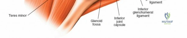
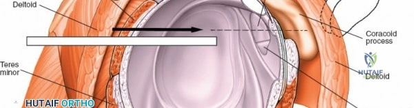
A thorough understanding of shoulder anatomy and biomechanics is indispensable for safe and effective arthroscopic intervention. The shoulder complex comprises four joints: the glenohumeral, acromioclavicular (AC), sternoclavicular, and scapulothoracic articulations. The glenohumeral joint, a ball-and-socket synovial joint, offers the greatest range of motion in the body, a characteristic facilitated by its inherent bony incongruence. Stability is primarily provided by dynamic stabilizers (rotator cuff muscles: supraspinatus, infraspinatus, teres minor, subscapularis) and static stabilizers (glenohumeral ligaments, labrum, joint capsule, negative intra-articular pressure).
Key anatomical structures visualized arthroscopically include:
* Glenohumeral Joint:
* Articular Cartilage: Covering the humeral head and glenoid, susceptible to chondral lesions.
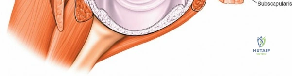
* Glenoid Labrum: A fibrocartilaginous rim that deepens the glenoid fossa, enhancing stability. Lesions (e.g., Bankart, SLAP) are common in instability and trauma.
* Glenohumeral Ligaments (Superior, Middle, Inferior): Capsular thickenings vital for static stability, particularly the inferior glenohumeral ligament complex with its anterior and posterior bands.
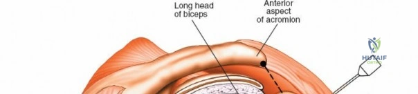
* Biceps Tendon (Long Head): Originates from the superior labrum (supraglenoid tubercle) and traverses the joint. Pathologies include instability, tenosynovitis, and tears.
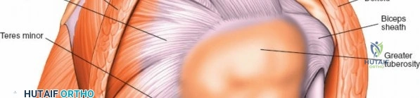
* Rotator Cuff Tendons: The articular surfaces of the supraspinatus, infraspinatus, and subscapularis are visible from within the joint, allowing assessment of partial articular-sided tears.
- Subacromial Space:
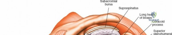
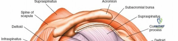
- Rotator Cuff (Bursal Surface): The supraspinatus, infraspinatus, and subscapularis tendons. Full-thickness tears and bursal-sided partial tears are observed here.
- Subacromial Bursa: A synovial lining that reduces friction between the rotator cuff and the overlying acromion/deltoid. Often inflamed (bursitis) or scarred in impingement syndromes.
- Acromion: The undersurface morphology (Type I, II, III) and osteophytes are crucial in subacromial impingement.
- Coracoacromial Ligament (CAL): Forms part of the coracoacromial arch.
- Acromioclavicular (AC) Joint: The undersurface of the distal clavicle and acromion are visible, allowing assessment of arthritis or osteophyte formation.
Biomechanically, the deltoid and rotator cuff act as a force couple. The rotator cuff depresses and compresses the humeral head into the glenoid fossa during arm elevation, preventing superior migration and subacromial impingement. This mechanism is known as concavity compression. When a rotator cuff tear occurs, particularly involving the supraspinatus and infraspinatus, this force couple is disrupted, leading to superior translation of the humeral head, altered kinematics, and progressive joint dysfunction.
Neurovascular anatomy is critical for safe portal placement and soft tissue release. The axillary nerve exits the quadrangular space and courses along the deep surface of the deltoid. It is typically located approximately 5 cm distal to the lateral border of the acromion, making standard lateral portals safe if kept superior to this zone. The suprascapular nerve traverses the suprascapular notch (innervating the supraspinatus) and the spinoglenoid notch (innervating the infraspinatus). Excessive medial dissection during rotator cuff mobilization or improper anchor placement at the superior glenoid neck can jeopardize this nerve.
Indications and Contraindications
Arthroscopic intervention is indicated for a wide spectrum of shoulder pathologies that have failed conservative management or present with acute, structurally significant lesions necessitating early surgical repair. The decision to proceed with arthroscopy must be based on a comprehensive clinical evaluation, advanced imaging, and a thorough understanding of the patient's functional demands.
Absolute indications include acute, traumatic full-thickness rotator cuff tears in active individuals, recurrent anterior glenohumeral instability with structural capsulolabral failure, and septic arthritis of the glenohumeral joint. Relative indications encompass chronic rotator cuff tears failing physical therapy, symptomatic SLAP lesions, refractory adhesive capsulitis, and calcific tendinitis unresponsive to corticosteroid injections and extracorporeal shockwave therapy.
Contraindications must be rigorously respected to prevent catastrophic outcomes. Absolute contraindications include active systemic or local soft tissue infection (unless the procedure is a therapeutic washout), severe medical comorbidities precluding anesthesia, and advanced glenohumeral osteoarthritis where arthroplasty is the definitive treatment of choice. Relative contraindications include massive, irreparable rotator cuff tears with advanced fatty infiltration (Goutallier stage 3 or 4) and severe proximal humeral migration, though partial repairs or superior capsular reconstruction may be considered in highly selected cases.
| Pathology Category | Operative Indications (Arthroscopy) | Non-Operative Indications |
|---|---|---|
| Rotator Cuff Tears | Acute full-thickness tears; Chronic tears failing >3-6 months conservative care; Significant weakness. | Asymptomatic tears; Partial-thickness tears <50%; Advanced fatty infiltration (Goutallier 3/4) with pseudoparalysis. |
| Glenohumeral Instability | Recurrent dislocations; First-time dislocation in high-risk (young, contact sport) athletes; Bony Bankart >15-20%. | First-time dislocation in older/low-demand patients; Voluntary multi-directional instability (MDI) without rehab trial. |
| Biceps Pathology | Type II/IV SLAP tears in young athletes; Refractory biceps tenosynovitis/subluxation; Pulley lesions. | Degenerative SLAP tears in older adults; Asymptomatic SLAP variants; Early tenosynovitis responding to injection. |
| Adhesive Capsulitis | Refractory stiffness >6-9 months despite aggressive physical therapy and intra-articular injections. | Freezing phase (highly inflammatory); Poor compliance with physical therapy protocols. |
| Acromioclavicular Joint | Refractory symptomatic AC joint osteoarthritis (distal clavicle excision); Acute high-grade AC separations (selected). | Asymptomatic radiographic AC arthritis; Low-grade (Type I/II) AC separations. |
Pre Operative Planning and Patient Positioning
Meticulous preoperative planning is the foundation of a successful arthroscopic shoulder procedure. High-resolution magnetic resonance imaging (MRI) without contrast is the standard modality for evaluating the rotator cuff, while MR arthrography provides superior sensitivity for labral pathology and capsular avulsions. Computed tomography (CT), particularly with 3D reconstruction and en face glenoid views, is essential for quantifying glenoid bone loss in cases of recurrent instability, guiding the choice between arthroscopic soft tissue repair and open bony augmentation (e.g., Latarjet procedure).
Anesthetic management typically involves a combination of general anesthesia and regional blockade. An interscalene nerve block provides excellent intraoperative muscle relaxation and significantly reduces postoperative opioid consumption. Controlled hypotensive anesthesia (maintaining mean arterial pressure between 60-70 mmHg) is frequently employed to optimize intra-articular visualization by minimizing capillary bleeding, provided there are no cerebrovascular or cardiovascular contraindications.

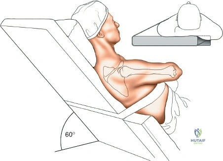
Patient positioning is a critical decision, with two primary options: the lateral decubitus position and the beach chair position.
Lateral Decubitus Position
The patient is placed on their non-operative side with the torso secured via a beanbag or rigid positioners. The operative arm is placed in balanced suspension with 10 to 15 pounds of traction, typically abducted to 45 degrees and forward flexed to 15-20 degrees.
* Advantages: Excellent joint distraction maximizing visualization of the inferior recess and glenoid neck; lower risk of cerebral hypoperfusion; continuous traction frees the surgical assistant.
* Disadvantages: Orientation is non-anatomic, requiring spatial adaptation; risk of neurapraxia to the brachial plexus if traction is excessive or prolonged; airway access is restricted for the anesthesia team.
Beach Chair Position
The patient is seated at an angle of 45 to 70 degrees, with the operative arm completely free for dynamic manipulation throughout the procedure.
* Advantages: Anatomic orientation of the shoulder girdle; excellent for subacromial work and rotator cuff repair; easy conversion to an open approach if necessary; unencumbered airway access.
* Disadvantages: Risk of cerebral ischemia (rare but catastrophic) due to the hydrostatic gradient between the heart and brain; requires an assistant to hold and manipulate the arm; limited visualization of the posteroinferior joint space.

Detailed Surgical Approach and Technique
The execution of advanced shoulder arthroscopy relies on precise portal placement, systematic diagnostic evaluation, and meticulous tissue handling. Fluid management is maintained via an arthroscopic pump system, typically set between 30 and 50 mmHg, with the ability to temporarily increase pressure for hemostasis.

Arthroscopic Portal Placement
Accurate portal placement dictates the trajectory of instruments and the ease of anchor insertion.
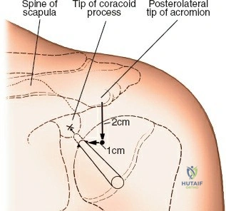
* Standard Posterior Portal: The primary viewing portal, established 2 cm inferior and 1-2 cm medial to the posterolateral corner of the acromion. It targets the "soft spot" between the infraspinatus and teres minor.
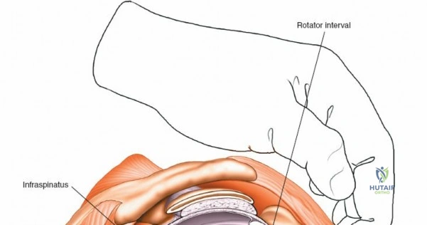
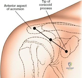
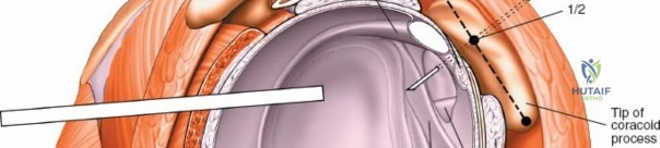
* Anterior Portals: Established under direct intra-articular visualization using an outside-in spinal needle technique. The anterosuperior portal is placed high in the rotator interval, just anterior to the biceps tendon, useful for viewing and fluid management. The anteroinferior portal is placed just superior to the subscapularis tendon, serving as the primary working portal for anterior labral repairs.
* Lateral Portal: Used primarily for subacromial viewing and working during rotator cuff repairs. Placed 2-3 cm lateral to the lateral edge of the acromion, bisecting the anterior and posterior borders.
* Accessory Portals: The Neviaser portal (suprascapular) is utilized for superior SLAP repairs. The Port of Wilmington (posterolateral) provides an orthogonal approach for inserting anchors into the posterosuperior glenoid. The 5 o'clock portal allows low anterior anchor placement for Bankart repairs.
Diagnostic Arthroscopy Sequence
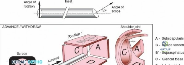
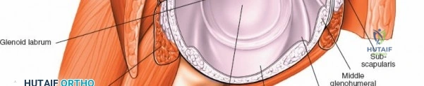
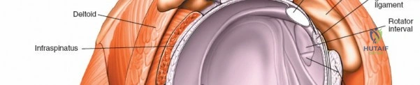
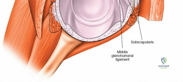
A systematic 15-point diagnostic tour ensures no concomitant pathology is overlooked. The sequence begins in the posterior recess, evaluating the articular surfaces of the humeral head and glenoid. The camera is advanced to inspect the superior labrum and biceps anchor. The long head of the biceps is pulled into the joint with a probe to assess for hidden tears or erythema. The rotator interval, including the superior glenohumeral ligament and coracohumeral ligament, is evaluated. The subscapularis tendon insertion is probed. The camera is then swept down the anterior labrum, visualizing the middle and inferior glenohumeral ligaments, and into the axillary pouch to assess for loose bodies or capsular avulsions (HAGL lesions). Finally, the articular surface of the supraspinatus and infraspinatus is inspected for partial articular-sided tendon avulsions (PASTA lesions).
Rotator Cuff Repair Principles
Following the intra-articular evaluation, the arthroscope is redirected into the subacromial space. A thorough bursectomy is performed to define the bursal anatomy and visualize the rotator cuff footprint. If indicated, an acromioplasty is performed to flatten a curved or hooked acromion (Type II/III).
Tear pattern recognition is critical. Crescent-shaped tears can often be repaired directly to bone. U-shaped or L-shaped tears require margin convergence sutures to close the longitudinal split before footprint fixation. Massive, contracted tears necessitate extensive soft tissue mobilization, including intra-articular capsular release, coracohumeral ligament release, and mobilization of the suprascapular nerve.
Footprint preparation involves lightly decorticating the greater tuberosity to expose a bleeding cancellous bone bed, optimizing the biological environment for tendon-to-bone healing. Fixation constructs have evolved significantly. While single-row repairs may be sufficient for small tears, double-row transosseous-equivalent (TOE) constructs are biomechanically superior for medium to large tears. TOE techniques utilize medial row anchors with sutures passed through the tendon, which are then crisscrossed over the bursal surface and secured with lateral row knotless anchors. This maximizes footprint contact area and contact pressure, theoretically enhancing healing rates.
Instability Repair Techniques
For anterior instability with a Bankart lesion, the anteroinferior labrum and capsule must be mobilized from the glenoid neck. An elevator is used to release the tissue until the subscapularis muscle belly is visible, ensuring adequate capsular shift. The glenoid rim is prepared with a burr or rasp.
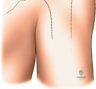
Suture anchors are placed on the articular margin of the glenoid. For a standard anterior repair, anchors are typically placed at the 5:30, 4:00, and 3:00 o'clock positions (right shoulder). Sutures are passed through the capsulolabral complex using a suture shuttling device, ensuring a healthy bite of capsule to achieve a superior and medial shift. The knots are tied, re-establishing the labral bumper and tensioning the inferior glenohumeral ligament. In cases with an engaging Hill-Sachs lesion but subcritical glenoid bone loss, a Remplissage procedure is performed concurrently, tenodesing the infraspinatus and posterior capsule into the humeral defect to prevent engagement over the anterior glenoid rim.
Complications and Management
While shoulder arthroscopy is generally safe, complications can occur, ranging from minor transient issues to severe, joint-threatening conditions. Proactive mitigation strategies and prompt recognition are essential for optimizing patient outcomes.
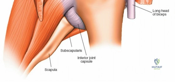
Neurological injury, though rare, is a devastating complication. The axillary nerve is at risk during inferior capsular release or if lateral portals are placed too distally. The musculocutaneous nerve can be injured during anterior portal placement or coracoid preparation. Most nerve injuries are traction neurapraxias that resolve spontaneously, but direct lacerations require microsurgical exploration and repair.
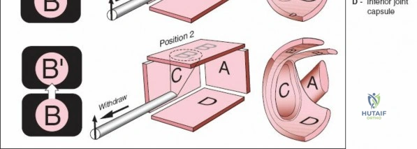
Fluid extravasation is ubiquitous in shoulder arthroscopy but can become clinically significant, leading to severe soft tissue swelling, airway compromise (especially in the lateral decubitus position),
Clinical & Radiographic Imaging